Improving protocols and procedures for strengthened radiation protection in interventional procedures
|
|
|
- Lesley Hamilton
- 5 years ago
- Views:
Transcription
1 Improving protocols and procedures for strengthened radiation protection in interventional procedures R. Loose German Commission on Radiological Protection (SSK) German Roentgen Society (DRG) Institute for Diagnostic and Interventional Radiology Nuremberg North

2 Frequency and collective dose of medical examinations in Germany 2009 Mammography one side Angiography/ Interventions Frequency others Collective effective dose others Dental Skeleton Gastro- / Urology Gastro-/ Urology Dental Mammography Skeleton Angiography/ Interventions Report to the German parliament 2010 on medical exposure


3 Fluoroscopic interventions in radiology and cardiology are the two most frequent procedures involving a significant radiation exposure of patients as well as an occupational exposure of the staff. Frequency 1. Cardiology 2. Radiology 3. Others Vascular surgery Gastroenterology Urology Other applications in operating rooms Critical pulmonary embolism Emergencyinterventions: The patient does not ask for the dose, but what about the staff?
4 Technical minimum requirements for all interventional fluoroscopy systems: Pulsed fluoroscopy Last image hold/run system Automatic exposure control (AEC) Selectable dose and/or image quality for fluoroscopy and angiography mode Removable grid Additional copper filter (children) Dose-area product meter (DAP/KAP) and fluoroscopy time C-arm system with under table x-ray tube (for monoplane system or first biplane tube) Basic protective shielding Contrast agent injector


5
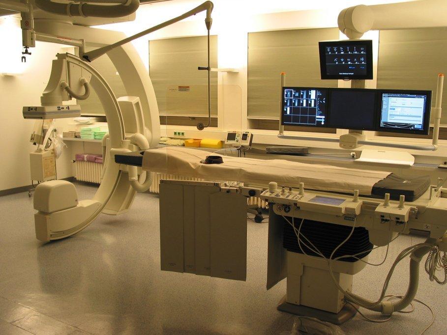
6 State of the art technique and protocols for interventional fluoroscopy systems: Display of all exposure parameters including thresholds for skin entrance dose Automatic contrast agent injector with programmable flow protocols DICOM store of exposure parameters (fluoroscopy and every single series) Flat panel detector Simulation of table movement, collimation and wedges without radiation Roadmapping, DSA overlay, store of fluoro loops Second monitor for reference images Third monitor for images of other modalities (CT, MRI, US, CBCT,.) Seamless protective under table shielding Additional over table shielding to reduce stray radiation from the patient Rotational angiography and/or cone beam CT (CBCT) for 3D visualisation Store of last image hold (LIH) instead of DSA series

7

8 Optimizing of run length in angio- and DSA-mode


9 Optimizing the frame rate and pulse in DSA mode and pulsed fluoroscopy LIH New SOP: dose reduction of 81%

10 Virtual insert of wedges

11 Virtual table motion


12 Tip of guide wire Roadmap


13 3D-angio / cone beam CT A.Dörfler, Erlange
14 Current and future developments:

15 2D/3D registration of CT/CBCT and DSA for planning, navigation and control of interventional procedures


 from 27")
16 Robot-systems: eccentric rotation with increased field of view (FOV) from 27 to 48 cm

17 Navigation (steering of catheters) with high magnetic fields in cardiology (no staff exposure)
 with low magnetic fields (reduced staff")
18 X-ray volume Magnetic volume Electromagnetic Navigation B.C. Meyer et.al. Radiologe 2009; 49: Navigation (tracking of catheters) with low magnetic fields (reduced staff and patient exposure) Fusion
19 New dynamic flat panel detectors (higher quantum efficiency) New dose tracking systems for skin entrance dose, erythema threshold and overlapping or new skin entrance fields
20 Radiation protection of staff (additional to general system requirements) Personal protective devices Good dosimetry


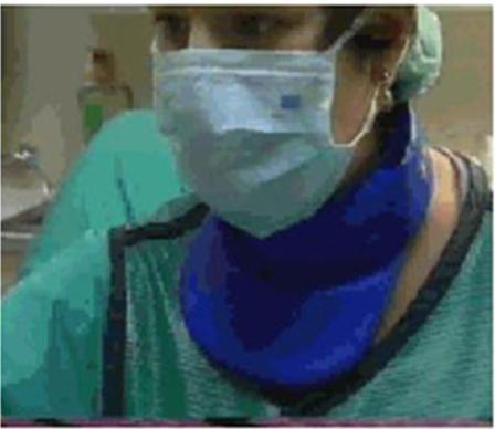

21 Thyroid collar 4x 0.5 mm lead equivalent ICRP: 20 msv/a for the eye lens!!!


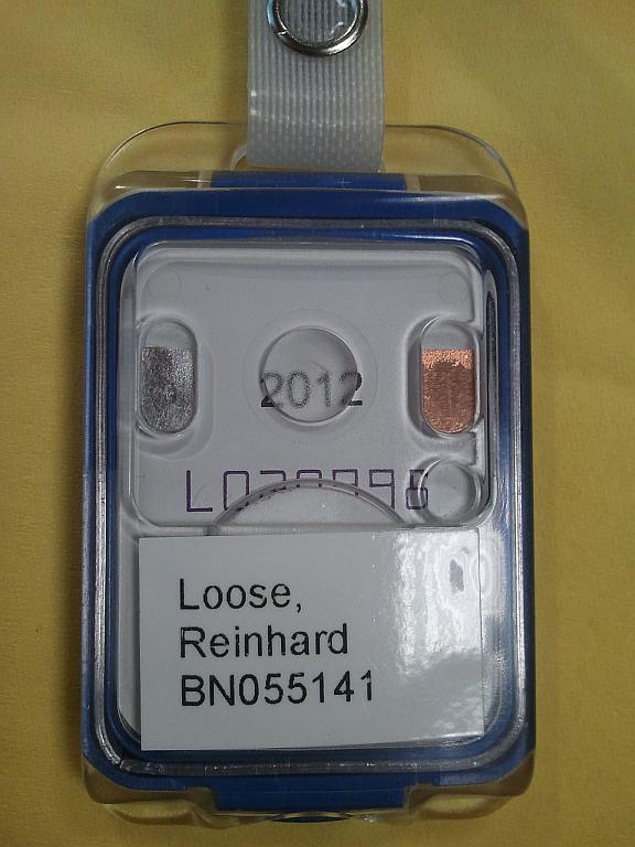
22 Standard dosemeters (film based)


23 Electronic dosemeters LCD-Display Battery Clip 1. August 2011!! Controlbutton Signal tone Beta-window THERMO FISHER SCIENTIFIC EPD Mk2


24 Electronic dosemeters with small flexible probe


25 Individual dosimetry when starting new procedures or changing procedures


26 FDG-PET Before SIRT After SIRT Patient with liver metastase after colon cancer FDG-PET before and 3 month after SIRT Y-90 Therapy with radioisotopes (e.g. Y-90 SIRT) Close cooperation with nuclear medicine


27 Different positions of TLD dosemeters
28 Radiation protection of patient and staff Justification (guidelines, referral criteria) Patient: informed consent and cooperation. training, training, training.
29 Training vs. fluoroscopy time Durchleuchtungszeit Fluoroscopy time [minutes] 10 Coronarangiographie PTCA 8 7, ,3 2,5 3,9 0 <75 Coro, <50 PTCA >300 Coro, >200 PTCA Erfahrung Procedures in Maßnahmen/Jahr per year Gleichmann 1993

30 Complications of arterial recanalization DGIR Report 2011 Every green or red bar indicates one institution Fortschr Röntgenstr 2012; 184:
31 Interventional Procedures Avoiding Radiation Injuries Information abstracted from ICRP Publication 85 Available at Task Group: J. Cardella, K. Faulkner, J. Hopewell, H. Nakamura, M. Rehani, M. Rosenstein, C. Sharp, T. Shope, E. Vano, B. Worgul, M. Wucherer
32 Avoiding Radiation Injuries Informed consent and records Patients are entitled to know the risks of radiation injury if likely to be high A written record should be kept if skin doses are estimated to be >3 Gy (1 Gy for repeated procedures) Not all skin reactions are due to radiation; e.g. contrast medium allergy Information abstracted from ICRP Publication 85
33 Follow-up Radiation skin injury may present late and the association not considered if no documentation All patients with estimated skin doses of 3 Gy should be followed up days after exposure A system to identify repeat procedures should be set up

34


35 The EC has announced the MARTIR CDROM in its WEB site allowing to download the full content:
36