SUPPLEMENTARY INFORMATION
|
|
|
- Janis Lucas
- 5 years ago
- Views:
Transcription

 c Testing the")

 LoxN OFP Expected Illegitimate b Test in vitro")


1 doi: /nature06293 SUPPLEMENTARY INFORMATION a Testing the incompatibility of Lox2272 and LoxP Construct Expected Lox2272 promoter (CMV) c Testing the incompatibility of LoxN with Lox2272 and LoxP Construct Illegitimate LoxN LoxP Lox2272 Outcome RFP Expected Illegitimate 1 Illegitimate 2 RFP Promoter (CMV) LoxN OFP Expected Illegitimate b Test in vitro no Cre M- LoxP OFP Cre mediated recombination Cre mediated recombination Outcomes Lox2272 Outcome M-RFP Outcomes Expected Outcome M-RFP M- M-RFP Illegitimate 1 Outcome M- Illegitimate 2 Outcome M- d Test in vitro no Cre + Cre RFP + Cre OFP M-RFP M- Supplementary Figure 1 Mutual incompatibility of Lox variants. a, A control construct was designed to detect promiscuous recombination of Lox2272 with the canonical LoxP sites. In this construct, recombination between the two Lox2272 sites triggers expression, while illegitimate recombination between LoxP and Lox2272 would switch on. b, In HEK cell stably expressing this control construct, RFP was expressed in absence of recombination. Cre recombination between the two Lox2272 led to expression. expression, which detects illegitimate recombination with the LoxP site, was not observed, indicating that Lox2272 is incompatible with LoxP. c, A novel Lox variant, LoxN, was designed. Incompatibility of LoxN with both Lox2272 and LoxP was simultaneously tested. In the control construct, recombination between the two Lox variants triggers M-RFP expression, while illegitimate recombination between LoxN and one of the other Lox sites would switch on M- or expression. d, In HEK cells stably expressing this construct, OFP is expressed in absence of recombination. Cre recombination induces recombination between the two LoxN variants, leading to M-RFP expression. M- or expression, which detects illegitimate recombination with the LoxP site, was not observed, indicating incompatibility of LoxN with both Lox2272 and LoxP. Scale bars: 50 µm. 1


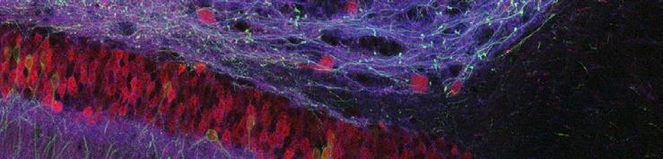
2 SUPPLEMENTARY INFORMATION doi: /nature06293 a b Cortex c Hippocampus d CA1 e Motor axon terminals Supplementary Figure 2 Combinatorial XFP expression in Brainbow mice. Additional examples of combinatorial expression in Thy1-Brainbow animals. Recombination was induced with CreERT2 and perinatal Tamoxifen injection. a-d, Cortex and hippocampus of Thy1-Brainbow-1.0 line L. b and d shows higher magnification of boxed regions in a and c. e, Motor axon terminals in a skeletal muscle of Thy1-Brainbow-1.0 line H. Scale bars a-d, 50 μm; e, 10 μm. 2


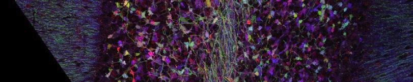


3 SUPPLEMENTARY INFORMATION doi: /nature06293 Hippocampus Superior colliculus Cerebellum a2 a3 b1 b2 b3 c1 c2 c3 d1 d2 d3 Chx10-Cre Islet1 Cre CAGGS-CreER + Tamoxifen no recombination a1 Supplementary Figure 3. Restriction of Brainbow-1 expression with specific Cre drivers Comparison of XFP expression in Thy1-Brainbow-1.0 mice line H under different recombination conditions a, In absence of recombination, RFP is visible in granule cells of the dentate gyrus (a1), scattered neurons in the superior colliculus (a2) and in the inner granular layer of the cerebellum (a3). No leaky expression is detected for the other XFP genes present in the Brainbow-1.0 construct (M- or ). b, Induction of recombination using CAGGS- CreERT2 mice leads to widespread expression of M- and in Tamoxifen injected animals. The membrane-tethered XFPs are primarily detected in neuronal processes, such as hippocampal mossy fibers (b1), axons arborizing in the colliculus (b2), parallel fibers and mossy fibers in the molecular and internal granule cell layer of the cerebellum (b3). c, Crossing Thy1-Brainbow-1.0 mice with Islet1Cre animals gives rise to a narrower pattern of recombination. In the dentate gyrus, M- and expression is restricted to scattered neurons (c1). Many axons aborizing in the superior colliculus show recombination (c2), as well as components of the cerebellar circuitry (c3). d, With Chx10-Cre driver mice, no or very little recombination is detected in the hippocampus and cerebellum (d1, d3). and M- are almost exclusively expressed in retinal ganglion axons, which arborize in the superior colliculus (d2). Scale bars: 100 μm. 3
4 doi: /nature06293 SUPPLEMENTARY INFORMATION Supplementary Figure 4 Use of color to resolve tracing ambiguities. In the dataset used for reconstruction of cerebellar circuitry (Fig. 5b), axon tracing throughout the volume was compared using one, two or three of the available color channels. The tracing power of the dataset was quantified by counting the number of ambiguities along the length of the axon. (Proximity to another axon of the same color caused ambiguity.) Increasing the number of color channels used for tracing increased color information and reduced the number of ambiguities. 4

5 doi: /nature06293 SUPPLEMENTARY INFORMATION Supplementary Figure 5. Color profiles of individual mossy fiber rosettes. a, Distribution in RGB space of individual pixels from four separate axons. The centroid of each cluster is represented as a large dot. b, Actual images from mossy fiber rosettes along each of the four axons represented in a. c, Axonal colors are represented in hue-saturation coordinates (binned). Each horizontal bar represents the centroid for a given axon s color. 341 axons are shown in total. 5

6 doi: /nature06293 SUPPLEMENTARY INFORMATION Supplementary Figure 6. Color consistency between axons and dendrites of a given neuron. a, Hippocampal granule neuron in dentate gyrus from Brainbow-1.0 line L. b, Cerebellar Purkinje cell from Brainbow-1.0 line J. Sampled regions from dendrite (triangle) and axon (circle) are indicated in a and b. c, d, Distributions in RGB space of individual pixels from sampled regions above. Dendritic and axonal distributions are superimposed. The centroid of each cluster is represented as a large triangle (dendrite) or circle (axon). 6
7 doi: /nature06293 SUPPLEMENTARY INFORMATION Supplementary Table 1. Incompatible Lox variants used to generate Brainbow-1 constructs Lox variant LoxP Lox2272 LoxN Sequence ATAACTTCGTATA GCATACAT TATACGAAGTTAT ATAACTTCGTATA GGATACTT TATACGAAGTTAT ATAACTTCGTATA GTATACCT TATACGAAGTTAT The 8-bp spacer which directs the specificity and the directionality of the recombination 12 is indicated in bold characters. Changes from the original LoxP sequence are underlined. Lox , which bears two changes in position 2 and 7 of the spacer, was confirmed to be incompatible with LoxP while efficiently mediating recombination with sites of identical sequences (Fig. 1a; Supplementary Fig. 1a). A new variant, LoxN, was designed based on the Lox2272 model, with different changes in position 2 and 7. This new variant was found to be incompatible with both LoxP and Lox2272 (Supplementary Fig.1b), while retaining comparable recombination efficiency (Fig. 1d, e). Sequences are displayed in the orientation used in the constructs, which avoids introducing start codons. 7
8 doi: /nature06293 SUPPLEMENTARY INFORMATION Supplementary Table 2. Thy1-Brainbow transgenic mouse lines # line XFPs subset size 1st 2nd 3rd 4 th basal after Cre type of expression cell types labeled Brainbow-1.0 RFP M- A dsred2 M-E M-mCerulean small medium combinatorial peripheral and central neurons B dsred2 M-E M-mCerulean large large exclusive non-myelinating Schwann cells (Schwann cells associated with neuromuscular junctions, autonomic ganglia and non-myelinated peripheral axons); irregular scattered motor neurons. C dsred2 M-E M-mCerulean small small combinatorial peripheral and central neurons D dsred2 M-E M-mCerulean small medium exclusive peripheral and central neurons E dsred2 M-E M-mCerulean small medium exclusive peripheral and central neurons F dsred2 M-E M-mCerulean large large combinatorial peripheral and central neurons G dsred2 M-E M-mCerulean large large combinatorial Bergmann glia of the molecular layer of the cerebellum; scattered neurons (few) G (Flp) dsred2 M-E M-mCerulean large large exclusive ibid. H tdimer2 M-E M-mCerulean large large combinatorial peripheral and central neurons including peripheral sensory neurons, cranial and spinal motor neurons (~75%), retinal ganglion cells, dentate gyrus granule cells, pyramidal neurons of CA1 and some cortical areas, inferior olive neurons and associated mossy fibers (~20%). H (Flp) tdimer2 M-E M-mCerulean large large exclusive ibid. I tdimer2 M-E M-mCerulean large large combinatorial peripheral and central neurons Brainbow-1.0 RFP CFP J dtomato mcerulean E large large combinatorial peripheral and central neurons K dtomato mcerulean E large large combinatorial peripheral and central neurons L dtomato mcerulean E large large combinatorial peripheral and central neurons (same types labeled as H; a few cerebellar Purkinje neurons) Brainbow-1.1 OFP M-RFP M- M Kusabira M-mCherry M-E M-mCerulean undetected large exclusive astrocytes of all areas of the brain and spinal cord; dentate gyrus granule cells; scattered neurons (few). Brainbow-2.0 RFP N tdimer2 M-ECFP large large combinatorial peripheral and central neurons O tdimer2 M-ECFP small small combinatorial peripheral and central neurons Brainbow-2.1 nuc-gfp RFP P hrgfpii-nls E tdimer2 M-mCerulean small small too sparse peripheral and central neurons Q hrgfpii-nls E tdimer2 M-mCerulean small medium combinatorial some neurons, astrocytes R hrgfpii-nls E tdimer2 M-mCerulean medium large combinatorial peripheral and central neurons (same types labeled as H; a few cerebellar Purkinje neurons) S hrgfpii-nls E tdimer2 M-mCerulean medium large combinatorial peripheral and central neurons A total of 25 Thy1-Brainbow transgenic mouse lines were generated. 9 lines not described here had either no detectable XFP expression, or did not transmit the Brainbow transgene to their progeny. Two lines (H and I) were initially derived from the same founder and were later segregated based on their distinct expression patterns. G (Flp) and H (Flp) are lines with reduced transgene copy number derived by crossing lines G and H with βactin-flp e mice (Fig. 4d). In line H, percentage of labeled cell types were estimated by counting the number of postsynaptic sites occupied by a labeled presynaptic axon. Recommended lines for the study of particular cell types are indicated in bold characters. M: membrane targeting signal; mxfp: monomeric variant of XFP; NLS: nuclear localization signal. 8