Modeling cytoskeleton self-assembly
|
|
|
- Allan McDowell
- 5 years ago
- Views:
Transcription
1 Modeling cytoskeleton self-assembly Dimitrios Vavylonis Department of Physics, Lehigh University BIOS 10 Lehigh University Oct 30, 2009


2 Cell organization How does the cell achieve internal organization? How is it regulated? Can we model cell structure and dynamics?
3 Thermal and viscous forces are very large on the scale of proteins v protein 9 m/ sec t = 0 t = 3 psec = sec in this time the protein travels nm << protein size ~ 4 nm The result of this is diffusion
 with equal probability at each step (a Monte Carlo method)")
4 Modeling diffusion: random walk on a lattice computer picks a random direction (up/down/left/right) with equal probability at each step (a Monte Carlo method) drunkard s walk

5 Brownian motion, continued x(t) 0 steps 100 steps 1000 steps x(t) = constant (D t) 1/2 Diffusion coefficient D for a protein in the cell ~ 4 m 2 /sec 5 m t = 0 fraction of a sec several sec

")
6 Polymerization of proteins to filaments actin monomers D ~D/2 ~D/3 actin filament + cross-links rigid, stationary cytoskeleton Kovar, Harris, Mahaffy, Higgs, Pollard Cell, (2006) Alberts et al, MBOC


7 Microtubule filaments and cell organization Alberts et al, MBOC
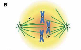
8 Search and capture of chromosomes by microtubules during mitosis dynamic instability dividing cell

9 GTP-cap model of dynamic instability GTP-tubulin cap GDP-tubulin Alberts et al, MBOC
10 GTP-cap toy model k+ c hydrolysis at blue/red interface, rate k hydro GTP-tubulin GDP-tubulin GTP-tubulin monomers diffuse in the cytoplasm at concentration c k - k rescue microtubule filament k cat Gillespie algorithm (a Monte Carlo method) 1. pick an event at random according to rate constants (polymerization, depolymerization, hydrolysis) 2. update configuration and time
11 Numerical simulations of the model large GTP-tubulin monomer concentrations small GTP-tubulin monomer concentrations example of length trajectory at small concentrations: subunits Time (sec)
 0 c* catastrophe and rescue region monomer")
12 Insights from simple model other trajectories average elongation rate subunits time (sec) 0 c* catastrophe and rescue region monomer concentration


13 Models of chromosome search and capture


14 Actin filaments and cell shape changes motile fibroblast (GFP-actin) Watanabe lab, Kyoto Univ. budding yeast Amberg Mol. Biol. Cell (1998) actin patches actin cables Neuron growth Forscher lab, Yale Univ. actin fission yeast Wu lab, Ohio State Univ. 2007

15

 ~5 m Watanabe and Mitchison, Science,")
16 Actin polymerization: driving force for cellular motions 5 nm pointed end barbed end Svitkina et al J. Cell Biol (1997) ~5 m Watanabe and Mitchison, Science, (2002) fibroblast EGFP-actin movie duration 156 sec

17 Actin network within a motile cell Svitkina et al. J. Cell Biol (1997)

18 Motility of cancer cells causes metastasis

19


20 Low concentrations of markers: actin speckles Naoki Watanabe, Kyoto University Numerical simulations of actin turnover based on analysis of speckle images (ongoing project)
 fission yeast cdc25-22 cell Zhou and Wang")

21 Actin Cytoskeleton in Cell Division GFP-actin kidney cell Pollard and Earnshaw, Cell Biology (2002) fission yeast cdc25-22 cell Zhou and Wang Mol. Biol. Cell 2008 CHD-GFP binds to sides of actin filaments spindle poles Spb1 Jian-Qiu Wu (Pollard lab, Yale Univ 2007) Vavylonis, Wu, et al. Science 2008

 molecules/node ~ 2 formin Cdc12p")

22 Contractile ring assembles from ~ 63 myosin II nodes in ~ 10 min Rlc1p-3GFP spinning disk confocal microscopy ~ 40 myosin II (Myo2p) molecules/node ~ 2 formin Cdc12p dimers/node Wu and Pollard, Science 2005 Vavylonis, Wu, Hao, O Shaughnessy, Pollard, Science 2008

 data: Wu (2007,")
23 Actin meshwork establishes connections among nodes 0 arc length s 2 R radial projection cdc25-22 cells red: nodes (Rlc1-RFP1) green: actin filaments (GFP-CHD) data: Wu (2007, 2008)


24 Search, capture, pull and release model actin filament polymerization actin filament capture ~ 0.2 m/sec r c ~ 100 nm lifetime of connections ~ 20 sec F 4pN traction on filaments between nodes lifetime of filaments ~ 20 sec Dynamic reestablishment of connections plasticity of network


25 Simulations with search, capture, pull and release Simulated radial projection red: nodes green: actin 0 30x time lapse, 2 R experiment: 20min model reproduces many observed features


26 Dependence on parameter values v pol 0.2 m/sec v pol 0.04 m/sec red: nodes green: actin filaments


27 Some mutant cells form clumps Wild type: robust ring formation ring Formin Mutant: clump formation clumps Hachet and Simanis, Genes and Development, 2008
28 Scaling arguments: t v c D d r d Q c r o Clump formation kinetics Q 2 A l * l * t * 1/ Q v t 0 * (Dt * r d / l r Monte Carlo Simulation of 2D bulk of nodes ) 0 1/ 2 B t clump r / v 1 t clump Q l d clump size ~ r 0 0 Node root mean square displacement r 0 * l 0.01 A: Slope ½ (diffusion) B: Slope 1 (clump formation) time(qt) * t t clump

29 Actin meshwork in the middle of a dividing yeast cell 4D confocal microscopy experiments (Jian-Qiu Wu, Ohio State)

 skeletonization (ridge point")
30 Systematic image analysis of actin in cells and in vitro Actin filament network in the middle of dividing cell Tracking of polymerizing actin filaments in vitro Li et al. (ISBI 2009, MICCAI 2009) skeletonization (ridge point detection)

31 Cell polarization and establishment of cell center wild type Pom1p Mid1p?? cytoplasmic concentrtation gradients? Padte et al. Curr. Biol 2006 Padte et al. Curr. Biol 2006 Celton-Morizur et al. J. Cell Sci Wu et al Dev Cell 2003


32 Formin For3p and actin cable assembly Martin and Chang, Dev. Cell, 8, 479 (2006) 1.7 m retrograde flow actin cables: bundles of actin filaments nucleated by formin For3p Kamasaki et al., Nature Cell Biol., 7, 916, (2005). EM: bundles of ~ 10 actin filaments short filaments ~ 100 subunits actin filament: 370 sub/ m Yang and Pon PNAS (2002) budding yeast cables nucleated by formins Bnr1p, Bni1p v ~ 0.3 m/sec ~ 110 actin subunits/sec
 ~ 50 depends on actin polymerization Wang and Vavylonis, PloS ONE")
33 3D actin cable turnover model (based on model proposed by Martin and Chang) ~ 50 depends on actin polymerization Wang and Vavylonis, PloS ONE (2008)
 comparison of simulated recovery to experiment:")
34 Fluorescence recovery after For3p photobleaching LatA: sequesters actin Martin and Chang (2006) comparison of simulated recovery to experiment:
35 Acknowledgments Nikola Ojkic, Matthew Smith, Tyler Drake Hui Wang, Alex Veksler, Eddy Yusuf Department of Physics, Lehigh University Xiaolei Huang, Tian Shen, Hongsheng Li Department of Computer Science, Lehigh University Naoki Watanabe Department of Pharmacology Kyoto University Jian-Qiu Wu Department of Molecular Genetics, Ohio State University Thomas Pollard Department of Molecular, Cellular and Developmental Biology, Yale University Support: HFSP, NIH, Lehigh University