chronic leukemia lymphoma myeloma differentiated 14 September 1999 Pr e- Transformed Ig Su rf ace Su rf ace Secre ted Myelom a
|
|
|
- Andrew Pope
- 5 years ago
- Views:
Transcription
1 Disease Usual phenotype acute leukemia precursor chronic leukemia lymphoma myeloma differentiated Pr e- B-ce ll B-ce ll Transformed B-ce ll Plasma cell Ig Su rf ace Su rf ace Secre ted Major malignant counterpart B- ALL CLL M acr oglobu - lin emi a Myelom a Major immu nological abnormalit y Gamm a globul ins Monoclonal IgM Monoclonal Ig or li ght chain 1
2 2

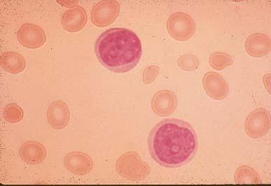
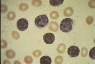
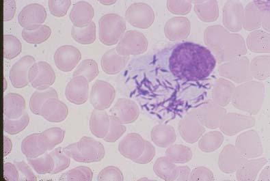
3 Chronic Lymphocytic Leukemia Most common leukemia Usual age > 50 yrs Increased proliferation and progressive accumulation of neoplastic, immunologically incompetent, clonal lymphocytes B cell origin > 99% 3
 Signs Lymphadenopathy (60%) Splenomegaly (50%) Hepatomegaly (<")
4 Clinical Features of CLL Highly variable presentation Asymptomatic, or vague, non-specific complaints Recurrent infection 10% (often pneumococcus) Signs Lymphadenopathy (60%) Splenomegaly (50%) Hepatomegaly (< 40%) 4

5 Laboratory Clinical Features of CLL blood and marrow lymphocytosis B cell monoclonality: κ vs λ surface light chain single Ig gene rearrangement hypoimmunoglobulinemia Prognosis Mean survival = months Range = few months to > 20 yrs 5
6 Immunological Abnormalities in CLL Disturbed Ab production Hypogammaglobulinemia (50%) bacterial infection Monoclonal Ig paraprotein in serum (10%) Autoantibodies (10%) Minor impairments in cell-mediated immunity Neoplastic lymphocytes Monoclonal surface Ig Abnormal response to Ig challenge Complications of CLL Recurrent infections Immune hemolysis Immune thrombocytopenia Progressive disease 6
7 Rai Staging System for CLL Stage Fea tures Median survival (years) I Lym pho cyto sis 13 II Lym pho cyto sis + lym pha denopathy 8 III Lym pho cyto sis + spleno mega ly 6 IV Lym pho cyto sis + an emia 1-2 V Lym pho cyto sis + thrombo cyto penia 1-2 Treatment of CLL No evidence that therapy prolongs survival Asymptomatic: watch and wait Symptomatic: Radiation for local complications Chemotherapy: fludarabine, alkylators, combinations Monoclonal antibodies (eg, Campath) Stem cell transplantation 7
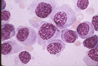
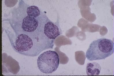
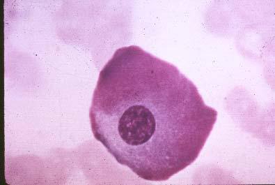
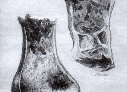
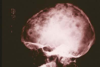
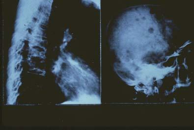
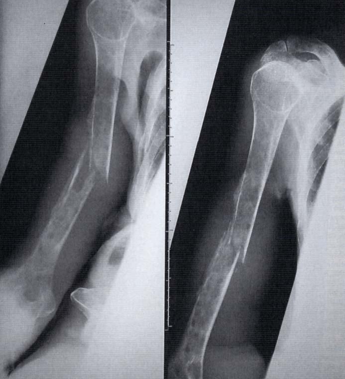
8 Multiple Myeloma Clonal malignancy of plasma cells Increasing incidence Blacks:whites 2:1 Age range yrs (peak age 70 yrs) Cause unknown (Environmental/Genetic factors) Classical Diagnostic Features of Myeloma Plasmacytosis in marrow Monoclonal protein in serum or urine Lytic disease of bone 8

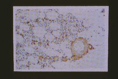
9 Marrow Plasmacytosis in Myeloma Plasma cells > 10% Usually much higher Often present in sheets Alternatively, biopsy-proven plasmacytoma Other causes of plasmacytosis: inflammation, cirrhosis, AIDS 9

10 10

11 11
12 12
 Other causes of monoclonal proteins (eg, CLL, lymphoma, benign monoclonal")
13 Diagnosis of Myeloma: Monoclonal Proteins 75-80% have serum monoclonal Ig (M-component, paraprotein, or spike on electropheresis) 10-20% make light chains only rapid renal excretion no paraprotein on serum protein electropheresis Non-secretory myeloma rare (< 1%) Other causes of monoclonal proteins (eg, CLL, lymphoma, benign monoclonal gammopathy) 13
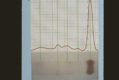
14 Apply serum or urine Protein Electropheresis (PEP): proteins separated according to charge and size Stain Scan Serum Protein Electrophereses in 2 Patients with Myeloma #1 #2 14
15 15

 Osteoblastic reaction minimal Hypercalciuria and")
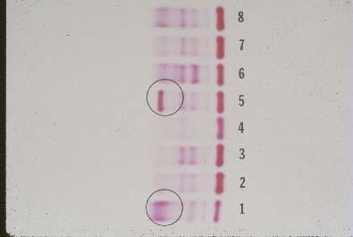
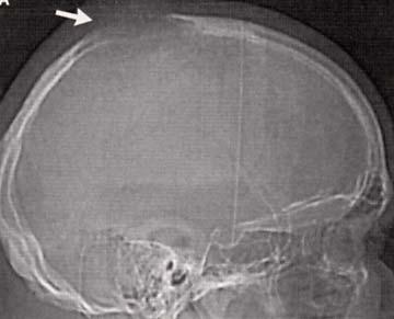
16 Immunofixation Electropheresis Bone Disease in Myeloma Unbalanced osteoclast activity Radiographic manifestations Osteoporosis almost invariable Usually multiple lytic lesions Axial skeleton involved (active marrow) Osteoblastic reaction minimal Hypercalciuria and hypercalcemia 16


17 17


18 18
19 19

20 20
21 Benign Monoclonal Gammopathy Monoclonal Ig as isolated finding More common than myeloma No bone disease, anemia, renal dysfunction Most remain stable About 10% eventually develop classical myeloma Myeloma at Presentation Early - asymptomatic, incidental diagnosis Paraprotein on electropheresis Mild marrow plasmacytosis Solitary plasmacytoma (10% of cases) Late - symptomatic Bone pain (usually lower back) Pneumococcal infection Systemic symptoms (eg, weakness, weight loss) Related to anemia, renal failure, hypercalcemia 21
 Encephalopathy and visual disturbances")
22 Hyperviscosity Syndrome Due to aggregating paraprotein Pathogenesis Circulatory insufficiency, abnormal hemostasis Manifestations Bleeding Dyspnea (congestion on CXR) Encephalopathy and visual disturbances 22
23 23

24 Immunological Features of Myeloma Monoclonal Ig and/or monoclonal light chain Levels of normal Ig s (hypogammaglobulinemia) Cellular immune responses usually preserved Bacterial infections common Early: S pneumoniae Late: S aureus, Gram negative rods 24

25 Amyloidosis in Myeloma Due to light chain deposition in tissues Incidence: λ amyloid > κ amyloid Organs commonly involved: Skin Tongue and GI Heart Peripheral nerves Kidneys Soft tissues No effective therapy, except?stem cell transplant 25


26 26
27 27
 Bortezomib (proteasome inhibitor) Stem cell transplantation 28")
28 Therapy for Myeloma Biphosphonates (pamidronate, zoledronate) Radiotherapy Corticosteroids and conventional chemotherapy Thalidomide (anti-angiogenesis) Bortezomib (proteasome inhibitor) Stem cell transplantation 28