12/6/12. Dr. Sanjeeva Srivastava IIT Bombay
|
|
|
- Donna Warren
- 5 years ago
- Views:
Transcription
 2 Equilibration of IPG strips 3 SDS-PAGE (second dimension) 4 Staining gel visualization")
1 Dr. Sanjeeva Srivastava IIT Bombay Gel-based proteomics 2-DE work-flow 1 Isoelectric focusing (first dimension) 2 Equilibration of IPG strips 3 SDS-PAGE (second dimension) 4 Staining gel visualization 1
2 2-DE work-flow 4 Staining gel visualization 5 Image analysis 6 Spot picking New methods for proteomics applications Fluorescent stain easily visualized with simple UV or blue-light transilluminators Endpoint stain, little background, sensitive Detects glycoprotein, low MW proteins Compatible with MS 2
3 Cy dyes, water-soluble derivatives of N-hydroxy succinimide that covalently binds the ε-amino groups of a protein s lysine residues Protein samples can be labeled with Cy dyes and mixed to run on a single gel Employed in difference in-gel electrophoresis, which eliminates problem of gel-to-gel variations Pro-Q diamond, a fluorescent dye capable of detecting phosphorylation Suitable for use with electrophoretic techniques and offer sensitivity in ng levels 3
4 Pro-Q diamond or other PTM detection stains can be combined with other staining procedures such as SYPRO Ruby Dual staining allows more than one detection protocol on a single gel Stain Comments Sensitivity Coomassie Blue Biosafe Coomassie Silver stain Silver stain plus SYPRO Ruby Most commonly used MS compatible MS compatible Easily visualized Non-hazardous MS compatibility an issue High sensitivity MS compatible High sensitivity MS compatible Linear over 3 orders of magnitude High sensitivity (Approximate) 40 ng 10 ng 1 ng 1 ng 1 ng 4
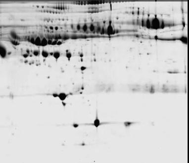
5 pi MW 5

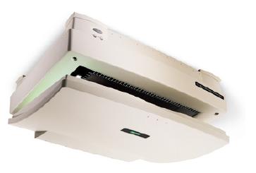

6 Molecular Imager Densitometer Typhoon Variable Mode Imager 6

7 2-D gels are scanned using a scanner and images are analyzed using various software These software enable Spot identification Comparison of gels Overlaying of images Cropping gels Statistical analysis Fil e Gel 1 Gel 2 Ed it Vie Report w s Gel1 - Control Tool s Image overlaying Gel2 - Treatment 7

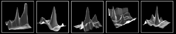
8 Crop tool Allows a specific defined region of gel to be cut out from the entire gel It helps in selection of regions with high spot density for further analysis Zoom tool Zoom tool expands a specific area of gel for further analysis Image overlaying To compare spot patterns on 2 different gels, separate images are overlaid to appear as single merged image Spots that coincide lie on top of each other while others retain their original position Spot analysis It is possible to obtain physical and statistical parameters for each spots on gels Allows gels comparison spot-by-spot basis 8


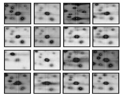

9 Healthy Control Disease Sample 9
10 Image Master 2D Platinum PDQuest Delta 2-D Dymension Ludesi 2-D gel image analysis Progenesis


11 User takes scalpel and makes a cut on particular spot in a gel and transfers it into a tube or 96 well plate along with water For each selected spot robotic arms moves and picks up individual spot and transfer it into the 96 well plate along with water 11
12 Traditional 2-DE: IEF and SDS-PAGE 2D-BN Gel Electrophoresis OFFGEL Electrophoresis 2-D Fluorescence Difference Gel Electrophoresis (DIGE) 12
13 OFFGEL electrophoresis separates proteins/ peptides according to their pi Separated components are recovered in liquid phase Compatibility with up or downstream techniques such as immunodepletion and LC/MS 13
14 hv Micro wells having sample A,B, C protein ph gradient Electrode IPG gel Protein-A: pi-4 Protein-B: pi-8 Protein-C: pi-9 IPG strip rehydrated and tightly sealed against frame of well in OFFGEL instrument Protein sample equally distributed in all the wells and a cover slip is applied to prevent evaporation hv Electrode ph gradient IPG gel Protein or peptides migrate though the gel when high voltage is applied 14
15 hv A ph=pi ph gradient B C Electrode IPG gel When ph = pi there is no protein migration Proteins separated based on pi can be removed in liquid phase OFFGEL fractionation can be combined with high-sensitivity on-chip electrophoresis of bioanalyzer This combination can enable 2-DE-type analysis with high resolution and high sensitivity Suitable for differential gene/protein expression applications 15
16 Staining techniques 2-D Image Analysis Spot picking Berg J., Tmyoczko J. & Stryer L., Biochemitry fifth ed., W. H. Freeman & company, ISBN: Nelson D. & Cox M.,Lehninger, Principles of Biochemistry fourth ed., W. H. Freeman and company. ISBN: X. Voet D. & Voet J., Biochemistry fourth ed., Wiley, ISBN: X. Saghatelian A. and Cravatt BF. Assignment of protein function in the postgenomic era. Nature chemical biology 2005, 1, Hanash S. Disease proteomics. Nature 2003, 422, Deborah Penque. Two-dimensional gel electrophoresis and mass spectrometry for biomarker discovery. ROTEOMICS - Clinical Applications. Volume 3, Issue 2, pages , No. 2 February 2009 Agilent 3100 OFFGEL Fractionator. Agilent Technologies 16