Supplemental Information
|
|
|
- Benedict Porter
- 5 years ago
- Views:
Transcription
1 Supplemental Information Intrinsic protein-protein interaction mediated and chaperonin assisted sequential assembly of a stable Bardet Biedl syndome protein complex, the BBSome * Qihong Zhang 1#, Dahai Yu 1, 3#, Seongjing Seo 1, Edwin M. Stone 2, and Val C. Sheffield 1 Supplementary Materials and Methods Antibodies and Reagents Rabbit polyclonal antibodies against human,,, and have been previously described. All antibodies were affinity purified and the specificities were verified in knockout mice or by RNAi in cell culture. Monoclonal anti-β-tubulin, polyclonal anti-β-actin, monoclonal anti-acetylated tubulin, monoclonal anti-γ-tubulin, rabbit anti-bbs8 and, thermolysin, and cycloheximide were purchased from Sigma (St. Louis, MO). Goat anti- (C-16) was purchased from Santa Cruz Biotechnology (Santa Cruz, CA). Rat anti-tcp1α and TCP1β were purchased from Stressgen Biotechnologies Inc (San Diego, California). Rabbit antiubiquitin antibody was from Abcam (Cambridge, MA). Smartpool RNAi against human, 0, 2, were from Dharmacon (Lafayette, CO). MG132 and MG115 were purchased from Calbiochem (San Diego, CA). Cell culture, transfection, and Immunofluorescence microscopy Skin fibroblast cells from a human patient with the common 0 mutation, c91fs95 and control skin fibroblast cells were purchased from Coriell Institute (Camden, New Jersey) and cultured in DMEM high glucose with 10% FBS. 293T cells were cultured in DMEM high glucose with 10% FBS, htert-rpe1 cells were cultured in DMEM/F-12 medium with 10% serum. All tansfections were performed using lipofectamine 2000 or RNAiMAX (Invitrogen, Carlsbad, CA). For RNAi transfection, a final concentration of nM RNAi was used. For immunofluorescence, cells were fixed in cold methanol for 5 min at C and were washed with 3 x PBS and blocked with blocking buffer (1% BSA in PBS). Primary antibodies were diluted in blocking buffer and incubated at room temperature for 1 hour. Cells were washed with 3 x PBS, blocked with blocking buffer, then incubated with Alexa 488 or Alexa 568 labeled secondary antibodies (Invitrogen, Carlsbad, CA). The slides were mounted with Vectashield mounting media containing DAPI (Vector Laboratories, Burlingame, CA). Immunoprecipitation Differentially tagged human BBS genes were co-transfected into 293T cells. Forty-eight hours after transfection, the cells were lysed in lysis buffer (1 x PBS, 1% Triton X-100, and protease inhibitor, Roche, Indianapolis, IN) and spun at 20,000 x g for 15 min at 4 0 C. The supernatants were cleared by incubation with protein G beads (Pierce, Rockford, IL). Cleared lysates were incubated with antibodies against corresponding tags for 2 hrs. Protein G beads were then added and incubated for another 2 hrs. The beads were washed four times with lysis buffer and the interactions were detected by Western blotting. 1
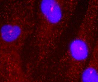
2 Western blotting Wild type and mutant testes were disrupted by TISSUEMISER (Fisher Scientific, Pittsburgh, PA) in lysis buffer (1 x PBS, 1% Triton X-100 and protease inhibitor, Roche, Indianapolis, IN). The disrupted tissues were freeze-thawed three times. The lysates were then centrifuged at 20,000 x g for 15 min and the concentrations of the supernatants were measured using the Bio-Rad Dc protein assay (Hercules, CA). Proteins were separated by electrophoresis using 4-12% NuPAGE Bis-Tris gels (Invitrogen, Carlsbad, CA) followed by transfer to nitrocellulose membranes and were detected by SuperSignal Dura extended substrate (Pierce, Rockford, IL). Site-directed mutagenesis Homozygote point mutations were generated by site-directed mutagenesis (Stratagene, La Jolla, CA) and verified by DNA sequencing. Supplementary Figure Legends Supplemental Figure S1. Summary of Homozygote point mutations identified in BBS patients, which were used in this study. Supplemental Figure S2. interacts with through. A. strongly interacts with and weakly with. Shown are co-ip assay of - with -tagged other BBSome subunits in 293T cells. B. interacts with through. Shown are co-ip assay of - with - and HA- in 293T cells. Supplemental Figure S3. Summary of the binding domains for PCM1 and BBIP10. A. truncation mutants were generated by PCR. GFP was added to the N-terminal of each truncated protein. These GFP- truncation mutants were co-transfected with HA-tagged PCM1 C-terminal fragment or -BBIP10. Co-IP results are summarized here. B. BBIP10 interacts with PCM1 only in the presence of. Supplemental Figure S4., BBS6, and 2 form a subcomplex. A. The interaction of BBS6 with 2 requires. Shown are the co-ip assays of -BBS6 with -2 in the presence of control sirna, sirna, or sirna. B. The interaction of with 2 requires BBS6. Shown are co-ip assay of -2 with - in the presence of control sirna or BBS6 sirna. C. Overexpression of BBS6 but not BBS6 mutants increases the interaction between and 2. Supplemental Figure S5. Loss of 0 does not affect primary ciliogenesis. Shown are immunofluorescent staining of skin fibroblast cells from a 0 patient with the common c91fs95 mutation using anti-acetylated tubulin as a ciliary marker (green) and (red). Similar percentages of cells contain cilia in and mutant cells. However, the intensity of staining is greatly decreased in the 0 mutant cells. 2
3 Supplemental Figure S6. Loss of 0 affects BBSome subunits protein levels and disrupts BBSome formation. A. Western blots of total protein from and 0c91fs95 skin fibroblast cells were probed with antibodies against BBSome subunits. Equal amount of proteins were loaded as demonstrated by BBS3, IFT88, and tubulin signals. B. Sucrose gradient analysis shows that loss of 0 disrupts BBSome formation. BBSome subunits are concentrated in fractions 12 and 13 of the sucrose gradient. BBSome subunits are totally absent in 0c91fs95 skin fibroblast cells. To demonstrate that the same amounts of proteins are loaded to the sucrose gradient, a non-specific band from antibody is shown. C. Loss of 0 does not affect the BBSome subunit mrna levels as shown by semi-quantitative RT-PCR. Supplemental Figure S7. protein stability requires BBS6, 0,, and. A. Western blots of protein samples from Bbs2, Bbs6, Bbs4, and Bbs1 M390R/M39R mutant mice testes, 0 mutant cell lines, or from RNAi knocked-down RPE1 cells demonstrate that protein stability requires BBS6, 0,, and, but not or. B. protein is not stable in the absence of. We introduced into Bbs7 null kidney cells by adenovirus expressing -. The stability of was examined by the protease thermolysin sensitivity assay. The half-life of in cells is significantly longer than in Bbs7 null cells. 3
4 Gene BBS5 Homozygous point mutations M390R V75G, L125R G277E, R295P G72S, T183A T211I, H323R G141R, A455T Figure S1 4

5 A B IP: - BBS5 BBS8 IB: IP: IP: IP: HA IP: IP: IP: HA lysates IB: 1 2 IB: HA 1: -/- 2: -/-/HA- Figure S2 5


6 A Figure S3 B G277E R295P 519 -BBIP10 HA-PCM1 -BBIP10 GFP- HA-PCM BBIP10 PCM1 IP: HA ? IP: HA total HA GFP 6


7 Figure S4 A B C -BBS6 / -2 Control sirna sirna sirna - / -2 Control sirna BBS6 sirna - / -2 control HA-BBS6 HA-BBS6 G52D HA-BBS6 T57A IP: IP: IP: IP: IP: IP: lysates lysates lysates 2 BBS6 7


8 ac-tub control 0c91fs95 Figure S5 8


9 0c91fs95 A B control c91fs BBS5 BBS8 BBS8 total from BBS3 IFT88 tubulin C 2 actin 0 BBS6 BBS8 0c91fs95 2 actin 0 BBS6 control BBS8 BBS5 BBS3 0c91fs95 BBS5 BBS3 control Figure S6 9

")


10 A B Bbs2 -/- Bbs6 -/- 0c91fs95 control RNAi thermolysin (min) Bbs7 -/- tubulin tubulin Bbs4 -/- Bbs1 M390R Percentage of protein remaining Bbs7 -/ Figure S7 10