Supplementary Figure Legends
|
|
|
- Edgar Davis
- 5 years ago
- Views:
Transcription
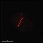
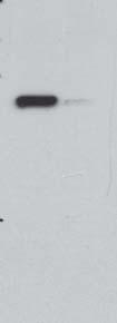
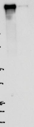
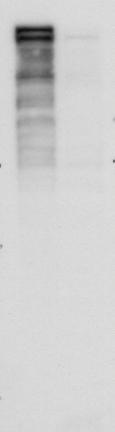
1 Supplementary Figure Legends Figure S1 gene targeting strategy for disruption of chicken gene, related to Figure 1 (f)-(i). (a) The locus and the targeting constructs showing HpaI restriction sites. The exons are represented by black boxes and the white bar indicates the probe region. Genomic fragments that are detected by the probe are shown as black bars. (b) Southern blot analysis of HpaI-digested genomic DNA from DT40 WT, +/- and -/- cells. (c) The immunoblots for wild-type and mutant in Fig.1g to show the proteins levels in wt and mut cells. Figure S2 E3 Ligase activity is required for mobility, related to Figure 2 (d). Cells were transfected with the wild type (YFP-) or a RING mutant (YFP-cs1), incubated for 24 hours. Comparable areas of fluorescence intensity were chosen for photobleaching (circled area). Three images were taken before hotobleaching. Fluorescence recovery was recorded in a microscope described in methods. Figure S3 is down-stream of DNA damage factors Brca1, RAP80, NBS1, MRE11, MDC1, γh2ax, RNF8 and RNF168 related to Figure 3. 1
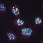
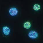
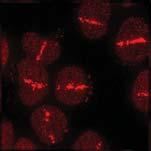
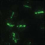
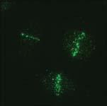
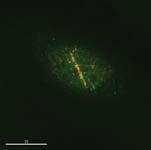
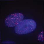
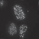
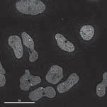
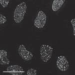
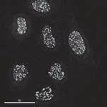
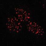
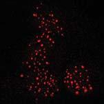
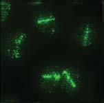
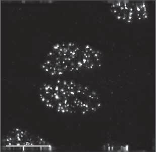
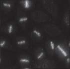
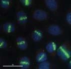
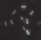
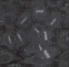
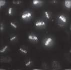
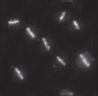
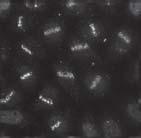
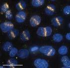
2 (a) and (b) Cells were treated with sirna to () or a non-target sirna () for 3 days, micro-irradiated in (a) or exposed to 3 Gy IR in (b), recovered for 1 hour, and immunostained with the indicated antibodies. Scale bars, 15 μm. Figure S4 Depletion of slows repair and delays recruitment of 53BP1 to DSB, related to Figure 3 (a)-(b) (a) 293 cells stably expressing GFP-53BP1 were treated with sirna to sirna (upper panel) or a non-target sirna (, lower panel) for 3 days, microirradiated and subjected to time-lapse microscopy immediately after treatment. Images were recorded at 30 seconds intervals. Scale bars, 5 μm. (b) Depletion of in (a) was confirmed by RT-PCR. 293 expressing GFP- 53BP1 were treated with sirna as in (a). Total RNA were extracted and subjected to RT-PCR analysis with GAPDH as a control. (c) depleted cells delay repair after DNA damage. HeLa or depleted HeLa cells (C6) were exposed to 10 Gy IR, incubated for the indicated time, and then immunostained with 53BP1 antibody. depleted cells showed slower recovery, related to Figure 3 (b). Figure S5 Recruitment of requires multiple DNA damage repair factors, related to Figure 3. 2
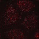
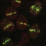
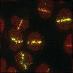
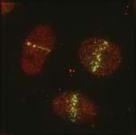
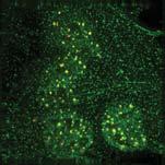
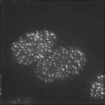
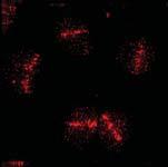
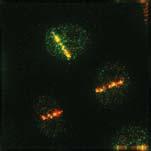
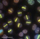
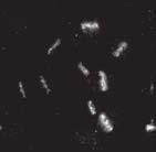
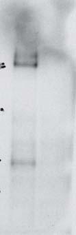
3 (a)-(c) Cells were treated with the sirnas indicated on the left for 3 days, microirradiated, recovered for 1 hour, and immunostained with the indicated antibodies. Scale bar, 30 μm. (b) The efficiency of sirna depletion in (a) and (c) was determined by western blotting using the indicated antibodies. The depletion of RNF8 was assessed by the assembly of Brca1 at DSB. The position of molecular weight markers is indicated. Figure S6 Comfirmation of depletion of the indicated genes by western blot, related to Figure 3, Figure 4 and supplementary Figure S3. HeLa cells were treated with the indicated sirna for three days and subjected to western blotting to assess protein level. The position of molecular weight markers is indicated. Figure S7 AsiSI mediated DNA cleavage leads to recruitment of SUMO-1 and SUMO-2 at DSB. (a)-(c) ChIP analysis was performed in AsiSI-ER-U2OS cells, before (0 h) and after 4OHT treatment (300 nm) for 4 or 8 hours, using antibodies specific to SUMO-1 and SUMO-2 or IgG as control. Enrichment of SUMO was determined by RT-qPCR using primers located 200 bp proximal to an AsiSI site on chromosome 1 at position (Chr1_6-200) (a), 100 bp proximal to an AsiSI site on chromosome 6 at position (Chr6_12-100) (b) or 2Mb 3
4 distal from an AsiSI site on chromosome 22 at position (Chr22-2 Mb) (c) as negative control. (d) and (e) Specificity of SUMO antibodies. AsiSI-induced recruitment of SUMO-1 and SUMO-2 at DNA double-strand breaks is abolished in response to Ubc9 depletion showing the specificity of the antibodies used for ChIP. AsiSI-ER-U2OS cells were transfected with non-target control () or sirna specific for Ubc9. ChIP analysis was performed before (untreated) and after 4OHT treatment (300 nm) for 4 h, using antibodies specific to SUMO-1 and SUMO-2 or IgG as control. Enrichment of SUMO was determined by RT-qPCR using primers located 200 bp proximal to an AsiSI site on chromosome 1 at position (Chr1_6-200) (d) or 100 bp proximal to an AsiSI site on chromosome 6 at position (Chr6_12-100) (e). (f and g) relates to Figure 5 g and h to show that no enrichment of CtIP or γh2ax was detected at a site that was distant from DSB breaks. Movie S1 The assembly of YFP- at laser track after DNA damage, related to Figure 2 (b) HeLa transiently expressing YFP- were recorded by time-lapse microscopy for 2 hours post micro-irradiation. Movie S2 4
5 Dissociation of YFP- from DSB as breaks are repaired, related to Figure 2 (b) Same cell and treatment as in Movie S1 to show the dissociation of YFP- from DSB 9 hours after micro-irradiation. Movie S3 Dynamic recruitment of SUMO2 at DSB after micro-irradiation, related to Figure 2 (e) and compared also with Figure 2 (b) HeLa stable cell line expressing YFP-SUMO2 was treated as Movie S1 and S2 to compare with the dynamics of recruitment. Movie S4 Dynamics of GFP-53BP1 recruitment at DSB, related to Figure 2 (b) 293 stable cell line expressing GFP-53BP1 was treated as for Movie 1, 2 and 3 5
6 Supplementary Table Antibodies Used in This Work Antibody Used Species Reference/ Suppliers β-actin WB Mouse SIGMA 53bp1 IF, WB Rabbit BETHYL Brca1 IF, WB Mouse Santa Cruz Fk2 IF Mouse BIOMOL IF Rabbit BETHYL IF Mouse Upstate Mdc1 IF, WB Rabbit Bethyl Mre11 IF, WB Mouse Abcam Nbs1 WB, IF Rabbit Novus RAD51 WB, ChIP Rabbit Santa Cruz RAP80 WB, IF Rabbit Bethyl IF Sheep In house IF Rabbit (Hakli et al. 2005) IF, WB Chicken In house RNF8 WB Goat NOVUS RNF168 WB Mouse Abcam, RPA70 WB, IF, ChIP Rabbit Cell Signaling Sumo-1 ChIP Sheep In house Sumo-2 WB, ChIP Sheep In house Sumo -2 WB, ChIP Rabbit ZYMED Ubc9 ChIP Sheep In house CtiP ChIP Rabbit BETHYL BrdU IF Mouse B-D FK2 IF Mouse BIOMOL Ubiquitin K63 IF Rabbit Millipore Ubiquitin K48 IF Rabbit Millipore Alexa Fluor-488 IF Anti mouse/rabbit Molecular Probes Alexa Fluor-568 IF Anti rabbit Molecular Probes Alexa Fluor-594 IF Anti mouse Molecular Probes Alexa Fluor-647 IF Anti mouse Molecular Probes Cye5 IF Anti sheep Jackson ImmunoResearch HRP WB Anti mouse- /rabbit/sheep- /chicken SIGMA Note: WB, western blot. IF, immunofluorescence staining. ChIP, Chromatin Immunoprecipitation.
7 SUPPLEMENTARY REFERENCE Hakli M, Karvonen U, Janne OA, Palvimo JJ SUMO-1 promotes association of SNURF () with PML nuclear bodies. Exp Cell Res 304:


8 Yin et al., Fig. S1 a b c 12kb +/+ +/- -/- wt mut 5kb tubulin 3kb


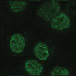
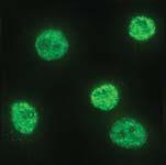
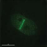
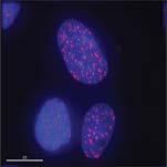
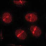
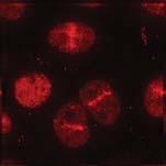
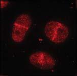
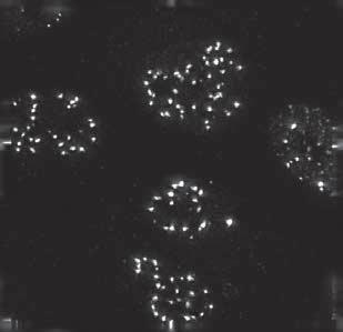
9 YFP-cs1 YFP-wt Yin et al., Fig. S2


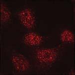
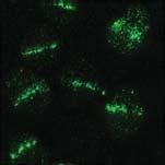

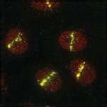
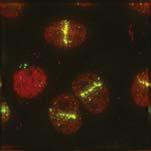
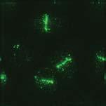
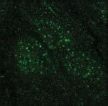

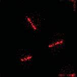
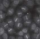
10 Yin et al., Fig. S3 IR, 3Gy micro-irradiation MDC1 MDC1 MDC1 MDC1 Mock Mock BRCA1 BRCA1 RAP80 RAP80 NBS1 NBS1 MRE11 MRE11 MRE11 MRE11 RNF8 RNF8 RNF168 RNF168 b


")

")
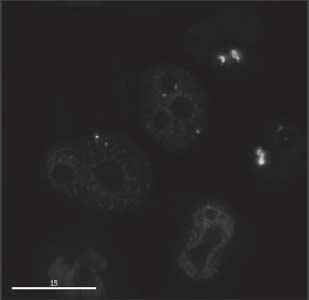
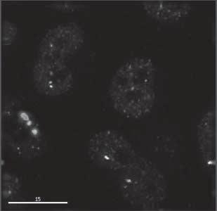
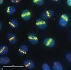
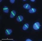
11 Yin et al., Fig. S4 a 293 stable cell line GFP-53BP1 b RT-PCR sirna GAPDH c HeLa (wt) Mock 1h 8h 24h C6 ( depleted) Mock 1h 8h 24h


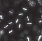

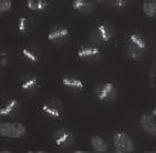


12 sinbs1 sirnf8 si53bp1 sibrca1 simre11 simdc1 MDC1 sibrca1-53bp1 sinbs1 sirna NBS1 BRCA1 kda Actin sirna simre11 Yin et al., Fig S5 NBS1 NBS1 BRCA1 BRCA1 BRCA1 BRCA1 53BP1 53BP1 MRE1 MRE1 kda kda MRE11 kda a b c 25O O kda si53bp O O Actin


13 Yin et al., Fig. S6 kda Anti kda Anti MDC β-actin sirna sinbs1 simdc1

14 a b c Yin et al., Fig. S7 d e f g