A Treasure Chest of Technological Innovations in Eye Care. Paul M. Karpecki, OD, FAAO Koffler Vision Group Lexington, KY
|
|
|
- Kathleen Bryant
- 5 years ago
- Views:
Transcription
1 A Treasure Chest of Technological Innovations in Eye Care Paul M. Karpecki, OD, FAAO Koffler Vision Group Lexington, KY
2 Paul M. Karpecki, OD, FAAO Financial Disclosures: AMO Alcon Labs Allergan Inc Bausch & Lomb Inc Cyanacon Ocusoft Eyemaginations Focus Laboratories Inspire Pharmaceuticals Ista Pharmaceuticals Konan Medical LCA Vision Inc. Pixel Optics Odyssey Medical OfficeMate Rapid Pathogen Screening Science Based Health TearLab Topcon TruVision Vmax WaveTec
3 Point-of-Care Diagnostics
 for an additional 3 seconds to ensure saturation of the")
4 RPS Adeno Detector Test Procedure Collecting the Sample 4. Dab the sampling pad inside the lower eyelid (palpebral conjunctiva) 4 6 times. Allow the sampling pad to rest against the conjunctiva (membrane on inside of the eyelid) for an additional 3 seconds to ensure saturation of the sampling pad with eye

5 RPS Adeno Detector Test Procedure Assembling the Detector 1. Locate the Test Cassette 2. Assemble the detector by gently placing the sampling pad of the Sample Collector into the sample transfer window of the Test Cassette body. Transfer Window 3. Press firmly where indicated until the detector is secure. Note: A double auditable click means the detector is properly assembled, transferring the sample to the test strip.
6 RPS Adeno Detector Test Procedure Running the Test 1. Open the buffer vial. Remove the Protective Cap from the Test Cassette. Do not allow any portion of the detector besides the absorbent tip to touch the buffer vial. Immerse the Assembled Detector s Absorbent Tip into the buffer vial for 15 seconds.

7 RPS Adeno Detector Reading & Interpreting the Results Positive Results: The Results Line and Control Line are RED in the result window, indicating that Adenovirus antigen is present. Results Line Control Line Control Line
8 Zirgan
9 Zirgan (Ganciclovir Ophthalmic Gel) 0.15% Product Background GAN9-008A
10 Zirgan (ganciclovir ophthalmic gel) 0.15% Indication Indication and Usage Zirgan is a topical ophthalmic antiviral that is indicated for the treatment of acute herpetic keratitis (dendritic ulcers).
11 Zirgan (ganciclovir ophthalmic gel) 0.15% Indication Dosage and Administration The recommended dosing regimen for Zirgan is 1 drop in the affected eye 5 times per day (approximately every 3 hours while awake) until the corneal ulcer heals, and then 1 drop 3 times per day for 7 days.
12 Ganciclovir Mechanism of Action Penetrates cell infected with the virus Phosphorylated within the cell to ganciclovir monophosphate by a viral thymidine-kinase Affinity for thymidine-kinase allows for specificity in its action Activation continues due to several cell kinases leading to formulation of ganciclovir triphosphate, which: Inhibits viral DNA polymerase Incorporates into viral DNA preventing replication
13 Properties of Zirgan Gel Polyfoil 5 gram tube with dropper fitting Gel formulation (due to carbomer-based vehicle) Allows for more prolonged contact time with the eye than oil-based formulations Aqueous gel allows for ganciclovir concentration of 0.15% Sufficient to ensure good tolerability and efficacy in treatment of superficial acute herpetic keratitis ph = 7.45 Osmolality = 300 mosmol Virgan 0.15% product monograph. Laboratoires Théa, Clermont-Ferrand, France. October 1998.
14 Zirgan Clinical Efficacy Results Results from a open-label, randomized, controlled, multicenter clinical trial evaluating ganciclovir ophthalmic gel 0.15% compared to acyclovir ophthalmic ointment 3% in patients with dendritic ulcers Clinical Resolution By Day 7 GCV 0.15% N = 77 ACV 3% N = (77%) 48 (72%)
15 Zirgan Clinical Efficacy Results Results from 3 randomized, single-masked, controlled multicenter clinical trial evaluating ganciclovir ophthalmic gel 0.15% compared to acyclovir ophthalmic ointment 3% in 213 patients with dendritic ulcers GCV 0.15% N = 57 ACV 3% N = 49 Clinical Resolution By Day 7 41 (72%) 34 (69%)

16

17
18 Fluramene Dye
19 Combination drop Fluoresceine and Lissamine dyes in one bottle Much higher potency and staining potential Saves time and is more sensitive

20

21
22 TearLab

23 TearLab Osmolarity Reader & Pens

24 TearLab Osmolarity Disposable Chip

25 TearLab Tear Collection
26 Osmolarity in the Diagnosis of Dry Eye Disease Clinical Test PPV Osmolarity 87% Schirmers 31% TBUT 25% Staining 31% Meniscus Height 33% Osmolarity is the gold standard test for Dry Eye 45 years peer reviewed research Osmolarity has been added to definition of Dry Eye Global marker of Dry Eye, indicating a Source: DEWS Report, Ocular Surface April 2007 Vol 5 No 2, & Tomlinson A, et. al., IOVS 47(10) 2006
 submission] Source: Kimberly MM et. al.")
27 TearLab < 2% coefficient of variation nanoliters Glucose 5.0% 5 microliters Cholesterol > 4.0% 20 microliters 20 µl 5 µl 50 nl Safe, simple collection No reports of corneal or conjunctival trauma in 468 eyes [TearLab FDA 510(k) submission] Source: Kimberly MM et. al., Clinica Chimica Acta 364 (2006), Volles DF et. al. Pharmacotherapy 18:1 (1998).,

28 Osmolarity in Diagnosis & Grading of Dry Eye
29 Tear Osmolarity Normals Variability Tight range (± 5 mosms/l) both in individuals and as a group Early/Mild cases Variation from one eye to the other and in response to normal environmental stress DED Subjects
30 Patient Education: TelScreen & Eyemaginations

31

32

33
34 QuickTime and a Motion JPEG OpenDML decompressor are needed to see this picture.
35 QuickTime and a Motion JPEG OpenDML decompressor are needed to see this picture.
36 Specular Microscopy & DSEK
37 Deep Lamellar Endothelial Keratoplasty Eliminate corneal sutures Eliminate corneal surface incisions Faster wound healing Smoother topography Stronger and more stable eye.

38
39
40

41 Normal ECD Low ECD Normal Endothelial Cell density declines with age Low ECD is a risk factor for corneal surgery

42 Normal ECD Polymegethism Normal Endothelial Cell density declines with age Change from uniform cell sizes to variability of cell sizes TypicalLargerSmaller

43 Normal ECD Pleomorphism Normal Endothelial Cell density declines with age Change from uniform hexagonal cell geometry to variability in cell shape Abnormal Normal

44 Corneal Guttata Discrete Guttata Moderate Guttata

 Stage 1 guttata Courtesy:")
45 Abnormal endothelium 35 YO female with 20 years of SCL wear Polymegethism (CV=62) Pleomorphism (Hex=51) Stage 1 guttata Courtesy: Craig Thomas, OD


46 Stage 3 Fuchs Endothelial Dystrophy Unmistakable with SM Courtesy: Craig Thomas, OD
47 Specular Microscopy Indicated in cases of Hereditary corneal dystrophy s with evidence of endothelial pathology on slit lamp examination ICD Code Screening device first if pathology is noted then full specular microscopy CPT Code National average of reimbursement $128
 DLEK with Descemetorhexis of Recipient (DSEK/DSAEK): Surgical Technique of stripping recipient Descemet s Corneoscleral incision and endothelium stripped from posterior cornea Endothelium")
48 (Diagram Courtesy of Dr. Ayad Farjo) DLEK with Descemetorhexis of Recipient (DSEK/DSAEK): Surgical Technique of stripping recipient Descemet s Corneoscleral incision and endothelium stripped from posterior cornea Endothelium replaced without sutures (endothelial cell pump dependent mechanism), and surface topography with minimal change Donor posterior stroma and endothelium added onto back surface of recipient cornea, adding tissue thickness


49 Endothelial Keratoplasty Post-op Day 1 Post-op Day 8

50 Endothelial Keratoplasty
51 DSEK Rapid recovery No induced cylinder No incisions or sutures
52 Current prospective results of hybrid DSAEK procedure in initial 100 cases Dislocation rate nearly equivalent to DLEK at 4%. Primary graft failure rate still higher than PK at 1% Visual recovery appears faster than DLEK Terry MA, Hoar K, Wall J, Ousley PJ. The histology of dislocations in endothelial keratoplasty (DSEK and DLEK): a laboratory-based, surgical solution to dislocation in 100 consecutive DSEK cases. Cornea 2006 (in press).

53 EK Endothelial Keratoplasty + Phacoemulsification in a Fuchs Patient
54 Anterior Segment Imaging: OCT & Scheimpflug

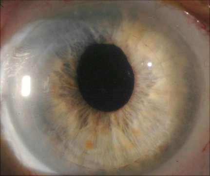
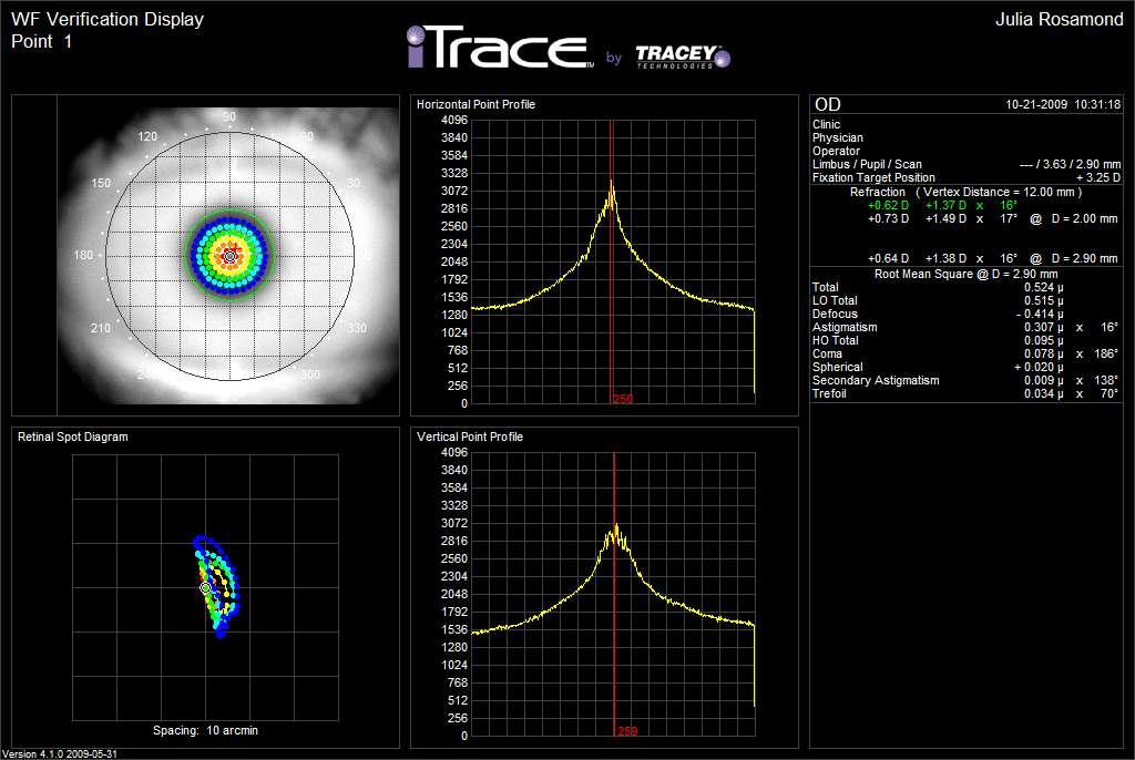
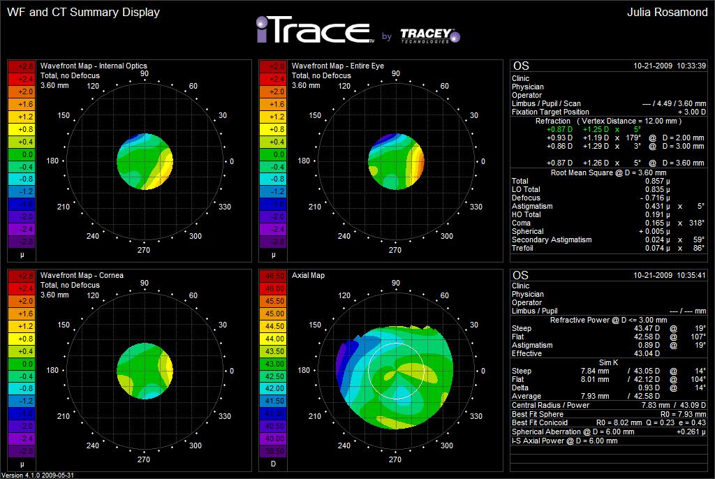
55 Scheimpflug Imaging
56 The Oculus Pentacam/Pentacam HR Pentacam, the screening and analysing tool for: Glaucoma (ant. segment) chamber angle, -depth, -shape, -volume, IOP corr. Cataract objective quantification, IOL calc. for post refractive patients Corneal refractive screening for refr. surgery, Intacs, implants, Flap/RBT, Cataract refractive piol, IOL calc/positioning, sph. abberations

57 Edge detection via software

58 4-maps refractive Application: Enhanced overview for refractive surgeons Hand-out to patients

59 IOL positioning
60
61 Visante OCT Applications Refractive Surgery and Anterior Segment Refractive Surgery Corneal laser refractive surgery Phakic IOLs Corneal refractive implants Anterior Segment Imaging and Surgery Corneal Imaging and Measurement Iris Imaging and Evaluation Trauma Assessment Crystalline Lens Imaging and Evaluation

62 Visante OCT Applications Post Op Enhancement request denied! bed < 250 microns image courtesy of Dr. J. Vukich

63 Visante OCT Applications Lamellar Keratoplasty lamellar keratoplasty image courtesy of Dr. L. Buratto

64 Visante OCT Applications Iris Lesions iris pigmented lesion image courtesy of Dr. J. Vukich

65 Iris Cyst (High Resolution) image courtesy of Dr. I. Ahmed

66 itrace of a patient with Keratoconus

67 A case of advanced keratoconus
68 Patient JER 60 y.o. very active Caucasian female Observed in clinic 12 months earlier Is an avid target and skeet shooter Noticing blur in eyes and wanted to improve vision but also having difficulty with soft contact lenses
69 Patient JER UCVA: 20/25-2 OD, 20/20-2 OS Rx: X 100 OD 20/ X 85 OS 20/20-
70 Patient JER Did not like how contact lenses would move and affect vision during the day Did not want to wear spectacles for potential fogging etc. Recommended overnight CRT rather than LASIK Patient tried CRT for ~2 months Complained of blurred vision OU
71 Patient JER 12 mo later Believed that the CRT warped her eye and vision has never been the same even with current SCL s itrace was performed prior to examination
72 Patient JER CC Diplopia and difficulty with target shooting CRT warped my eyes UCVA: 20/30-2 OD, 20/25-2 OS Rx: X 100 OD 20/ X 100 OS 20/20-
73

74
75
76

77 Patient JER - SLEx
78 Collagen Cross Linking (CXL)
79 UVA -Riboflavin 30 minutes of drops and UV radiation exposure Strengthen collagen bonds Early results are promising as far as corneal integrity improvements but side effects (pain etc.) are relatively unknown and long term data does not exist

80 Ectasia Diagnosis and Management Post-op
81 VMax phoropter and Electronic Eyewear
82 Vision Care Applications Phoropters of the future: VMax Technology
83 Snellen is identification: Need more sensitive testing: Point Spread Function (PSF)
 rather than Snellen")
84 VMax Technology Based on point spread function (PSF) rather than Snellen recognition
85 VMax Technology VMax Technology Objective and Subjective features Measures down to 0.1D Statistical increase in VA in pilot study Spectacles developed to match the technology
86 Revolutionary New Spectacle Technologies Electroactive optics Microelectronic controls Composite lens materials Integrated eyewear systems

87 Presenting PixelOptics Eyewear!
88 Frame Looks and weighs same as today s fashion frames Houses micro-electronics Available in several materials and styles


89 1Micro-Electronic st Engineering Control (prototype) Prototype
90 Focus Activation Range finder Tilt switch Touch switch
91 How Do Electro-active Thin layer of Liquid Crystal (LC) sandwiched between two layers of Transparent Electrodes Lenses Work? Array of voltages in patterned electrode creates structured electric field Cigar-shaped LC molecules rotate with change in voltage This rotation alters index of refraction Edge-on view of Layers
92 Example: EA Element Turned ON Low-Index edge Hi-Index center Low-Index edge E/A Element ON Incoming Variable Converging Wavefrontspeed of lightwavefront Variable speed of light causes change in wavefront shape Huygen s Principle explains change in direction of rays The result is positive lens power
93 Example: EA Element Turned OFF E/A Element OFF Constant index center to edge Flat Wavefront Entering Constant speed of light Flat Wavefront Leaving Wavefront emergences unchanged Result is zero add power
94 Benefits of Electro Active Lenses Active focus Clearer vision Distance field 2X wider than PAL* Intermediate up to 10x wider than PAL* Near field 2X wider than PAL* Up to 100% less distortion* *Based on equivalent add powers
95
96 Lenses Thin, light (1.67 index) Look cosmetically the same as today s state-of-the-art lenses Can be surfaced/finished the same as today s ophthalmic lenses Compatible with customary lens treatments
97 3-D Technology & Intraoperative Aberrometry


98 New Advances in Microsurgery: High-Definition 3D Surgical Visualization System


99 TrueVision 3D HD camera mounted to a conventional microscope Live surgical field displayed on a 46 3D HD flat panel Allows head-up display surgery with additional case info

100 3D HD Visualization in Cataract Surgery at the Eye Institute of West Florida Experience of over 2000 cases operating directly from a 3D HD video display. Advantages: Excellent resolution and image quality Depth of field Surgeon ergonomics Retained access to microscope oculars Intraoperative instruction 3D video/photo recording & playback Full view for OR staff 3D patient education
101 Interactive 3D Surgical Guidance Software For Capsulorrhexis and Limbal Relaxing Incisions Using a High-Definition 3D Visualization System Note: Investigational Use Only

102 Investigational Use: 3D Overlay Surgical Guidance Software LRI Guidance: Preoperative 3D slit lamp photo capture Note: Software Under Investigational Use Only

103 Investigational Use: 3D Overlay Surgical Guidance Software LRI overlay template on a tracked eye Note: Software Under Investigational Use Only

104 Investigational Use: 3D Overlay Surgical Guidance Software Capsulorrhexis Guidance: OR image registration Note: Software Under Investigational Use Only
105 Investigational Use: 3D Overlay Surgical Guidance Software Capsulorrhexis Guidance: Overlay template confirmation Note: Software Under Investigational Use Only
106 QuickTime and a decompressor are needed to see this picture.

107 WaveTec Vision Fresh Technology for the OR
108 ORange Used in the OR on a Range of applications and patients Intraoperative wavefront aberrometer: Tabot-Moire interferomtery Attaches to the bottom of surgical microscope Allows surgeon to take on-demand images of the eye in the OR Provides refraction data to guide surgeon decision-making real-time


109 ORange in the OR ORange has a small footprint
110 ORange Applications Refractive Measurements Phakic Refraction Irregular astigmatism? Aphakic Refraction True corneal power and refraction of the eye True refractive astigmatism as compared to estimated cylinder based on corneal toricity from topography Which is the best magnitude and axis to treat with LRI s or Toric IOLs In development: Spherical Aberration determination
111 ORange Applications Refractive Measurements (con t) Pseudophakic Refraction Spherical Equivalent (SE) IOL confirmation on the table IOL exchange? Intraoperative Residual Cylinder Intra-op LRI enhancements Toric IOL repositioning Potential? Intacs surgery, PKP surgery to minimize resid cyl

112 LRI Refractive Information

113 TORIC Refractive Information Based on the ORange analysis, the surgeon rotates the toric IOL to the recommended position An additional ORange measurement confirms if placement is correct or if an adjustment is required
114 Femto-Phaco
115 Study Purpose To evaluate the safety and efficacy of femtosecond laser incisions in the lens and lens capsule prior to standard and refractive cataract surgery 60 patients laser capsulotomy 60 patients manual capsulorrhexis Measured diameter accuracy Determined ease and completion of capsule removal Porcine eye SEM capsule edge quality

116 Laser Capsulotomy


117 Laser Capsulotomy Results Perfect centration Precision diameter: < ± 0.25 mm No radial tears Easy and complete removal of capsule No adverse events
118


119 Capsulotomy SEM Manual Capsulorrhexis LenSx Capsulotomy Porcine eyes 10x and 300x
120 Methodology Lens Treatment 61 patients 26 patients softer lenses Cylinder laser pattern Liquefied 35 patients harder lenses Cross laser pattern Fragmented eliminated the need for lens sculpting Collected phaco power and time on 21 patients 54 Patients Traditional phaco collected phaco time and power Nuclear density matched to patients in laser group

121 Laser Lens Treatment Lens Liquefaction Cylinder pattern

122 Laser Lens Treatment Lens Fragmentation Cross pattern
123
124 Future lenses may include gel technologies and thermodynamic materials QuickTime and a YUV420 codec decompressor are needed to see this picture.
125 The importance of good communication QuickTime and a YUV420 codec decompressor are needed to see this picture.
126 Conclusions Many exciting advances in technology Important as gatekeepers to be aware of the technologies Patients are more educated than in the past and expect to see doctors who know the answers to their eye care questions and can communicate that knowledge
127 THANK YOU!