Detection of protein expression
|
|
|
- Peter Potter
- 5 years ago
- Views:
Transcription
1 Detection of protein expression by immunocytochemistry Dennis Brown, Ph. D. Program in Membrane Biology/Renal Unit MGH East
2 Level of Resolution? Level of Resolution? General area in a tissue Which cell type? Organelle (membrane, cytosol, organelle, nucleus?) Colocalization Sidedness on membrane
3

 GFP (PC) - green V-ATPase (IC)")
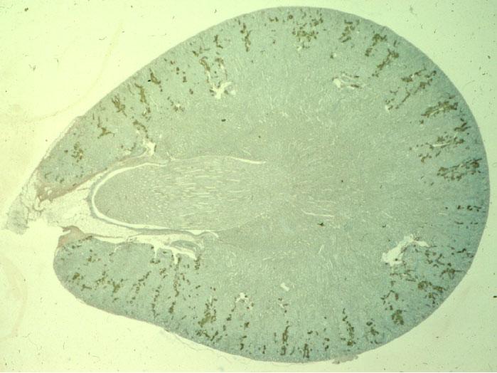
4 Detection of GFP in a transgenic mouse kidney using thick vibratome section (left) and 5 µm cryostat section (right) GFP (PC) - green V-ATPase (IC) - red IC
5 Double immunofluorescence staining - 5 µm cryostat section PC - AQP Basolateral IC - PP Apical

6 Golgi localization of Arf1 GTPase in LLC-PK1 cultured cells


7 Immunogold showing sidedness of epitopes External domain antibody Lumen Cytosol
8 Tissue/cell preparation - effects of fixation 1. No fixation - snap freezing 2. Light fixation Acetone/methanol Precipitating fixatives (Carnoy s ( Carnoy s, Bouin s solutions) 2-4% paraformaldehyde 3. Intermediate fixation Paraformaldehyde/lysine/periodate 4% paraformaldehyde/0.1% glutaraldehyde 4. Stronger fixation 1-4% glutaraldehyde (for EM structure)
9 Embedding/sectioning procedures Embedding/sectioning procedures 1. Light Microscopy Frozen sections - 5 µm cryostat or 1 µm semithin semithin Paraffin-embedded sections Resin-embedded embedded sections (Epon ( Epon,, hydrophilic resins) Whole mounts, cell cultures on coverslips/filters Thick vibratome sections 2. Electron Microscopy Frozen sections - ultracryomicrotomy Resin embedded sections Epon peroxidase or gold Resin embedded sections - K4K, HM20, LR resins Epon-embedding after pre-embedding embedding labeling with

10 Double immunofluorescence staining - 5 µm cryostat section PC - AQP Basolateral IC - PP Apical

11 SEMITHIN (1 MICRON) CRYOSTAT SECTION - AQP2/AE1

12 Whole-mount of vas deferens stained for proton pumps


13 Detection of cytosolic CaBP by immunogold on K4M sections

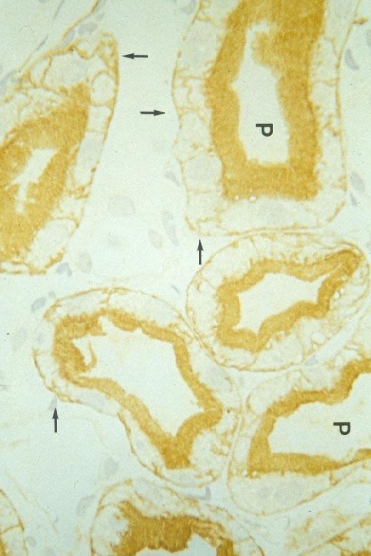
14 AQP2 on apical surface of principal cells using antibody against the external domain of AQP2 Pre-embedding gold labeling of tissue Epon embedding Note absence of intracellular label (gold)
15 Which technique do I use? Which technique do I use? 1. Light microscopy Fluorescence Peroxidase or other enzymatic method Amplification techniques (e. g., NEN-tyramide kit) 2. Confocal microscopy Conventional laser confocal (stacks, rotations) Dual-photon confocal (increased tissue depth) Spinning disk confocal (live cell imaging) 3. Electron microscopy Immunoperoxidase Immunogold
 AE1 (blue) Proton pump (red) V-ATPase (red) Proton-pump And FITC-dextran")
16 Triple immunofluorescence staining - 5 µm cryostat section FITC-dextran (green) AE1 (blue) AE1 (blue) Proton pump (red) V-ATPase (red) Proton-pump And FITC-dextran (yellow)

17 Immunoperoxidase Immunofluorescence
18 Tyramide amplification to detect antigens in different cells using antibodies raised in the same species 1st primary used very dilute, then amplified using FITC kit 2nd primary used at normal dilution and detected with IgG-CY3
19 Tyramide amplification HRP activity Ty-F + any protein H2O2 Conventional Indirect IF 2 antibody 1 antibody F F antigen FF F F F F F F F F F F F F HRP HRP F F F F F F Activated Ty-F is stable for only a short time - restricted diffusion


20 Immunogold labeling of proton pumps on microvilli - internal domain (Lowicryl-K4M Embedding)


21 Calbindin 28 in Principal cells of rat kidney Immunogold on K4M
22 Hints/Tips/Troubleshooting Increasing weak signals Concentration of antibody Brighter fluorophore Longer incubation Amplification Digital enhancement
23 (Adobe Photoshop levels command) Digital Enhancement


24 300 dpi 600 dpi 1200 dpi Effect of initial image Resolution on enlargement quality
25 Hints/Tips/Troubleshooting Antigen Retrieval Techniques Sodium dodecyl sulfate Microwaves plus acidic buffer Protease digestion Detergents (TX-100, saponin)

26 Antigen retrieval using pre- Treatment of tissue sections With SDS Syntaxin 3 staining in kidney With SDS No SDS Brown et al. (1996) Histochem Cell Biol,105: p261
27 Hints/Tips/Troubleshooting Reducing Background Quench aldehydes (NH4Cl) Protein/IgG blocking solutions High salt PBS (2.7% NaCl) Centrifuge antibodies Sharp microtome blades Other secondary reagents


28 Use of Evans Blue counterstain in immunofluorescence

29 Use of Photoshop levels for background removal


30 Digital Deconvolution - removes haze and sharpen image
31 Hints/Tips/Troubleshooting Controls Omit primary antibody Pre-immune serum Antigen/peptide inhibition Affinity purification of IgG Western blotting Other antibodies vs same protein Knockout mice
32 Thank You Thank You Mary McKee Peg McLaughlin Robert Tyszkowski Richard Bouley Sylvie Breton Christopher Brown Vladimir Marshansky Ivan Sabolic Tian-Xiao Sun Alfred Van Hoek Program in Membrane Biology Core