Food microbiology. Dr. Krisch Judit
|
|
|
- Primrose Osborne
- 5 years ago
- Views:
Transcription

1 Food microbiology Dr. Krisch Judit TÁMOP C-12/1/KONV
2 Most probably number (MPN) by Hoskins Make ten-fold dilution series Inoculate from all dilutions into a separate tube of an all-purpose broth medium. Incubate the tubes Observe for the presence or absence of growth. Create a key number Determine the MPN from the Hoskins table
3 MPN

4 Plate count methods Pour plate method
5 Pour plate method Make ten-fold dilution series Add 1 ml cell suspension from all dilutions into sterile petri dishes. Pour the appropriate medium into the Petri dish and mix it well with the solution Incubate the dishes Count the number of colonies (should be between 30 and 300) Determine the original cell number by the formula: colony count x dilution

6 Plate count methods
7 Spread plate method Make ten-fold dilution series Add 0.1 ml cell suspension from all dilutions to petri dishes containing the appropriate, solidified medium Spread the cell suspension over the medium surface with a glass spreader Incubate the dishes Count the number of colonies (should be between 30 and 300) Determine the original cell number by the formula: colony count x 10 x dilution
8 Cell number enumeration Colony forming unit (CFU)/1ml or 1g (From all single cell a colony will be formed) MPN: value from the table x dilution Pour plate: number of colonies x dilution Spread plate: number of colonies x 10 x dilution

9 Spread plate method
10 MPN is used for the estimation of total germ count, coliform count Poured plate is used for the enumeration of yeasts and facultative anaerobs Spread plate is used for the enumeration of aerobes and molds
11 Optimum temperature Optimum temperature for microorganisms is different Psychrophiles (0-25 C) - can grow in refrigerator Mesophiles (30-37 C) most microbes Termophiles (25-80 C) Bacterial spores can survive boiling (100 C)
12 Effect of heat treatment Mechanism of action of high temperatures: Denaturation of proteins Damage of cell membrane Cell death Mechanism of action of low temperatures: Inhibition of key enzymes in cell metabolism Growth reduction effect
13 Types of heat treatment in food industry Pasteurization kill vegetative forms mild (62-65ºC 30 min) fast (72-76 ºC s) very fast (85 ºC 1-2 s) ultra (132 ºC 1 s) Sterilization (121ºC 1,5 atm 20 min) kill all cells including bacterial endospores
14 Investigation on the effect of heat treatment Used microorganisms E. coli non spore forming B. subtilis spore forming Enumerate the cell count of the bacterial suspensions using the spread plate method Treat E. coli at 80 C and take samples at every 5 min. Determine the cell number of the samples using the spread plate method. Treat B. subtilis at 100 C and take samples at every 5 min. Determine the cell number of the samples using the spread plate method.
15 Investigation on the effect of heat treatment Create two tables with the number of survivals Create a time-kill curve with your data
16 Effect of disinfectants Aim: elimination of pathogens in vegetative form Disinfectants used in food industry Halogenic - Hypochlorous acid (HClO), iodine Peroxides - H 2 O 2, Peracetic acid CH 3 COOOH Quaternary ammonium compounds
17 Investigation on the effect of Hypochlorous acid and a quaternary ammonium compound Used microbes E. coli B. cereus Used disinfectants: Hypo (HClO) 2 % First (quaternary) 2% Solutions to stop the reaction: Na-tiosulfate Tween 40
18 Investigation on the effect of Hypochlorous acid and a quaternary ammonium compound Add 2-2 ml bacterium suspension to 18 ml disinfectant In every 5 min take 1 ml from the solution and add 9 ml stopper solution to it Make decimal dilutions from above described samples Estimate the number of survivals by the spread plate method
19 Effect of UV radiation UV cause DNA damage in the cell Microorganism have a repair mechanism When the time of UV radiation is too long or too intensive the damage will be irreversible
20 Effect of UV radiation Used microorganism E. coli Spread 0.1 ml E. coli suspensions on TGE mediums. Treat the plates under UV lamp for 5, 10, 15, 20 min Incubate the plates for 7 days at 37 ºC Count the survivors on the plates
21 Effect of osmolytes on the growth of microbes Osmotic pressure of solutions Isotonic normal material transport through the cell wall Hypertonic water goes out from the cell, the cell shrinks Hypotonic water goes into the cell, the cell swells, can burst
22 Effect of sucrose on the growth of yeasts Some yeasts are osmotolerant Used yeast: S. cerevisiae, S. diastaticus, Z. rouxii Used sucrose solutions: 5, 10, 20; 40 % Add 0.1 ml yeast cell suspension to 5 ml sucrose solution Incubate the tubes for 7 days at 30 ºC Count the cells in the tubes by spread plate method
23 Effect of salt on the growth of bacteria Some bacteria are halotolerant Used bacteria: E. coli, B. subtilis, B. cereus Used salt solutions: 2.5, 5.0, 7.5; 10 % Add 0.1 ml bacterium cell suspension to 5 ml salt solution Incubate the tubes for 7 days at 37 ºC Count the cells in the tubes by spread plate method
24 Optimal ph for the growth of microorganisms ph effect the membrane function ph effect also the function of enzymes Optimal ph Bacteria: Fungi: 5-7 (but in some cases can grow below ph 2)
25 Effect of ph on the growth of microorganisms Used microbes: E. coli, B. subtilis, B. cereus; S. cerevisiae ph solutions (Mcllvain-buffer): 2.2, 4.0, 6.0; 8.0 Add 0.1 ml microbe cell suspension to 5 ml buffer solution Incubate the tubes for 7 days at 30 ºC Count the cells in the tubes by spread plate method
26 Total germ count 1 Weight out aseptically 10 g or 10 ml from the examined food Solid food should be masticate Add it to 90 ml sterile physiological saline (PS) Mix well Make a tenfold dilution series (5-6 dilution)
27 Total germ count 2 Inoculate 1 ml from all dilution into 3-3 tubes containing TGE broth Incubate the tubes (24 h, 37 ºC) Observe for the presence or absence of growth turbidity, color change. Create a key number: take into account the last dilution where all tubes were positive then the subsequnt 2 dilutions and write the number of positive tubes
28 Total germ count 3 Key number is: 320 From the Hoskins table you can find the MPN value: 0,93 Multiply this value with the dilution factor of the first member of the key number: 0,93 x 10 2 CFU/g
29 Determination of aerobic spore forming bacteria 1 Members of the genus Bacillus can be found in this group B. subtilis, B. cereus, B. megaterium causing food spoilage and poisoning At 80 ºC vegetative cells die but the spores survive After incubation the spores will germinate and all cells in the broth comes from the germinated spores
30 Determination of aerobic spore forming bacteria 2 Weight out aseptically 10 g or 10 ml from the examined food Solid food should be masticate Add it to 90 ml sterile physiological saline (PS) Mix well Make a tenfold dilution series (5-6 dilution)
31 Determination of aerobic spore forming bacteria 3 Inoculate 1 ml from the first 3 dilutions into TGE broth Heat the tubes at 80 ºC for 20 minutes Incubate the tubes (24 h, 37 ºC) Observe for the presence or absence of growth - turbidity. Create a key number as described above
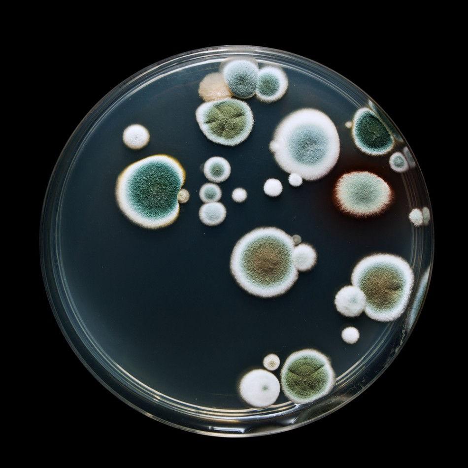
32 Determination of anaerobic spore forming, H 2 S producing bacteria 1 Members of Clostridium genus can be found in this group From sulfite they can form H 2 S and if Fe-citrate is present in the broth FeS will be formed FeS gives black color to the broth Anaerobic environment is created by adding an oil layer on the surface of the broth RCM broth is used containing cystein (sulfur containing amino acid) and Fe-citrate
33 Determination of anaerobic spore forming, H 2 S producing bacteria 2 Weight out aseptically 10 g or 10 ml from the examined food Solid food should be masticate Add it to 90 ml sterile physiological saline (PS) Mix well Make a tenfold dilution series (5-6 dilution)
34 Determination of anaerobic spore forming, H 2 S producing bacteria 2 Inoculate from the first two dilutions to RCM broth Heat the tubes at 80 ºC for 20 minutes Close the tubes with a paraffin layer Incubate the tubes (24 h, 37 ºC) Observe for the presence or absence of growth turbidity and/or black coloration. Positive: turbidity non H 2 S forming anaerobes; black color - H 2 S forming anaerobes
35 Determination of yeasts and molds Weight out aseptically 10 g or 10 ml from the examined food Solid food should be masticate Add it to 90 ml sterile physiological saline (PS) Mix well Make a tenfold dilution series (3 dilution)
36 Determination of yeasts Pour plate method Yeasts are able to grow under semi-anaerobic conditions. Use malate extract (ME) agar with antibiotic to prevent the growth of bacteria. Add 1 ml from each dilution to sterile Petri dishes. Pour lukewarm (40 C) ME agar on the inoculum (about 15 ml) and mix it carefully by swirling. After the agar is solidified invert the Petri dish and incubate it for 24 h at 30 C.
37 Determination of molds Spread plate method Molds are strictly aerobes. Can grow only on the surface of a medium. Use malate extract (ME) agar with antibiotic to prevent the growth of bacteria. Drop 0.1 ml suspension from all dilution on prepared ME medium and spread it evenly on the surface of the agar. Incubate for h at 30 C.
38 Determination of cell count Count plates with colonies Yeasts: colony number x dilution factor Molds: colony number x 10 x dilution factor


39 Yeasts and molds counting
40 Determination of Salmonella In the Salmonella genus Gram negative rod shaped bacteria can be found. There are two species S. enterica and S. bongori, 6 subspecies and a high number of serovars. Salmonella species are pathogens causing food poisoning. Determination of Salmonella is the duty of official laboratories. In the European Union no Salmonella is allowed in 25 g food.
41 Determination of Salmonella Step 1 Sometimes the number of salmonella is very low. Before determination we have to make an enrichment step. Weight out aseptically 25 g or 25 ml from the examined food. Add it to 225 ml pre-enrichment broth (Trypticasein Soy Broth (TSB). This broth is not selective, it is used for the recovery of injured cells. Incubate for 24 h at 37 C.
42 Determination of Salmonella Step 2 Selective enrichment of Salmonella species in Rappaport Vassiliadis Broth. Malachite green and Mg chloride inhibit the growth of other bacteria (Shigella). Add 10 ml from the TSB broth to 90 ml Rappaport Vassiliadis Broth. Incubate for 24 h at 37 C.
43 Determination of Salmonella Step 3 Selective determination of salmonella species on Rambach Agar. This selective medium is inhibitory for many microorganisms other than Salmonella. Streak a loop from the enrichment broth on Rambach Agar. Incubate for 24 h at 37 C. Salmonella colonies are red, coliforms form blue and other bacteria colorless colonies.


44 Rambach Agar Red colonies: Salmonella spp. Blue colonies: E. coli Author: CHROMagar, France
45 Verification of the presence of Salmonella on TSI Agar Triple Sugar Iron (TSI) Agar is used. Contain: 1% lactose, 1% sucrose, 0.1% glucose; phenol red as ph indicator and compounds for the production of hydrogen sulfid. An agar slant is made with aerobic and anaerobic areas. Positive colonies from Rambach agar are picked up on a needle and then TSI slant is inoculated by first stabbing the butt down to the bottom, and then streaking the surface of the slant.
46 TSI Agar Interpretation of the results Many enterobacteria ferment glucose with the production of acids which will change the color of the medium. However, glucose will used up soon and the organisms in the presence of oxygen are forced to catabolize peptones and amino acids for their energy supply. Alkaline end-products from these substances will revert the ph of the slant to an alkaline ph and thus change the color of the agar slant back to red (after hours).
47 TSI Agar Interpretation of the results Organisms such as Salmonella spp. or Shigella spp. and other organisms which attack glucose but do not ferment lactose or sucrose will produce an alkaline slant and acid butt in TSI slants in 18 to 24 hours. If the glucose is metabolized to CO2, the gas will be seen as bubbles or cracks in the agar butt. If hydrogen sulfide is formed during growth, a gray or black streak of iron sulfide is seen. Organisms which attack lactose and/or sucrose, such as Escherichia, will produce acid slants and acid butts usually with the formation of gas.
48 TSI Agar Interpretation of the Agar Butt results Yellow color Red color Black color Bubbles or cracks Glucose fermentation No glucose fermentation H 2 S production Gas production during glucose, lactose fermentation
49 TSI Agar Interpretation of the results Agar surface Yellow Red Lactose and/or sucrose fermentation No lactose and/or sucrose fermentation Salmonella positive: agar butt: yellow with black, gas formation, red surface.


50 Bacteria on TSI agar slants From left to right: Control; P. aeruginosa, E. coli, Salmonella Typhimurium, Shigella flexneri
51 TSI reactions Microorganism S. Paratyphi A S. Typhimurium S. Paratyphi B S. Enteritidis Sh. dysenteriae Ent. aerogenes E. coli Citrobacter spp. Klebsiella spp. Pr. vulgaris Agar putt Y+G Y+G Y+G Y+G Y Y+G Y+G Y+G Y+G Y+G Agar surface R R R R R Y Y Y Y Y Formation of H 2 S Y= yellow color (acid production) Y+G= yellow color +gas production R= red color, no change in color (no acid production) or alkaline ph revert yellow color back to red += H2S production, black color K. pneumoniae Y/Y+G R - -= no H2S production Ps. aeruginosa R R -
52 Determination of Enterobacteriaceae 1 Gram negative bacteria, members of the Salmonella, Proteus, Klebsiella, Serratia, Citrobacter, Escherichia, Enterobacter, Shigella can be found here Enterobacteria Enrichment (EE) broth is used in the first step Brilliant green and ox-bile inhibit the growth of Gram-positive bacteria
53 Determination of Enterobacteriaceae 2 Weight out aseptically 10 g or 10 ml from the examined food Solid food should be masticate Add it to 90 ml sterile physiological saline (PS) Mix well Make a tenfold dilution series (5-6 dilution)
54 Determination of Enterobacteriaceae 3 Inoculate 1 ml from all dilution to 3-3 tubes containing EE broth Incubate the tubes (24 h, 37 ºC) Observe for the presence or absence of growth - turbidity Create a key number as described above Based on the MPN table add the cfu/g or ml
55 Determination and identification of glucose fermenting Enterobecteriaceae Use VRBG Agar VRBG = Violet Red Bile Glucose agar will exclude lactose fermenting coliform bacteria. Bile salts and crystal violet inhibit the growth of Gram-positive cocci. Glucose fermenters produce red colonies with redpurple halos in the presence of Neutral Red, the ph indicator.
56 Determination of glucose fermenting Enterobecteriaceae Streak from the positive tubes a loopful inoculum on VRBG agar Incubate the plates for 24 h at 37 ºC Pink colonies with a reddish halo verify the presence of glucose fermenting Enterobacters.
57 Streaking plate technique
58 Streaking plate technique 1. Streak a line with the loop containing the inoculum at the top end of the agar plate until 1/3 of the plate is covered 2. Sterilize the loop in the flame, cool it and move it in a zig-zag horizontal pattern over the straight line. 3. Rotate the plate about 60 degrees and streak a new line 4. Sterilize the loop again and repeat step 2 5. Rotate the plate about 60 degrees and streak a new line 6. Sterilize the loop again and repeat step 2 7. At the end you will get individual colonies

59 E. coli on VRBG Agar
60 Determination of coliforms 1 The coliform group includes aerobic and facultative anaerobic, Gram-negative, non-spore forming bacilli that ferment lactose and form acid and gas at 35 C within 48 hours. Brilliant Green Bile Broth (BBL) is used to confirm the presence of coliforms. Oxbile and Brilliant Green inhibit Gram-positive bacteria and many Gram-negative bacteria, other than coliforms. During lactose fermentation gas production occur. This is detected by Durham tubes.
61 Determination of coliforms 2 Weight out aseptically 10 g or 10 ml from the examined food Solid food should be masticate Add it to 90 ml sterile physiological saline (PS) Mix well Make a tenfold dilution series (5-6 dilution)
62 Determination of coliforms 3 Add Durham tubes to the BBL broth. Inoculate 1 ml from all dilution to 3-3 tubes containing BBL broth Incubate the tubes (24 h, 37 ºC) Observe for the presence or absence of coliforms turbidity with bubble formation. Create a key number from the positive tubes as described above Enumerate cell count from the MPN table

63 Brilliant Green Bile Broth From left to right: Uninoculated BBL broth Non-coliform bacterium Coliform bacterium
64 Verification of the presence of E. coli Indole is generated by reductive deamination from the amino acid tryptophan. E. coli is able to produce indol.
65 Verification of the presence of E. coli From the coliform positive tubes add 1 ml to tryptophan containing broth. Incubate for 24 h at 44 ºC. Add 5 drops of Kovac's reagent to the culture broth. Positive result: red/pink layer at the top of the tube.

66 Positive indole test

67 Determination of coliforms from water 1 Membrane Filtration (MF) method is used Filter 100 ml sample through a membrane filter with pore size of 0.45 µm.
68 Determination of coliforms from water 2 Place the filter on Chromocult Coliform Agar Incubate for 24 h at 37 ºC Count the colonies with the different colors Blue colonies: E. coli, pink colonies: coliforms Using other chromogenic media you can get different colony colors.

69 Determination of coliforms from water
70 Determination of Enterococci 1 Gram positive cocci, Enterococcus faecalis and Enterococcus facium are indicators of fecal contamination Enterococcus selective broth is used in the first step Sodium azide inhibit the growth of other bacteria
71 Determination of Enterococci 2 Weight out aseptically 10 g or 10 ml from the examined food Solid food should be masticate Add it to 90 ml sterile physiological saline (PS) Mix well Make a tenfold dilution series (5-6 dilution)
72 Determination of Enterococci 3 Inoculate 1ml from all dilution to 3-3 tubes containing Enterococcus selective broth Incubate the tubes (24 h, 37 ºC) Observe for the presence or absence of growth - turbidity Create a key number as described above Enumerate cell count from the MPN table
73 Verification of the presence of Enterococci on Chromocult Enterococci Agar Growth of Enterococci is stimulated. They cleave the chromogenic substrates in the medium resulting in red colonies. Sodium azide and ox bile inhibit most accompanying microbial flora. Non-Enterococci produce colourless, blue/violet or turquoise colonies.
74 Verification of the presence of Enterococci Streak from the positive tubes a loopful inoculum on Chromocult Enterococci agar Incubate the plates for 24 or 48 h at 37 ºC Red colonies verify the presence of Enterococci.


75 Chromocult Enterococci Agar Aerococcus faecalis Aerococcus viridans
76 Determination of Staphylococcus aureus
77 Determination of Staphylococcus aureus Staphylococcus aureus is a gram positive coccus. The coagulase positive strains causing hemolysis are considered to be human pathogens. The methycillin resistant S. aureus (MRSA) can cause even death.
78 Baird Parker agar Baird Parker Agar is used for the isolation and differentiation of coagulase-positive staphylococci in food. The medium contains lithium and tellurite, which inhibit most of the contaminating microflora, while glycine and pyruvate enhance Staphylococci growth..
79 Baird Parker agar Staphylococci can reduce tellurite to telluride, which results in grey to black coloration of the colonies. With the addition of egg yolk, the medium becomes yellow slightly opaque. A clear halo develops around colonies from coagulase positive Staphylococcus aureus
80 Determination of Staphylococcus aureus 1 Weight out aseptically 10 g or 10 ml from the examined food Solid food should be masticate Add it to 90 ml sterile physiological saline (PS) Mix well Make a tenfold dilution series (3 dilution)
81 Determination of Staphylococcus aureus 2 Inoculate Baird Parker Agar with 0.1 ml suspension from all dilutions Spread the inoculum over the medium Incubate for 24 h at 37 C. Count the black colonies with a clear halo around. CFU/g= colony count x 10 x dilution factor

82 S. aureus on Baird Parker Agar Author: Matthias M.