Jill L. Caudill M.Ed. SCT(ASCP) Program Director Cytotechnology Program Mayo Clinic Rochester, MN
|
|
|
- Nicholas Hoover
- 5 years ago
- Views:
Transcription
 Program Director Cytotechnology")
1 Jill L. Caudill M.Ed. SCT(ASCP) Program Director Cytotechnology Program Mayo Clinic Rochester, MN
2 None
3 o o o o o History and rationale for changes in our practice Fluorescence in situ hybridization (FISH) Assistance with FNA - EBUS Tissue review for molecular testing Additional roles for Cytotechnologists
4 2000 ogyn onon-gyn oeus FNA odigital Image Analysis 2011 o FISH o FNA prescreens o ER/PR and Her2 review o Specimen problem solving o Circulating Tumor Cells o Tissue identification o AFB screens o Test Development o Education and Quality o EBUS FNA

5 Number of Specimens 120, ,000 80,000 60,000 40, ,000 0 Non-GYN FNA GYN Other Specimen Type
6 Number of Path Reviewed Cases GYN Cases and HPV Results Cytology Monthly Case Volume Trends FNA Non-GYN 3000 GYN Path Review HPV Results Reported GYN Jan-10 Feb-10 Mar-10 Apr-10 May- 10 Jun-10 Jul-10 Aug-10 Sep-10 Oct-10 Nov-10 Dec-10 Jan-11 Feb-11 Mar-11 Apr-11 May Month
7 Test Volumes Additional Roles of Cytotechnologists Tumor Identification AFB CellSearch ER/PR/HER2 FISH DIA Automated HER2 Implemented CellSearch Test Implemented Tumor Identification and AFB Screening Implemented ER/PR/HER2 CT review Implemented DIA Test Implemented FISH Test Implemented Year
8 Molecular Volumes Volumes 100% 90% 80% 70% 60% 50% 40% 30% 20% 10% 0% Year CTC Breast IHC FISH DIA
9 2011 Tissue Review Volumes Jan Feb Mar April May June July August Sept
10 In 2005 our laboratories split into two labs: Cytopathology and Molecular Cytology and Imaging There are shared CTs between the two labs In 2011 molecular cytology and molecular anatomic pathology laboratories were combined
11 Created Roles for Cytotechnologists that make sense for the individual practice (driven by need to save pathologist time and understanding the unique skills of the cytotechnologist) Developed Cytotechnology curriculum that supports the expanded cytotechnology roles
12 FISH Testing: UroVysion- urine specimens-2000 Cytotechs read in cytogenetics 2000 Molecular Cytology lab (processing and reads) Biliary Brush specimens-2004 Bronchial Brush specimens-2006 Esophageal Brush, Barrett s neoplasia-2011
13 Why Cytotechnologists are Well Suited for FISH Analysis We re good at systematic scanning Normal appearing cells Abnormal appearing cells - FISH normal Abnormal appearing cells - FISH abnormal
14 We re used to evaluating nuclei DAPI Stain DAPI counterstain helps highlight artifacts, but more importantly allows visualization of a cell s nuclear morphology Thus, the filter used to assess the DAPI stain is used for scanning purposes
15 Normal cells should have benign morphological features like smooth nuclear borders and homogenous chromatin Abnormal cells usually have atypical morphological features such as enlarged nuclei, mottled chromatin, and irregular nuclear borders Normal Abnormal

16 Signal patterns for abnormal cells are recorded from left to right in the following order: # of chromosome 3 copies # of chromosome 7 copies # of chromosome 17 copies # of 9p21 gene region copies p


17 Definition = having an extra copy or copies of more than one different chromosome Example signal patterns: CEP 3 CEP 7 CEP 17 9p Polysomy

18 Cytotechnologists notice subtle differences Signals may be splitting/fragmented and hard to count (especially common with the red probe)

19 Background debris, bacteria, intensely stained signals, etc may pick up probe and thus appear on another or all filters


20 Two cells located in close proximity may appear to be one cell with an abnormal signal pattern
21 Support of leadership Organizational structure of lab Development team


22
23 Cytotechnologists and others Research and validation of testing New vocabulary required when crossing border into more clinical lab testing Controls critical Working with vendors
24 Probe Targets 5p15 CEP 6 7p12 8q24 Green Blue Red Gold


25 Polysomy Polysomy Tetrasomy Tetrasomy
26 Clinician sends a bronchial brushing Orders FISH reflex FISH diagnosis of: Negative Cytology diagnosis of: One slide used for FISH testing Tetrasomy Polysomy Routine cytology is performed 2 slides Negative Atypical Suspicious Positive Positive biopsy or TBNA (Hyper-tetrasomy) No further testing Signed out as Positive No further testing
27 +500 tests performed Cost Cytology ~$250 FISH ~$550 Reimbursement Created a database with clinical info and followup
28 Retrospective studies Voss JS, Kipp BR, Halling KC, Henry MR, Jett JR, Clayton AC, Rickman OB. Fluorescence in situ hybridization testing algorithm improves lung cancer detection in bronchial brushing specimens. Am J Respir Crit Care Med 2010;181: FISH increases the detection of lung cancer for indeterminate peripheral nodules < 2 cm Cytology: sensitivity = 3% FISH: +44% > 2 cm Cytology: sensitivity = 26% FISH: +28%


29
30

31

32
33 Test Path Analysis Time Reduction Expense Reduction FISH 96% 71% Breast IHC 86% 67% CTC 71% 41% Tissue Review (first year) 33% Not calculated at this point
34 Cytotechnologist FISH Workflow Match Paperwork and Slides 43 minutes per case FISH Analysis Capture Images for Permanent File Enter FISH Interpretation into LIS
35 Pathologist FISH Workflow Review Signal Patterns 2 minutes per case Review Representative Images Review Patient Clinical Information Verify/Release Report in LIS
36 *Average CT and Pathologist Salaries taken from most recent ASCP survey and Physician Salary Survey: Modern Healthcare;2009, Vol 39, With CT analysis Without CT analysis CT time (min) Pathologist Time (min) N per year Salary Cost* $149, $531,127 Savings -$375,598
37 *Average CT and Pathologist Salaries taken from most recent ASCP survey and Physician Salary Survey: Modern Healthcare;2009, Vol 39, IHC- ER, PR, HER 2 Analysis CT time (min) Pathologist Time (min) N per year Salary Cost* With CT analysis Without CT analysis $91, $274,980 Savings -$183,480
38 * Average CT and Pathologist Salaries taken from most recent ASCP survey and Physician Salary Survey: Modern Healthcare;2009, Vol 39, Circulating Tumor Cell Analysis CT time (min) Pathologist Time (min) N per year Salary Cost* With CT analysis Without CT analysis $16, $27,992 Savings -$11,485

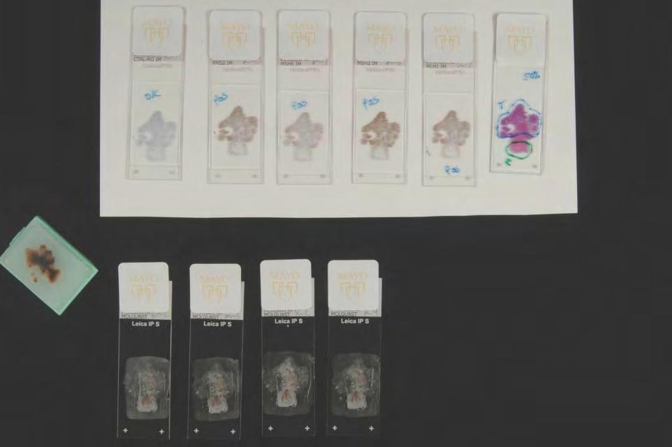
39
40 Identify training initiatives, objectives, and resources Familiarize participants with theories, principles, and terminology Microscopy 50 cases as second reviewer 50 cases as first reviewer Concordance requirement

41 Previously 4-6 months to complete while working Established online module within Cytotechnology Program Working cytotechnologists now expected to enroll in module to complete training and work in FISH area

42
43 Control slides Monitor assay performance Assess signal characteristics Any abnormal case other than high percentage positive, must be reviewed by 2 nd tech QC program Similar to cytology GYN QC 5% of CTs cases/month Documentation Proficiency Testing CAP


44 2011
45 New in fall of 2011 EUS GI FNA adequacy since 2000 AM and PM rotations one CT each EBUS AM rotation only

46 Since 2009 AP Research rotation
47 Specimen/Tumor Test Lung (EGFR) KRAS ALK 1 Melanoma Melanoma/GIST Colon & Endometrium BRAF KIT MLH1 MSH2 MSH6 PMS2 MSI KRAS
48 2011 Tissue Review Volumes Jan Feb Mar April May June July August Sept

 MLH 1 Interpret Pos/Neg IHC Cytology Sequencing if loss")
49 Specimen/ Tumor Test CT Role Technique(s) Performing lab Comments/Rationale EGFR none sequencing Genzyme Mutation analysis determines likeliness of responding to EGFR targeted therapies Lung KRAS Block Selection Tumor marking % tumor Real time PCR - sequencing Molecular Genetics EGFR TKIs not successful/some adverse effects - different protocol required ALK 1 Block Selection Tumor marking FISH (also IHC and RT-PCR possible) Cytogenetics Drug protocol -different from EGFR/KRAS drugs Melanoma BRAF Block Selection Tumor marking % tumor PCR fragment size analysis Molecular Genetics New drug Vemurabenib Melanoma/ GIST KIT Block selection; % whole slide indication sequencing Molecular Anatomic Pathology KIT inhibitors (Gleevac) MLH 1 Interpret Pos/Neg IHC Cytology Sequencing if loss by IHC; MLH1 hypermethylation and BRAFV600 can also help determine somatic vs risk of germline Colon and Endometrium HNPCC MSH 2 Interpret Pos/Neg IHC Cytology MSH 6 Interpret Pos/Neg IHC Cytology PMS Mismatch 2 Repair Genes Interpret Pos/Neg IHC Cytology MSH2/MSH6 loss - probably HNPCC - send to sequence MSH2/MSH6 loss - probably HNPCC - send to sequence PMS2 loss only- sequence, likely HNPCC; If MLH1/PMS2 - either somatic change or germline mutation in MLH1 (see MLH1) MSI Block selection; Tumor marking; Normal control marking; % PCR fragment size analysis Molecular Genetics Can be done for somatic/germline determination or prognostic factors. MSI High (and MLH1 hypermethylation) likely HNPCC (lower stage, better prognosis and indications for drug protocols) MLH 1 Block selection; Tumor marking; Normal control marking; % PCR - hypermethylation Molecular Genetics Done when MLH1 IHC is negative; hypermethylation suggests somatic change (not HNPCC)
50 Case Review: -Review orders and paperwork -Ensure proper tissue/block sent -May need to select best block and order recuts -Review slides and mark appropriately -Review IHC cases and indicate Pos/Neg


51


52

53

54

55 4x 60% tumor with all the pieces KRAS case


56 TBNA ALK case Cell blocks can be more challenging 70% tumor 30% lymphs instructions to Cytogenetics describing tumor nuclei 20x

57 20x
58


59
60 Tissue review with pathologist Sit with Education specialist QA Quarterly review CAP Proficiency

61 QA/QC KRAS w block selection Circling & % EE 76
62 Performance comparisons on slides EE DD KK EE DD KK 16 28


63 QA/QC MLH1HM/BRAF #2 DD II KK LL Marking and percentage comparisons


64 QA/QC MLH1HM/BRAF #2
65 Avoid lymphoid aggregates Avoid fat for normal Avoid tumor floaters around normal continues to be problematic. Now only needs to be done for MSI not MLH1HM
66 Establishing trust with new non-cytology pathologists Not familiar with our skills Learning curve for both Rapidly changing testing environment
67 Set up education Vendor information/training Background resources If tissue review with pathologist to assist in setting up necessary primary lecture/scope work Pathologist accessible for questions Training protocol Thresholds for concordance QA/QC plan
68 Calculation being reset to be similar to those used for FISH and other tests Compare time savings

69 IHC Quantitation ER/PR Her2 Circulating Tumor Cells AFB reads

70 Colon Cell Search System Breast Prostate
71

72 Expanded Cytotechnologist Role? Changed Pathologist-Cytotechnologist Relationship? Still work as team New group of pathologists to work with Created Visibility for Cytotechnologists in Department/Institution? Impact on resource allocation? Yes Yes YES!
73 We have not worked outside the current regulatory environment No change in CPT codes/billing Pathologist does final review Cytotechnologist as an Independent Practitioner Much bigger than our single practice can change
74 Trying to keep pace with changing times Relieved that our path is in line with society and market analysis conclusions Positive upbeat about future of cytology and the role that cytotechnologists will play Use our practice needs (pathologist shortage, economic press of practice, demand for new improved technologies) to define education curriculum for cytotechnologists.
75 Career Ladder Cytotechnologist Senior Cytotechnologist Lead Specialist Education/Training Focus Pathologist Assisting Focus Quality Focus Development Technologist Assistant Supervisor Supervisor
76 Schizophrenic GI less/more Biliary cytology/fish Esophageal cytology/fish Colon tissue review More education/training as new tests come up Decisions on how to provide education to working cytotechnologists as well as students
77 Use Cytotechnologists in Expanded Roles Provide cost effective service Reduce burden on Pathologists Preserve the field of cytotechnology in effect preserving the application of morphologic assessment on numerous aspects of laboratory testing Enhance satisfaction for cytotechnologists


78 Our Decade of Change ( ): Optimize the current scope of practice without additional formal education on the job training Looking Ahead from 2010 Expand Cytotech role with novel educational tools

79 We don t do blood We won t give up our microscopes We only look at tissue with cytology cases


80

81