EFFECT ASSESSMENT OF SYNTHETIC GLUCOCORTICOIDS TO THE SELECTED FRESHWATER INVERTEBRATE AND VERTEBRATE SPECIES
|
|
|
- Chloe Jennifer Hodges
- 5 years ago
- Views:
Transcription
1 EFFECT ASSESSMENT OF SYNTHETIC GLUCOCORTICOIDS TO THE SELECTED FRESHWATER INVERTEBRATE AND VERTEBRATE SPECIES A THESIS SUBMITTED IN FULFILMENT OF THE REQUIREMENTS FOR THE DEGREE OF DOCTOR OF PHILOSOPHY NAVDEEP BAL B. Tech, M.S. SCHOOL OF SCIENCE, COLLEGE OF SCIENCE, ENGINEERING AND HEALTH RMIT UNIVERSITY JULY, 2017
2
3 DECLARATION I, Navdeep Bal, declare that this thesis contains my original work which is submitted as a requirement in full for the degree of Doctor of Philosophy. I certify that except where due acknowledgement has been made, the work is that of the author alone; the work has not been submitted previously, in whole or in part, to qualify for any other academic award; the content of the choose an item is the result of work which has been carried out since the official commencement date of the approved research program; any editorial work, paid or unpaid, carried out by a third party is acknowledged; and, ethics procedures and guidelines have been followed. NAVDEEP BAL JULY, 2017 i
4 RESEARCH ABSTRACT Anthropogenic activities in the aquatic environment have resulted in an unmanaged disposal of pharmaceuticals and personal care products (PPCPs), which is a cause for concern. An emerging class of aquatic contaminants that scientists are becoming aware of are glucocorticoids (GCs) from the category of steroid hormones. GCs in their natural form as cortisol controls metabolic, reproductive, immunity and stress related responses in mammals and teleosts. However, highly potent synthetic GC drugs like dexamethasone (DEX), prednisolone (PDS) were developed for human medication in order to treat severe inflammatory diseases and to minimize the side effects of GCs since there were problems with the high doses and clinical GC resistance. Recently GC activity (based on reporter gene assays) have been detected in effluents from hospitals and sewage treatment plants (STPs) in Australia. Since these treated STP effluents are released into rivers, lakes or creeks, the biological effects of GCs on aquatic organisms are of concern, yet information on their effects is very limited. The present study aimed at effect assessment of synthetic GCs to selected freshwater invertebrate and vertebrate species such as crustaceans (Ceriodaphnia dubia), gastropod (Physa acuta), and early life stage (ELS) of fish species, Maccullochella peelii and Macquaria ambigua. Multigenerational (F0-F3) PDS and DEX exposures were conducted to determine the effects on the life-history traits and at population level of C. dubia. There was a positive trend of increased toxicity followed by reduced life history traits such as fecundity, brood size, time to first brood and intrinsic rate of population increase and body growth (length and area) of C. dubia in the case of both studied chemicals. Exposed species exhibited more sensitivity to ii
5 DEX than PDS chemical and calculated EC50 (95% CI) values in F3 generation were 18.2 µg/l (CI: ) for DEX and µg/l (CI: ) for PDS. The preliminary investigation of 42 d PDS exposure to P. acuta showed premature and delayed embryonic hatching at µg/l and 125 µg/l PDS respectively. Lower calcium (Ca) concentrations and impairment of shell development in juveniles at 62.5 µg/l, resulted in shell thinning. Further to this, the multigenerational PDS exposure (126 d) presented a deep insight into the developmental malformations, in particular, shell structure and ontogenesis suppression in almost all predominant life-cycle stages (egg, juvenile and adult) of P. acuta snails. LOEC (lowest observed effect concentration) for multigenerational PDS exposure was at 4 µg/l as measured from biological, molecular, biochemical and morphological indices in exposed F0-F2 generations. Long-lasting anomalies in epigenetic phenomena resulted in transmission of toxic effects into the produced progeny. In addition to multigenerational invertebrate studies, the present research has provided a great insight into GC toxicity to ELS of two Australian native freshwater fish species, Murray cod and golden perch. The three studied GC drugs (PDS, DEX and hydrocortisone-hc) were found to have toxic effects of varied magnitude on the exposed larvae as evident from measured biological, cellular, histological, morphological and behavioural (swimming and feeding) endpoints during 14 d exposure period. Golden perch larvae were observed to be more sensitive than the Murray cod larvae when exposed to the same drug concentration in identical exposure conditions. Survival EC20 (95% CI) values for DEX, PDS and HC were 0.03 µg/l ( ), PDS: 103 µg/l ( ), HC: 281 µg/l ( ) respectively for 7 dph (days post hatching) golden perch larvae and 0.22 µg/l ( ), PDS: 164 µg/l ( ), HC: µg/l ( ) respectively for Murray cod. iii
6 Further to preliminary GC investigation to fish ELS, two independent ELS exposure bioassays were conducted to determine the effects of chronic PDS exposures on different developmental stages of Murray cod using newly hatched and unexposed 14 dph larvae. Glucocorticoid induced-osteoporosis (GIOP) like symptoms were observed in PDS exposed 35 dph larvae. Histopathological analysis of the morphogenesis of eye structure exhibited cataract-like phenotypes in PDS treated larvae. Ca and phosphorous (P) concentrations were significantly reduced at and 15.6 µg/l respectively. Larvae dependent on external food exhibited the greatest sensitivity towards PDS toxicant and their sensitivity was greater than that of early yolk-sac larvae. This finding was evident in the multiple physiological, morphological and behavioural parameters studied. The present study has further investigated the influence of temperature and synthetic GCs on the organogenesis of golden perch larvae. Survival and yolk-sac depletion rate of exposed 7 dph larvae at the extreme higher and lower test temperatures (30 o C and 15 o C) and GC (PDS and DEX) exposures were significantly different to those exposed to GCs at the other two test temperatures (20 and 25 o C). In addition to short term exposure, chronic 21 d PDS exposure at 21 o C significantly affected the biological, craniofacial development and behavioural activities (feeding activity and touch-evoke response) of the exposed golden perch. Cell death (apoptosis) in the 96 hph PDS exposed larvae was found to be increasing with an increased drug dose. Short term exposure of fish ELS to higher or lower temperatures in combination with additional chemical stressors like synthetic GCs; or chronic exposure to GCs at median temperatures, could potentially imbalance the population and ecosystem and limit the capacity of fish populations to respond effectively to changing conditions. The present study has highlighted the importance of multigenerational and chronic ELS exposure studies of invertebrates and vertebrates in identifying the responses of stressed non- iv
7 target aquatic organisms under long-term exposure of GC compounds. Furthermore, worst case screening level risk assessment through calculation of HQs showed PDS and DEX as high risk contaminants and revealed that there is a potential ecological risk associated with release of these GC chemicals into the aquatic ecosystems. Both Murray cod and golden perch fall under a category of endangered or threatened Australian freshwater fish species. Results of the present study cumulatively provide a reference point to utilize freshwater Australian native species to monitor GC pollution. The continual and uncontrolled exposure of anthropogenic GC chemicals in an aquatic ecosystem may result in reduction in local biodiversity by reducing total number of species which become sensitive to a chemical stressor over a period of subsequent generations and thereby affecting them at their population level. v
8 ACKNOWLEDGEMENT First and foremost I give thanks to the Almighty Lord for all the blessings he has bestowed upon me and made this Ph.D. possible. I thank my beloved parents for their continued faith, support and understanding throughout my academic career. Special thanks to my loved ones who stood always with me and became an integral part of this journey. I would like to express my sincere gratitude to my supervisors Prof. Dayanthi Nugegoda (RMIT) and Dr. Anupama Kumar (CSIRO) for their substantial and continued advice, support and encouragement. I am grateful to RMIT University and CSIRO for the fee help and research opportunities. I would also like to thank staff of ecotoxicology team (Hai Doan, Debra Gonzago and Adrienne Gregg) at CSIRO for their help and advice. I am thankful to Dr Jun Du for her time and help with the analytical chemistry work. A special thank you to microscopy and histology staff of Adelaide University for training on different instruments. Thank you to Cathy Fiebiger, Caroline Johnston and John Gouzos (CSIRO) for assisting with ICP-AES analyses. NAVDEEP BAL vi
9 TABLE OF CONTENTS DECLARATION... i RESEARCH ABSTRACT... ii ACKNOWLEDGEMENT... vi LIST OF TABLES... xiii LIST OF FIGURES... xvi LIST OF ABBREVIATIONS... xxv LIST OF PUBLICATIONS... xxx CHAPTER Background introduction Glucocorticoids as an emerging contaminant Therapeutic effects Administration and pharmacokinetics Mode of action Environmental presence of glucocorticoids Glucocorticoid-induced toxicity: a journey from humans to aquatic species Aim and objectives of the present study Test species used in the present study Ceriodaphnia dubia (cladocera) Physa acuta (class gastropoda) Fish early life stage Murray cod (Maccullochella peelii, mitchell, 1838) Golden perch (Macquaria ambigua, richardson, 1845) Thesis structure vii
10 References CHAPTER Introduction Materials and methods Test organisms and culture maintenance Algae culturing Test chemicals Experimental design for toxicity bioassays Acute test Multi-generational toxicity experiment Statistical analyses Results Physical parameters and toxicant concentrations in the test solutions Acute toxicity test Multigenerational experiment Fecundity Average time to first brood Mean brood size Body length Intrinsic rate of population increase Discussion Acknowledgement References Supplementary information CHAPTER Introduction Materials and methods Culturing of test specimens Embryo toxicity test viii
11 3.2.3 Post-hatching juvenile toxicity test Quantifying shell thickness using scanning electron microscope Inductively coupled plasma (ICP) analyses Chemical analyses of the water in the testing beakers Data analyses Results Water quality and physical conditions Water chemistry analyses Embryo toxicity test Biological effects Post-hatching juvenile exposure test Biological effects SEM detection ICP analyses Discussion and conclusion Acknowledgement References Supplementary information CHAPTER Introduction Materials and methods P. acuta cultures Experimental design of the multigenerational bioassay Quantifying calcium concentrations Global DNA methylation assay Shell thickness and structure analysis Statistical analyses Results Physicochemical parameters and chemical analyses of test solutions ix
12 4.3.2 Multigenerational PDS exposure to different life stages of P. acuta Adult snails (F0adult) to egg masses (F1egg-masses) Egg masses (F1egg-masses) to juveniles (F1juvenile) Juveniles (F1juvenile) to adult snails (F1young adult) Egg masses (F2egg-masses) to juveniles (F2juvenile) Shell structure Calcium quantification Global DNA methylation Discussion and conclusion Acknowledgement References Supplementary information CHAPTER Introduction Materials and methods Test chemicals Test species Bioassay performance Histopathological analyses Behavioural analyses Water quality and analytical data acquisition Statistical data analyses Results Water quality and chemistry during the exposures Biological traits Morphometric analyses Behavioural activity Histopathological analyses Discussion x
13 Acknowledgement References Supplementary information CHAPTER Introduction Materials and methods Culture and maintenance of fish larvae Experimental structure and GC exposure bioassays Behavioural analyses Micro CT scan Histological analyses Biochemical analyses Statistical data analyses Results Water quality and chemistry during the exposures Physiological parameters Biochemical analyses Micro CT scan Histopathological analyses Discussion Acknowledgement References Supplementary information CHAPTER Introduction Materials and methods Larvae collection Test chemicals xi
14 7.2.3 Exposure bioassays Morphometric analyses Alcian blue staining to quantify cartilage development Acridine orange staining to detect apoptosis Behavioural analyses Chemical analyses and water quality Statistical data acquisition Results Physicochemical parameters and chemical analyses of test solutions Temperature and GC exposure bioassays Biological parameters Yolk-sac depletion Morphological impairment Long-term 21 d PDS exposure Biological parameters Apoptosis analysis Craniofacial cartilage development Behavioural analysis Discussion Acknowledgement References Supplementary information 253 CHAPTER General discussion Major findings and summary Risk assessment- HQ approach Concluding remarks and future perspectives References xii
15 LIST OF TABLES CHAPTER 1: Table 1.1: Comparative physio-chemical characteristics of commonly prescribed corticosteroids 7 Table 1.2: Adverse effects associated with corticosteroids therapy Table 1.3: Summary of published studies demonstrating the effects of glucocorticoid exposures to the aquatic species...18 CHAPTER 2 Table 2.1: Comparison of effect concentrations of dexamethasone (DEX) and prednisolone (PDS), expressed in mg/l units and 95% confidence interval (CI) on Ceriodaphnia dubia after 48 h acute exposures 66 Table 2.2: Comparison of effect concentrations of dexamethasone (DEX) and prednisolone (PDS) on reproduction (cumulative number of neonate daphnid-1) of Ceriodaphnia dubia after multigenerational exposure over a four consecutive generations (F0-F3). Results of effect concentration (EC10, EC50, LOEC and MATC) and 95% confidence intervals (CI) are expressed in µg/l.. 68 Table S2.1: Physicochemical properties of test chemicals prednisolone and dexamethasone.. 88 Table S2.2: Average water quality parameters and measured chemical concentrations of test solutions at 0 and 24 h time intervals during multi-generational exposure of prednisolone (PDS) and dexamethasone (DEX) using Ceriodaphnia dubia as a test organism. Values expressed in terms of mean±sd and n= CHAPTER 3: Table S3.1: Measured concentrations of prednisolone (PDS) in test solutions by LCM- MS/MS (mean±sd) (n=18) and average water quality parameters measured on alternate days at 0 h and 48 h for 42 d of PDS exposure (mean±sd) (n=21) 125 xiii
16 Table S3.2: Thickness measurement (µm) of shells of freshwater snail, Physa acuta by SEM following 42 d prednisolone (PDS) exposure. Each column represents showing mean± SD of four different replicates per treatment group (p<0.001) (n=4). 127 CHAPTER 4: Table S4.1: Average water quality parameters and measured test concentrations in test solutions at 0 h and 48 h time intervals of multigenerational prednisolone (PDS) exposure (mean±sd). ND denotes not detected CHAPTER 5: Table 5.1: Comparison of effect concentrations of dexamethasone (DEX), prednisolone (PDS) and hydrocortisone (HC) on survival trait of Murray cod (Maccullochella peelii) and golden perch (Macquaria ambigua) larvae after 7 d exposures. Results of effect concentration (EC20, EC50) and 95% confidence intervals (CI) are expressed in µg/l Table 5.2: Scoring of different developmental abnormalities observed in Murray cod, Maccullochella peelii (3a) and golden perch, Macquaria ambigua (3b) larvae following 14 d glucocorticoid exposure. Malformations scored on a scale of 0 to 5 are demonstrated as regular (normal development with zero abnormalities), mild ( 1), medium ( 3) and severe ( 5). All the malformations are scored in comparison to larvae from control treatments Table S5.1: Average water quality parameters and measured chemical concentrations in test solutions at 0 h and 48 h time intervals of glucocorticoid (hydrocortisone-hc, prednisolone-pds and dexamethasone-dex) exposures and data is expressed as (mean±sd) and ND denotes not detected CHAPTER 6: Table S6.1: Average water quality parameters and measured test concentrations in test solutions at 0 h and 48 h time intervals of multigenerational prednisolone (PDS) exposure (mean±sd). ND denotes not detected 225 CHAPTER 7: xiv
17 Table S7.1: Average water quality parameters and measured chemical concentrations in test solutions at 0 h and 48 h time intervals of glucocorticoid (prednisolone-pds and dexamethasone-dex) exposures and data is expressed as (mean±sd) CHAPTER 8: Table 8.1: No observed effect concentration (NOEC) data for synthetic glucocorticoids from the present study. The figures in red denotes the lowest NOEC for PDS and DEX used to derive PNEC xv
18 LIST OF FIGURES CHAPTER 1: Figure 1.1: Schematic representation of exposure and fate of pharmaceutical and personal care compounds into an aquatic system and their consequent effect on to the nontarget aquatic wildlife species Figure 1.2: Pictorial representation of life-cycle stages and reproduction cycle of Ceriodaphnia dubia Figure 1.3: Pictorial representation of life-cycle stages and reproduction of Physa acuta Figure 1.4: Pictorial representation of life-cycle stages and reproduction cycle of Murray cod, Maccullochella peelii Figure 1.5: Pictorial representation of life-cycle stages and reproduction of golden perch, Macquaria ambigua Figure 1.6: Pictorial representation of distribution of Murray cod (dashed lines) and golden perch (yellow to red dots) on an Australian map Figure 1.7: Flowchart illustration of thesis structure CHAPTER 2: Figure 2.1: Pictorial representation of experimental design of the multigenerational study...64 Figure 2.2: Relationship between reproduction (cumulative number of neonates per daphnid) and time to first brood, days of Ceriodaphnia dubia in four test generations (F0- F3) during multigenerational dexamethasone (DEX) and prednisolone (PDS) exposure. Data is represented as mean±sd and triangular symbols represent the value of test concentrations of PDS and DEX Figure 2.3: Mean brood size of individual broods of respective four generations of Ceriodaphnia dubia exposed to different concentrations of dexamethasone (DEX) and prednisolone (PDS). Control carriers (water and solvent) subjected to zero toxicant concentration. Each bar represents mean±sd (n=10) and one-way ANOVA Holm-Sidak s multiple comparison test for statistically significant differences from control treatment at p<0.05 denoted by * symbol...72 xvi
19 Figure 2.4: Comparison of growth (body length) between the F0 generation and F1-F3 generations of Ceriodaphnia dubia during dexamethasone (DEX) (A) and prednisolone (PDS) (B) exposures. Horizontal bars represent standard deviations Figure 2.5: Intrinsic rate of population increase (r) of four successive generations of Ceriodaphnia dubia subjected to different concentrations of and dexamethasone (DEX) (A) and prednisolone (PDS) (B). Values represent r as mean % control and controls were set to 100% solid line in Figures A and B Figure S2.1: Effects of dexamethasone (DEX) and prednisolone (PDS) on reproduction of Ceriodaphnia dubia. A, B: A, B: Cumulative number of neonates per daphnid in DEX and PDS treatment respectively (mean±sd); C, D: Average time to first brood in days in DEX and PDS treatment respectively (mean±sd). Control carriers (water and solvent) subjected to zero toxicant concentration. * indicates a significant difference with the control performed using one-way ANOVA analysis (p<0.05) (n=10) Figure S2.2: Growth (adult body length in mm) of four generations of Ceriodaphnia dubia exposed to different concentrations of dexamethasone (DEX) (A) and prednisolone (PDS) (B). Control carriers (water and solvent) subjected to zero toxicant concentration. Each bar represents mean±sd and one-way ANOVA Holm-Sidak s multiple comparison test for statistically significant differences from control treatment at p<0.05 (n=10) Figure S2.3: Growth (adult body area in mm2) of four generations of Ceriodaphnia dubia exposed to different concentrations of dexamethasone (DEX) (A) and prednisolone (PDS) (B). Control carriers (water and solvent) subjected to zero toxicant concentration. Each bar represents mean±sd and one-way ANOVA Holm-Sidak s multiple comparison test for statistically significant differences from control treatment at p<0.05 (n=10) CHAPTER 3: Figure 3.1: Embryonic toxicity of Physa acuta following exposure to prednisolone (PDS). Endpoints depicted are survival (A), heart rate (B), percentage hatching (C) and growth (D). Each bar represents the mean±sd of six replicates with two egg masses per replicate (p<0.001) (n=6); SD= standard deviation xvii
20 Figure 3.2: Embryonic developmental stages and deformation in Physa acuta embryos after exposure to prednisolone (PDS). A: Morula/ Gastrula (2nd day), B: Trochophora (4th day), C: Veliger (6th day), D: Hippo (8th day), E: Grown embryo before hatching, F/G: Deformation of shell (at µg/l of PDS) and H: Decayed embryo/cessation of development during PDS exposure Figure 3.3: Percentage embryonic developmental stages of Physa acuta following exposure to prednisolone (PDS) at different time intervals; A: Day 4, B: Day 7, C: Day 9. D= Day 12 and E: Day 14 (n=6) Figure 3.4: Post-hatching toxicity of Physa acuta following exposure to prednisolone (PDS). Endpoints shown are survival (A) and growth rate (B). Each bar represents the mean± SD of six different replicates per each treatment group (p<0.001) (n=6) Figure 3.5: Focus imaging of Physa acuta shells after 42 d exposure of prednisolone (PDS) to assess shell thickness by scanning electron microscopy (SEM). A: water control; B: 15.6 µg/l; C: 62.5 µg/l and D: 1000 µg/l of PDS Figure 3.6: Calcium concentrations in shells of Physa acuta juveniles following 42 d exposure to prednisolone (PDS). Each bar represents the mean±sd of six different replicates per treatment group (p<0.001) (n=6); SD= standard deviation 114 Figure S3.1: X-ray diffraction graph showing qualitative elemental composition in Physa acuta following 42 d prednisolone (PDS) exposure. Elements indicated in the graph are calcium (Ca), phosphorus (P), sulfur (S), magnesium (Mg), carbon (C) and oxygen (O) CHAPTER 4: Figure 4.1: Schematic representation of prednisolone (PDS) exposure and sampling conditions in the present multigenerational toxicity bioassay using the freshwater snail, Physa acuta as a test species Figure 4.2: Mean percentage of eggs surviving per snail (A) and Mean egg masses per snail (B) during 14 d prednisolone (PDS) exposure in F0adult of freshwater snail, Physa acuta; p< (n= 5).141 Figure 4.3: Percentage developmental stages of Physa acuta embryos during 14 d prednisolone (PDS) exposure. Data columns representing percentage xviii
21 development on different exposure days (4-14) of F1egg-masses exposed embryos (n=5) Figure 4.4: Concentration-response profile for percentage survival (A), hatching (B), shell length (C), and shell thickness (D) of exposed Physa acuta during multigenerational prednisolone (PDS) exposure, expressed as a percentage maximum response. Data points with error bars represent mean±sd (n=5). The variable slope of four parametric curves for individual exposed generation/lifecycle specific stage was generated by nonlinear regression. The horizontal axis is on a logarithmic scale representing log (dose) values of nominal PDS concentration (µg/l) Figure 4.5: Visual representation of embryonic development in control (normal) and prednisolone (PDS) treated F2egg-mases Physa acuta snails following a multigenerational exposure regimes. Abbreviations denoted as FM: foot malformation; SM: shell malformation and MM: mouth malformation Figure 4.6: SEM micrographs of Physa acuta snail shell structures of F1 generation adults. Pictures showing shell structures of control group (A-C) and prednisolone (PDS) exposed snails (D-E), where A: outer periostracum layer (OPL); B: Inner nacreous layer (INL); C: Middle prismatic layer (MPL); D, E: abnormally formed MPL Figure 4.7: Relationship between calcium concentration and growth trait of F1young adult (A) and percentage global DNA methylation in exposed snails (B) following multigenerational prednisolone (PDS) exposure. Data is expressed as mean±sd and error bars indicate standard deviations Figure S4.1: Dose response curves of prednisolone (PDS) on life-cycle stages (F0adult- F2juvenile) of Physa acuta snails: A, B, C: Percentage survived, hatched and abnormal snails respectively (mean±sd); D: Growth (shell length) (mean±sd). Symbol * indicates a significant difference with the control performed using oneway ANOVA analysis (p<0.001) (n=5). Control carriers (water and solvent) subjected to zero toxicant concentration CHAPTER 5: Figure 5.1: Percentage survival of Murray cod, Maccullochella peelii and golden perch, Macquaria ambigua larvae following 7 d exposure to dexamethasone (DEX) (A), xix
22 prednisolone (PDS) (B) and hydrocortisone (HC) (C). Data is expressed as mean±sd and asterisk symbol * denotes significant difference with the control, performed using one-way ANOVA analysis (p<0.05); ppt and ppb represents ng/l and µg/l respectively Figure 5.2: Percentage increment in growth rate of Murray cod, Maccullochella peelii and golden perch, Macquaria ambigua larvae following 7 d exposure to dexamethasone (DEX) (A), prednisolone (PDS) (B) and hydrocortisone (HC) (C). Data is expressed as mean±sd and asterisk symbol * denotes significant difference with the control, performed using one-way ANOVA analysis (p<0.05); ppt and ppb represents ng/l and µg/l respectively Figure 5.3: Relationship between percentage growth arrest and yolk-sac volume of 7 dph larvae of Murray cod, Maccullochella peelii and golden perch, Macquaria ambigua larvae following 7 d exposure to dexamethasone (DEX) (A), prednisolone (PDS) (B) and hydrocortisone (HC) (C). Data is expressed as mean±sd. Grey coloured boxes show data of golden perch larvae while white coloured ones show Murray cod Figure 5.4: Percentage feeding rate of Murray cod, Maccullochella peelii and golden perch, Macquaria ambigua larvae following 14 d exposure to dexamethasone (DEX) (A), prednisolone (PDS) (B) and hydrocortisone (HC) (C). Data is expressed as mean±sd and asterisk symbol * denotes significant difference with the control, performed using one-way ANOVA analysis (p<0.05); ppt and ppb represents ng/l and µg/l respectively Figure 5.5: Percentage swimming rate of Murray cod, Maccullochella peelii and golden perch, Macquaria ambigua larvae following 14 d exposure to dexamethasone (DEX) (A), prednisolone (PDS) (B) and hydrocortisone (HC) (C). Data is expressed as mean±sd and asterisk symbol * denotes significant difference with the control, performed using one-way ANOVA analysis (p<0.05) Figure 5.6: Histopathological sections of the whole Murray cod larvae subjected to glucocorticoid exposure. C1-C2: Craniofacial development and yolk-sac size of control larvae, T1-T4: dexamethasone (DEX) treated larvae at 2.5 μg/l and T5- T8: DEX treated larvae at 62.5 μg/l CHAPTER 6: xx
23 Figure 6.1: Biological endpoints measured during prednisolone (PDS) exposure on different larval developmental stages of Murray cod, Maccullochella peelii following two different exposure regimes (AC and BC). Individual figures represent experimental results as (A, B): percentage survival of larvae in ELS-AC and BC respectively; (C): percentage feeding rate in ELS-AC and BC; (D): specific growth rate in ELSAC and ELS-BC. Each bar represents the mean±sd and asterisk symbol * denotes statistically significant values at p< Figure 6.2: Calcium and phosphorous concentrations in control and prednisolone (PDS) treated Murray cod larvae, Maccullochella peelii following 35 d exposure. Each bar represents the mean±sd and asterisk symbol * denotes statistically significant values at p< Figure 6.3: Quantitative parameters showing (A) bone volume vs total volume and (B) bone strength vs total volume of control and prednisolone (PDS) treated Murray cod larvae, Maccullochella peelii following 35 d exposure. Each bar represents the mean±sd and asterisk symbol * denotes statistically significant values with p< Figure 6.4: Micro CT graphs of vertebral column of Murray cod larvae, Maccullochella peelii following 35 d prednisolone (PDS) exposure. (1): Whole body scanned 3D constructed image of control larvae, (2, 3) treated larvae at 125 µg/l and 500 µg/l PDS respectively, (4, 5) vertebral column of at 15.6 µg/l and 500 µg/l PDS respectively, (6) craniofacial development of control larvae, (7-8) abnormalities in craniofacial profile of treated PDS treated larvae at 125 µg/l, (9) at 500 µg/l and (10) data file showing 3D reconstructing of 2D scanned images Figure 6.5: Histological sections indicating morphological and cellular changes in the eyes of exposed Murray cod, Maccullochella peelii (7 and 35dph) following prednisolone (PDS) exposure. (A1): 7dph control and (A2 and A3): PDS treated larvae at 125 and 500 μg/l respectively, (B1): 35 dph control larvae, (B2-4): PDS treated larvae at 62.5, 125 and 500 μg/l respectively. Abbreviations denoted as C, cornea; L, lens; R, retina; GCL, ganglion cell layer; IPL, inner plexiform layer; INL, inner nuclear layer; OPL, outer plexiform layer; ONL, outer nuclear layer; RPE retinal pigment epithelium) xxi
24 Figure 6.6: Morphological changes in the craniofacial profile of exposed Murray cod, Maccullochella peelii (35dph). (1): Control larvae, (2): PDS treated larvae of 62.5 μg/l treatment, (3-5) 125 and 500 μg/l treatment respectively. Arrows indicate malformations to head (blue) and jaw (green) in treated larvae Figure 6.7: Histological sections indicating morphological and cellular changes in the facial muscle of Murray cod, Maccullochella peelii following 35 d prednisolone (PDS) exposure. (1): Control larvae and (2-4) PDS treated larvae of 62.5, 125 and 500 μg/l treatment respectively CHAPTER 7: Figure 7.1: Percentage survival of golden perch, Macquaria ambigua larvae following 7 d exposure to four different temperatures (15, 20, 25 and 30 o C) and two glucocorticoid chemicals, prednisolone (PDS) and dexamethasone (DEX). Data is expressed as mean±sd and asterisk symbol * denotes significant difference with the control, performed using one-way ANOVA analysis (p<0.05); ppt and ppb represents ng/l and µg/l respectively Figure 7.2: Rate of yolk-sac depletion of 120 hph golden perch, Macquaria ambigua larvae following exposure to four different temperatures (15, 20, 25 and 30 o C) and two glucocorticoid chemicals, prednisolone (PDS) and dexamethasone (DEX). Data is expressed as mean±sd and an asterisk * denotes those that are significantly different to the control, performed using one-way ANOVA analysis (p<0.05) Figure 7.3: Morphological development of control larvae of golden perch, Macquaria ambigua following exposure to four different temperatures (15, 20, 25 and 30 o C). Green arrows indicate eye development and pigmentation, white arrows for yolkac edema and red arrows for malformed vertebral column Figure 7.4: Percentage survival (A) and specific growth rate of golden perch, Macquaria ambigua larvae following 21 d exposure to prednisolone (PDS). Data is expressed as mean±sd and an asterisk * denotes those that are significant different from the control, when tested using one-way ANOVA (p<0.05) Figure 7.5: Fluorescent microscopic analysis of 96 hph golden perch, Macquaria ambigua larvae following prednisolone (PDS) exposure and stained with acridine orange. xxii
25 Control larvae (C1) and PDS treated larvae of 25 μg/l (T1), 100 μg/l (T2-T4) and 200 μg/l (T5, T6) treatments. White arrows indicate apoptosis Figure 7.6: Cartilage developmental abnormalities in golden perch, Macquaria ambigua larvae following 21 d prednisolone (PDS) exposure. Ventral (A) and dorsal (B) view of alcian stained heads, picturing craniofacial skeleton of control and treated larvae. C1, C1 : Control 7 dph larvae; C2, C2 : control 21 dph; T1, T2: 7 dph larvae with disarranged cartilage at 25 µg/l PDS; T1, T3: severe disarrangement at 200 µg/l PDS in 7 dph; T4, T2 : 21 dph treated larvae at 100 µg/l PDS Figure 7.7: Morphological abnormalities in organogenesis of golden perch, Macquaria ambigua following prednisolone (PDS) exposure, showing malformations in development of yolk-sac, craniofacial, tail and food ingestion. A: 7 dph and B: 21 dph larvae Figure 7.8: Feeding rate (A) and time-lapse images of behavioural touch-evoke response (B) prednisolone (PDS) exposed golden perch larvae, Macquaria ambigua larvae CHAPTER 8: Figure 8.1: Conceptual diagram showing multigenerational glucocorticoid (prednisolone and dexamethasone) exposure effects on Ceriodaphnia dubia. Unshaded region represents the present study effects (indicated by red arrows) and shaded region (light red) represents possible affected biological organizational levels in the predicted future Figure 8.2: Conceptual diagram showing multigenerational glucocorticoid (prednisolone) exposure effects on Physa acuta. Unshaded region represents the present study effects (indicated by red arrows) and shaded region (light red) represents possible affected biological organizational levels predicted in the future Figure 8.3: Conceptual diagram showing glucocorticoids (hydrocortisone-hc, prednisolone- PDS and dexamethasone-dex) exposure effects on early life stage of Murray cod, Maccullochella peelii and golden perch, Macquaria ambigua. Red arrows indicate effects on 7 dph larvae and yellow arrows on 14 dph larvae. Abbreviations denoted as: dph= Days post hatching, LOEC= Lowest observed effect concentration xxiii
26 Figure 8.4: Conceptual diagram showing chronic glucocorticoid (prednisolone) exposure effects on Maccullochella peelii. Unshaded region represents the present study effects (indicated by red arrows) and shaded region (light red) represents possible affected biological organizational levels in the predicted future. Abbreviations denoted as: BS= Bone strength, BV= Bone volume, dph= Days post hatching Figure 8.5: Conceptual diagram showing chronic glucocorticoid exposure effects on golden perch, Macquaria ambigua. Unshaded region represents the present study effects (indicated by red arrows) and shaded region (light red) represents possible affect affected biological organizational levels in the predicted future xxiv
27 LIST OF ABBREVIATIONS µg/l µs/cm AEC AES ANOVA API AR ATC bpm C Ca CaCO3 CAS CI CSIRO CuSO4.5H2O d DEX DEX-EQ DN DO dpf Microgram per Litre Micro Siemens per Centimetre Animal Ethics Committee Atomic Emission Spectrometry Analysis of Variance Active Pharmaceutical Ingredient Androgen Receptor Anatomical Therapeutic Chemical Beats per Minute Carbon Calcium Calcium Carbonate Chemical Abstracts Service Confidence Interval Commonwealth and Scientific Research Organisation Copper (II) Sulfate Pentahydrate Day Dexamethasone Dexamethasone Equivalent Deoxyribonucleic Acid Dissolved Oxygen Days Post Fertilization xxv
28 dph DW EDC ELS EPBC FITC g/mol GCL GR h HC HNO3 HPI HPLC ICP INL INL IPL LCMS LOD LOEC log Kd log Kow Days Post Hatching Dry Weight Endocrine Disrupting Chemical Early Life Stage Environment Protection and Biodiversity Conservation Fluorescein Isothiocyanate Grams per Mole Ganglion Cell Layer Glucocorticoid Receptor Hour Hydrocortisone Nitric Acid Hypothalamic Pituitary Interrenal High Pressure Liquid Chromatography Inductively Coupled Plasma Inner Nacreous Layer Inner Nuclear Layer Inner Plexiform Layer Liquid Chromatography Mass Spectrometry Limit of Detection Lowest Observed Effect Concentration Solid-water Distribution Coefficient Octanol-water Partitioning Coefficient xxvi
29 LogP LogS LOQ MATC mc MDB MEC Mg MHW Mm mmol/kg MPL MRM MS N ng/l nmol/kg NOEC NSAID NSW O O C Partition Coefficient between Aqueous and Lipophilic Phases Logarithm (base 10) of the Solubility Limit of Quantification Maximum Acceptable Toxicant Concentration Methyl Content Murray Darling Basin Measured Environmental Concentration Magnesium Moderately Hard Water Milli Meter Milli Molar Per Kilogram Middle Prismatic Layer Multiple Reaction Monitoring Mass Spectrometry Population Size Nano Gram Per Litre Nano Molar Per Kilogram No Observable Effect Concentration Nonsteroidal Anti-Inflammatory Drug New South Wales Oxygen Degree Celsius OECD ONL Organisation For Economic Co-Operation And Development Outer Nuclear Layer xxvii
30 OPL OPL P PDS pg pka Ppb PPCP ppm ppt PR r RPE rpm S SD SEM SPE STP TIC USEPA UV-VIS VTG Outer Peristracum Layer Outer Plexiform Layer Phosphorus Prednisolone Picogram Water-Solubility Coefficient Parts Per Billion Pharmaceutical And Personal Care Product Parts Per Million Part Per Trillion Progesterone Receptor Intrinsic Rate Of Increase Retinal Pigment Epithelium Revolutions Per Minute Sulfur Standard Deviation Scanning Electron Microscope Solid Phase Extraction Sewage Treatment Plant Total Ion Current United States Environmental Protection Agency Ultraviolet Visible Vitellogenin xxviii
31 Wk WWTP Week Waste Water Treatment Plant xxix
32 LIST OF PUBLICATIONS 1. Bal, N., Kumar, A., Nugegoda, D., Assessing multigenerational effects of prednisolone to the freshwater snail, Physa acuta (Gastropoda: Physidae). J. Hazard. Mater. 339: Bal, N., Kumar, A., Du, J., Nugegoda, D., Multigenerational effects of two glucocorticoids (prednisolone and dexamethasone) on life-history parameters of crustacean Ceriodaphnia dubia (Cladocera). Environ. Pollut. 225: Bal, N., Kumar, A., Du, J., Nugegoda, D., Prednisolone impairs embryonic and post-hatching development and shell formation of the freshwater snail, Physa acuta. Environ. Toxicol. Chem. 35(9): Bal, N., Kumar, A., Nugegoda, D., The comparative toxicity of three differently potent glucocorticoids to early life stages of two freshwater Australian fish, Macquaria ambigua and Maccullochella peelii. J. Hazard. Mater. (To be submitted) 5. Bal, N., Kumar, A., Nugegoda, D., Early life-stage exposure to prednisolone alters phenotypic traits and causes osteoporosis-like-symptoms in the freshwater fish, Murray cod (Maccullochella peelii). Environ. Sci. Tech. (To be submitted) 6. Bal, N., Kumar, A., Nugegoda, D., Organogenesis in Australian native freshwater fish, Macquaria ambigua: influence of temperature and exposure to synthetic glucocorticoids. Environ. Sci. Tech. (To be submitted) xxx
33 CONFERENCE PRESENTATIONS 7. Singapore: Presented oral presentation at SETAC (Society of Environmental Toxicology and Chemistry) Asia /Pacific 16-19th Sept, Presented on Multigenerational effects of two glucocorticoids (prednisolone and dexamethasone) on life-history parameters of crustacean Ceriodaphnia dubia (Cladocera). 8. Sydney, Australia: Presented oral presentation at International conference on emerging contaminants (EmCon 2016) and micro pollutants (WiOW2016) in the environment on 20-23rd Sept, Presented on Assessing multigenerational effects of prednisolone to the freshwater snail, Physa acuta (Gastropoda: Physidae). 9. Adelaide, Australia: Presented at 9th SETAC Asia /Pacific 14-17th Sept, 2014 (Poster presentation). Presented on Impact of glucocorticoids on aquatic species- a knowledge gap from an Australian perspective. 10. Melbourne, Australia: Presented at Higher Degree by Research Student Conference 17th Oct, 2014 (Poster presentation). Presented on Impact of glucocorticoids on aquatic species- a knowledge gap from an Australian perspective. xxxi
34
35 LITERATURE REVIEW CHAPTER Background introduction Pharmaceuticals and personal care products (PPCPs) are a class of chemical compounds widely used in human health care, cosmetic care, feedlot and agricultural practice (Daughton and Ternes, 1999; Daughton, 2004). It was estimated by Daughton (2004) that PPCPs contain more than 3,000 individual bioactive chemicals that are used in the formulation of large number of end-use products. According to the Australian complementary healthcare council annual report, the pharmaceutical industry had exports of $3.89 billion in (API, 2014; Sansom et al., 2015). As a consequence of extensive therapeutic use of these chemicals, there comes the concern for the fate and release of PPCPs in an aquatic environment. This presence was as a result of discharge from various waste streams such as waste from untreated municipal sewages, untreated residues from hospitals, and residues from pharmaceutical manufacturing, livestock farming and erroneous disposal of unused or expired medications into the waste waters (Chang et al., 2007, Fan et al., 2011, Ammann et al., 2014, Isobe et al., 2015). Studies have determined the fate of these contaminants in an aquatic system which depends upon their degree of natural attenuation and physico-chemical characteristics such as water-solubility coefficient, octanol-water partition coefficient (log Kow), dissociation constant (pka), etc. (Lapworth et al., 2012; Pal et al., 2010). Studies have also postulated that the pharmaceutical chemicals are not completely removed during sewage treatment processes and are present in various environmental samples at concentrations ranging from µg/l to ng/l (Kugathas et al., 2012; Jia et al., 2015; Chang et al., 2009). This exogenous waste on continual discharge into the water bodies can be bio accumulated and interfere with the ecology of aquatic biota. Figure 1.1 illustrates the potential sources of release and fate of PPCPs in an aquatic ecosystem. 1
36 The review report by Boxall and co-authors (2012), emphasised the need to assess the effects of PPCPs from treated effluent and their receiving environment in Australia and to study their adverse effects on native fauna by adopting well-targeted comprehensive investigations. Studies have reported that approximately 50 of the most commonly used drugs (such as antibiotics, nonsteroidal anti-inflammatory drugs (NSAIDs), steroidal, antidepressants drugs, etc.) are likely to be present in wastewater from sewage treatment plants (STPs) and are found in the Australian riverine environment at ng/l to µg/l levels (Khan and Ongerth, 2004; Roberts et al., 2015). Very limited monitoring and systematic survey have been conducted in Australia to-date, but pharmaceutical exposure from both urban and rural industries is known. 2

37 Figure 1.1: Schematic representation of exposure and fate of pharmaceutical and personal care compounds into an aquatic system and their consequent effect on to the non-target aquatic wildlife species. 3
38 1.2 Glucocorticoids as an emerging contaminant In relation to the presence of different types of PPCPs in the aquatic environment, an emerging class of aquatic contaminants that scientists are becoming aware of are steroid hormones. Steroid hormones are mainly classified into five groups depending upon the binding affinity of the receptor to which they attach. Corticosteroids comprising glucocorticoids (GCs) and mineralocorticoids, from the class of steroid hormones, contain both synthetic and naturally produced hormones. Among other classes of steroid hormones such as estrogens, androgens; corticosteroids, in particular GCs, are the most widely prescribed drugs worldwide (Runnalls et al., 2010; Macikova et al., 2014; Kadmiel et al., 2013; Lamb, 2007). In US, medicines containing synthetic GCs as the main ingredient ranked in the top 100 most prescribed drugs (Drugs, 2013) and estimated consumption of GCs was 1.2% among the US population during the (Overman et al., 2013). Pharmaceutical data from UK revealed that in clinical use, 64% of total prescribed were steroids, and these prescriptions were nearly 14 fold higher than estrogens and androgens (Runnalls et al., 2010). According to data from the Australian crime commission, the number of steroid seizures has increased seven fold between 2004/05 (50) and 2013/14 (357) (ACIC, 2015) Therapeutic effects: Natural glucocorticoids, such as cortisol and cortisone, are known for their role in controlling energy supply and lowering the responses to inflammation (Frerichs and Tornatore, 2004). However, the synthetic analogues of these natural GCs came into light when cortisol levels in the body were not enough to cure severe or chronic inflammation. The potential role of GCs in regulating physiological functions in mammals have been well documented in several scientific reports. GCs play a key role in regulating a number of processes such as immunological (anti-inflammatory, anti-allergic, immunosuppression, asthma and autoimmune disorders); metabolic (energy and glucose 4
39 metabolism), stress response and adaptation; developmental (bone development, replication and differentiation, lung maturation, hippocampal, remodelling axons and dendrites); fluid homeostasis; arousal and cognition (memory); circadian cell cycle rhythm; eye and skin diseases in the vertebrates (Dickmeis et al., 2007; Canalis and Delany, 2002; Almawi et al., 2002; Kugathas and Sumpter, 2011). GCs are also administered for short term prenatal therapy to enhance fetal survival and to reduce intraventricular haemorrhage in preterm babies (Sloboda et al., 2005), while long term GC administration is provided for symptoms incurred during pregnancy (Pacheco et al., 2007). They are also used in veterinary medicines as growth promoters to increase muscle size in animals (Duchatel et al., 1993). In addition, these compounds are used by professional sport persons owing to their anti-inflammatory, euphoric and mood elevating properties (Ciriaco et al., 2013) Administration and pharmacokinetics: Synthetic GCs such as prednisone, prednisolone (PDS), dexamethasone (DEX), are used extensively for their uncompetitive stronger anti-inflammatory properties in comparison to their natural counterparts (Pelt, 2011; Liu et al., 2013). However, these synthetic GC drugs also differ from each other in levels of their pharmacokinetic and pharmacodynamic properties. PDS is an active metabolite of the drug prednisone (synthetic corticosteroid) and has activities of both glucocorticoids and mineralocorticoids making it useful in treating liver failure patients who are unable to metabolise prednisone in their bodies (Czock et al., 2005). DEX has been reported to have a longer acting glucocorticoid activity and five to six times more potency than PDS (Zoorob and Cender, 1998; Inaba and Pui, 2010; He et al., 2014). Furthermore, DEX is considered to have little to no mineralocorticoid activity while PDS has half of that activity (Inaba and Pui, 2010). It has been established that varied GC activities of DEX and PDS correspond to their different molecular structures (Inaba and Pui, 2010; He et al., 2014). The higher potency of DEX at lower doses is often explained by its fluorine atom at the 9α position which slowed 5
40 down its metabolic activity and thereby increases its glucocorticoid activity (Inaba and Pui, 2010; Muller et al., 2007). Halogen groups such as chlorine and fluorine are introduced in the synthesis of artificial GC chemicals to increase their potency and stability in the human system (Kugathas and Sumpter, 2011). In addition, DEX has been reported to exert stronger reporter gene activation than their natural counterpart s cortisol when tested at similar test concentrations (Takeo et al., 1996; Greenwood et al., 2003), and activated both variants of GC receptors in rainbow trout fish (in vitro exposure) (Bury et al., 2003). Several studies have demonstrated that potencies of GCs and their binding to GR (glucocorticoid receptor) in fish in similar manner as described for other mammalian receptors (Kugathas and Sumpter, 2011; Cruz et al., 2013). The comparative physico-chemical properties of widely prescribed corticosteroids are presented in Table Mode of action: GCs perform the biological actions in an organism by signalling through the GR, which is present in every cell in the humans. This intracellular GR is liganddependent transcription factor (Evans, 2005), which is critical for the functioning of homeostatic and metabolic processes. GCs are able to induce cellular responses via both genomic and nongenomic mechanisms. GCs play their role as anti-inflammatory compounds by interfering with the transcription of inflammatory enzymes in the body (receptordependent processes) and have ability to inhibit inflammation even at the cell stage (Barnes, 2006). GCs undergo site specific interaction with GR inside the cell and builds a hormonereceptor complex (GC-GR complex) which leads to regulation of gene expression and production of different types of proteins (Newton, 2000; Kugathas and Sumpter, 2011). 6
41 Table 1.1: Comparative physio-chemical characteristics of commonly prescribed corticosteroids Compound Chemical formula and structure Molecular mass (g/mol) Relative Relative glucocortic mineralocortic oid activity oid activity Intrinsic solubility in water Plasma half-life Equivalent dose (mg) LogP CAS No. Melting point ( o C) Vapor Pressure (mm Hg at 25 o C) Hydrocortisone C21H30O X10-13 Prednisone C 21H 26O X
42 Prednisolone C21H X10-13 Methylprednisolone C22H30O X10-14 Fludrocortisone C21H29FO X
43 9
44 Betamethasone C 22 H 29 FO Log P: partition coefficient of a molecule between an aqueous and lipophilic phases, usually octanol and water; CAS: Chemical Abstracts Service (Source: Team, 2017; Pubchem.ncbi.nlm.nih.gov. 2017; Drugbank.ca. 2017; Nlm.nih.gov, 2017; Chem.nlm.nih.gov. 2017) 10
45 1.3 Environmental presence of glucocorticoids Studies have reported the environmental detection of GC chemicals around the world due to various anthropogenic disturbances or lack of complete removal during wastewater treatment processes (Piram et al., 2008; Khan and Ongerth, 2004; Kugathas et al., 2012; Schriks et al., 2010). Countries like US, Europe, Japan, and Australia have detected the presence of GCs in various environmental samples at concentration ranging from ng/l to µg/l (Jia et al., 2015; Mehinto et al., 2015; Van der et al., 2008; Macikova et al., 2014; Suzuki et al., 2015; Leusch et al., 2014, 2016; Roberts et al., 2015; Ternes et al., 2003; Scott and Sloman, 2004; Piram et al., 2008). GC activity (in particular DEX-EQ) was detected at ng/l levels in surface waters of Netherlands (Schriks et al., 2010). In addition, hospital wastewater collected prior to sewage treatment in Netherlands showed the presence of GCs at levels up to 1918 ng/l PDS, 545 ng/l prednisone, 472 ng/l cortisone, 301 ng/l cortisol, etc. (Schriks et al., 2010). Based on a modelling approach, water samples from the river Thames in the UK were found to contain total GCs at concentrations ranging between ng/l (Kugathas et al., 2012) and GC activities were detected in effluent samples from WWTPs in the Arizona region of the USA at ng/l (Chang et al., 2009). In France, effluents of an industrial WWTP (pharmaceutical manufacturing plant discharge) and receiving river water downstream samples showed high concentrations of GCs, i.e. 23 µg/l (DEX), 0.3 µg/l (prednisone) and µg/l (6α-methylprednisolone) (Creusot et al., 2014). GCs such as triamcinolone, flumethasone, PDS, DEX, etc., were detected at concentrations ranging from < ng/l in the river water samples of the Danube river in Europe (Tolgyesi et al., 2010). Likewise Europe, river water samples from the Beijing region of China were reported to contain total GCs up to 52 ng/l (Chang et al., 2009). Presence of synthetic GCs such as PDS and betamethasone valerate were also detected in STP effluents in Japan (Kitaichi et al., 2010). 11
46 GCs (both natural and synthetic) and their metabolites have also been detected in livestock excrements. Metabolites of cortisol (11oxoaetiocholanolone) were detected in the range of nmol/kg in lactating cows and reached to 202 mmol/kg after transportation stress in cow faeces (Mo stl et al., 2002; Palme et al., 2000). A study by Isobe and Suzuki has showed that the GC-like activities detected in effluent samples were equivalent to cumulative activities of GCs they contained (Isobe et al., 2015; Suzuki et al., 2015). Some of these detections were reported only for a few ng/l concentrations of the individual GCs in effluents and wastewaters. However, a few recent studies have shown that wide usage of synthetic GCs in numerous products could result in GR agonist activities in environmental samples ranging from tens to hundreds ng/l (Macikova et al., 2014; Isobe et al., 2015; Suzuki et al., 2015; Nakayama et al., 2014; Piram et al., 2008). Some other in vitro studies have showed GR agonist activities in the effluents and wastewaters (Van der et al., 2008; Schirks et al., 2010; Bain et al., 2014; Macikova et al., 2014). Studies have also shown that during the effluent removal process from STPs, the synthetic GC prednisolone removal was lower in comparison to natural GCs such as cortisol and cortisone. Chang et al. (2007) reported that this varied efficacy of GCs removal is informative in determining the status of sewage discharges containing multiple types of GCs. 1.4 Glucocorticoid-induced toxicity: A journey from humans to aquatic species Scientific studies have revealed that any disturbance in GC action inside humans or prolonged exogenous intake, resulted in a number of adverse effects such as osteoporosis (bones), cataract (eye), diabetes and impaired glucose metabolism (Odermatt et al., 2006; Rutkowski, 2001). A few cases have been reported where prenatal intake to excess GCs resulted in cleft palate, gastroschisis, constrained fetal growth and stillbirths to premature 12
47 births (Abbott, 1995; Mandl et al., 2006; AHFS, 2000). These teratogenic effects of GCs incurred to progeny continued to their adulthood where instances of disorders related to cardiovascular, neuroendocrine have been reported (Seckl, 2004). The adverse effects associated with corticosteroid therapy in humans are presented in Table 1.2. Table 1.2: Adverse effects associated with corticosteroids therapy Affected system Key organ Disorder/adverse effect Dermatologic Skin and soft tissue Cushingoid appearance Psoriasis Eczema Oedema Hirsutism Striae Skin thinning Alopecia Hypertrichosis Neurologic Brain Neuropathy Pseudomotor cerebri Cardiovascular Heart Arterial hypertension Congestive heart failure Premature atherosclerotic Arrhythmias Perturbations of serum lipoproteins Psychological Mental or behavioural Insomnia Dysphoria Intracranial hypertension Decreased libido 13
48 Musculoskeletal Muscles and bones Cognitive difficulty Psychosis Osteoporosis Rheumatoid arthritis Myopathy Asceptic necrosis Growth retardation Endocrine Placenta, skin, liver, kidneys, stomach, intestine, pituitary gland. Adrenal cortex suppression Diabetes mellitus Hypothyroidism Immune Different organs and systems Immunosuppression Lymphocytopenia Delayed wound healing Higher susceptibility to infections Ophthalmic Eye Glaucoma Posterior subcapsular cataract Keratitis Uveitis Exophthalmos 14
49 Metabolism Liver/Pancreas Obesity Hyperglycaemia Fluid retention Impaired glucose metabolism Negative potassium, calcium nitrogen balance Insulin resistance Beta-cell dysfunction Cushingoid appearance Metabolic alkalosis Reproductive Ovaries and testis Gonadal virilisation Preterm birth Intrauterine growth retardation Amenorrhea Gastrointestinal Liver Gastritis Peptic ulcer Pancreatitis Steatohepatitis Visceral perforation Digestive haemorrhage Renal Kidney Fluid volume shifts Hypokalaemia Vascular Blood vessels (arteries and veins) Thromboembolism Vasculitides Arteriosclerosis 15
50 As GCs are known to impair glucose metabolism in humans (Rutkowski, 2001), similar effects were observed when external cortisol administration to fish increased the plasma glucose concentrations (Vijayan et al., 1996). In another study, fish exposed to DEX (synthetic GC) had significantly lowered the leukocyte counts (Pickering et al., 1987), an effect similar to that found in humans. Studies have reported that reduced leukocyte counts (lymphocytopenia) resulted from short to long term stress in fish (Pickering and Pottinger, 1989, Runnalls et al., 2010). A growing body of aquatic studies has reported that extended exposure of aquatic species to anthropogenic GC chemicals resulted in growth reduction (Lorenz et al., 2009), immunosuppression (Salas et al., 2012; Kugathas and Sumpter, 2011), decreased regenerative capacity (Mathew et al., 2007), osteoporosis like symptoms (De Vrieze et al., 2013; Pasqualetti et al., 2015), increased plasma glucose levels (Kugathas et al., 2013), reduced fecundity (LaLone et al., 2012), sex change (fish masculinization) (Kugathas et al., 2013, Grillitsch et al., 2010, Hattori et al., 2009). Apart from these mentioned vertebrate studies, only a single study is available on GC exposure effects in crustaceans (invertebrate). PDS and DEX have been reported to inhibit the reproduction and population growth at concentrations 0.23 mg/l and 0.05 mg/l respectively in crustaceans C. dubia and Daphnia magna (DellaGreca et al., 2004). A summary of published studies to date is presented in Table 1.3, showing GC-induced toxicity effects to non-target aquatic species. Given the detection of GC activity in the treated effluents, further studies are warranted to assess their potential ecological risk in the Australian riverine environment. There is a need to conduct long-term to multigenerational exposure studies as short term or single parental exposures are found to be limited in addressing the real ecological scenario where organisms face toxicant exposures on a long-term basis. Furthermore, if the species have a short reproduction cycle then this long-term exposure is very likely to continue and effect 16
51 the progeny lifecycle too. Limited overseas studies have reported the GC toxicity to aquatic species and none of them has reported the long-term or multigenerational exposure effects. Ecotoxicological data on GC toxicity is lacking from a perspective of Australian aquatic species. Taking into account the potential research gaps, the present study focussed on effect assessment of selected GCs to freshwater invertebrate and vertebrate species. 17
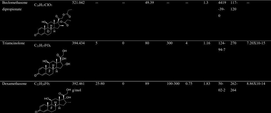
52 Table 1.3: Summary of published studies demonstrating the effects of glucocorticoid exposures to the aquatic species. Drug Common name and Taxon Binomial name Exposure time Drug concentrations Key finding Reference Prednisolone Zebrafish Danio rerio 72 hpf 0.1, 1,10 µg/l Alterations to the ontogeny and behaviour of embryos Dexamethasone Zebrafish Danio rerio 120 hpf 100 um Reduction in cortisol levels, delayed physical development, lesser locomotor ability and changes in gene expression. McNeil et al., 2016 Wilson et al., 2016 Beclomethasone dipropionate Dexamethasone Fathead minnow (Fish) Crucian carp (Fish) Pimephales promelas 21 d 10, 100, 1000 ng/l Perturbation in blood glucose homeostasis, immune depression and skin androgenisation Carassius auratus 9 d 200 µg/fish Higher pathogen susceptibility, lower lysozyme activity and expression of immune genes Margiotta- Casaluci et al., 2016 Qi et al., 2016 Dexamethasone, prednisolone and triamcinolone Zebrafish Danio rerio 120 h (embryos), 4 d (adult) 50 nm, 5nM, 50 pm, 500 pm Cortisol Zebrafish Danio rerio 5 d 25 µg cortisol (g body mass)-1d-1 Perturbation in the expression of GC genes in larvae and adult male liver Maternal stress induced higher embryo cortisol content and alteration in gene expression Chen et al., 2016 Faught, 2016 Cortisol Zebrafish Danio rerio 72 hpf 32 pg Cardiac edema and altered gene expression Nesan and Vijayan, 2016 Dexamethasone Trahira/ wolf fish/ tiger fish Hoplias malabaricus 12 doses and fed twice every wk 0.03, 0.3, 3 µg/kg Induced haematological changes and immunotoxin effects Ribas et al.,
53 Dexamethasone Rainbow trout (Fish) Oncorhynchus mykiss 42 d 3, 30, 300, 3000 ng/l Effect on CYP450-mediated reactions (EROD, BCFOD, BQOD and PNPH) and induction of CYP3A-like protein. Burkina et al., 2015 Beclomethasone dipropionate Rainbow trout (Fish) Oncorhynchus mykiss Dexamethasone Zebrafish Danio rerio 120 hpf (embryos), Dexamethasone Trahira/ wolf fish/ tiger fish 14 d 100, 1000 ng/l Increase in blood glucose, oxidative stress. Almroth et al., d (adult) Hoplias malabaricus 12 doses and fed twice every wk 100 µm Perturbations in GR activity during embryonic development impacted the Wilson et al., 2015 structure, function and molecular composition of adult heart. 0.03, 0.3, 3 µg/kg Caused oxidative stress and disturbed antioxidant system in the gonads of exposed male species Guiloski et al., 2015 Clobetasol propionate, clobetasone butyrate Common carp (Fish) Cyprinus carpio 21 d 1 µg /L Protein degradation and increase in serum free amino acid levels Nakayama et al., 2014 Prednisolone Zebrafish Danio rerio 21 d 9 µg/l Osteoporosis-like phenotype in regenerating scales Beclomethasone dipropionate Fathead minnow (Fish) Pimephales promelas 21 d 0.1, 1 and 10 µ g/l Increase in plasma glucose concentration, decrease in blood lymphocyte count, induction of male secondary sexual characters and decrease in plasma VTG concentrations in female fish Dexamethasone Zebrafish Danio rerio 120 hpf 100 µm Alterations in physiology, behavioural activity and reduction in whole embryo cortisol Cortisol and dexamethasone Pacific salmon (Fish) Oncorhynchus spp. 5 wk 10, 50 ng/ml Increase in in vitro multiplication of parasite haemoflagellate Cryptobia salmositica, and additive effect acting via the interaction of GC-like receptor. De Vrieze et al., 2013 Kugathas et al., 2013 Wilson et al., 2013 Li et al.,
54 Dexamethasone Fathead minnow (Fish) Pimephales promelas 21 d 500 µg/l Reduction in cumulative fecundity, spawning frequency, female plasma estradiol levels and an increase in plasma VTG protein levels. LaLone et al., 2012 Dexamethasone Fathead minnow (Fish) Pimephales promelas 28 d 500 µg/l Increase in deformed gill opercula and reduction in weight and length compared with control fry. LaLone et al., 2012 Dexamethasone Fathead minnow (Fish) Pimephales promelas Egg stage to 28 dph µg/l Decrease in survival at 577 µg /L Overturf et al., 2012 Prednisolone and beclomethasone dipropionate Fathead minnow (Fish) Pimephales promelas 21 d 1 µg/l Increase in plasma glucose and reduction in leucocytes Overturf et al., 2012 Dexamethasone Senegalese sole (Fish) Solea senegalensis 72 h and 14 d 0.1, 1 and 10 ppm Increase in susceptibility to pathogen, reduction in growth, activation of expression of genes involved in the innate immune system and cellular stress defence Salas-Leiton et al., 2012 Dexamethasone Chinook salmon (Fish) Oncorhynchus tshawytscha 7 d 50 mg/g Reduction in resting plasma cortisol levels and internal cell atrophy McQuillan et al., 2011 Cortisol metabolite 5a-androstan Japanese medaka Oryzias latipes 21 d 0.1, 1, 10, 100 µg/l Masculinization of female fish Grillitsch et al., ,11,17-trione (Fish) Dexamethasone Rainbow trout (Fish) Oncorhynchus mykiss 48 h 50 and 100 mg/kg body weight Increase in EROD and MROD activity Smith and Wilson,
55 Corticosterone, aldosterone and dexamethasone African clawed frog (Amphibian) Xenopus laevis 21 d 100nM (Aldo), 10, 100 and 500Nm (Cortisone and DEX). Reduction in whole body length, hind limb length, mrna expression in tail tissues Lorenz et al., 2009 Dexamethasone Rainbow trout (Fish) Oncorhynchus mykiss 5 wk 300 mg/kg of feed at 1% body weight Inhibition in innate immune response to parasite resulting in higher infections. Lovy et al., 2008 Dexamethasone and hydrocortisone Zebrafish Danio rerio 96 hpf 100 mg/l equivalent to DEX: µm, HC: µm Alteration in craniofacial development and increase expression and activity of matrix metalloproteinases Hillegass et al., 2008 Dexamethasone and hydrocortisone Cortisol Zebrafish Danio rerio 72 hpf 1, 10 and 100 mg/l Atlantic salmon (Fish) equivalent to DEX: 2.55, 25.48, µm, HC: 2.76, 27.59, Induction of craniofacial abnormalities, altered somitogenesis, blood pooling and pericardial, yolk sac edema and increased MMP-13 mrna µm Salmo salar 6 d 50, 100 mg/kg Reduction in growth, yolk-sac volume and utilization and induced morphological malformations. Hillegass et al., 2007 Eriksen et al., 2006 Prednisolone and dexamethasone Rotifer Brachionus calyciflorus 24 h 100mg/500mL 50% mortality at 22.3 mg/l PDS and mg/l DEX. DellaGreca et al., 2004 Prednisolone and dexamethasone Water-flea (Crustacean) Daphnia magna 24 h 100mg/500mL No effect at 85 mg/l PDS and 50% mortality at 48.3 mg/l DEX. DellaGreca et al., 2004 Prednisolone and dexamethasone Alga Pseudokircheneriella Subcapitata 3 d 100mg/500mL No effect at 160 mg/l PDS and 100 mg/l DEX Della Greca et al.,
56 Prednisolone and dexamethasone Shrimp (Crustacean) Thamnocephalus platyurus 24 h 100mg/500mL 23% mortality at 140 mg/l PDS and 50% mortality at mg/l DEX. DellaGreca et al., 2004 Prednisolone Dexamethasone Water-flea (Crustacean) Atlantic salmon (Fish) Ceriodaphnia dubia 7 d 100mg/500mL Affected reproduction at 0.23 mg/l DellaGreca et al., 2004 Salmo salar 48 h 10, 60, 200 µg/l Increase in susceptibility to infections with parasite Gyrodactylus derjavini. Nielson and Buchmann, 2003 Cortisol Rainbow trout (Fish) Oncorhynchus mykiss 5 d 50mg/Kg Negatively affected the molecular and biochemical response, gene expression alteration in the GC responsive genes. Vijayan et al., 2003 Dexamethasone Rainbow trout (Fish) Oncorhynchus mykiss 14 d 100 µg/fish Increase in susceptibility to infections with parasite Gyrodactylus derjavini Mikailov. Lindenstrom and Buchmann, 1998 Dexamethasone Rainbow trout (Fish) Oncorhynchus mykiss 96 h 2 mg/kg body weight Reduction in CYP450 proteins in the liver Lee et al.,
57 1.5 Aim and objectives of the present study Aim: Effect assessment of synthetic glucocorticoids to the selected freshwater invertebrate and vertebrate species. The following objectives are designed to address the research aim. Each objective is further divided into sub-objectives and designed to test the subsequent hypotheses (null or alternate). Objective 1. To assess the impact of sub-acute and multigenerational glucocorticoid exposures on selected freshwater invertebrates. Sub-objective 1.1: To evaluate multigenerational effects of two glucocorticoids (prednisolone- PDS and dexamethasone- DEX) on life-history parameters of crustacean Ceriodaphnia dubia (Cladocera). Objective 1.1 is aimed to test the subsequent hypothesis: Hypothesis 1.1a (Null): Exposure to synthetic GC chemicals (PDS and DEX) over a period of successive generations does not result in adverse effects to the exposed C. dubia. Hypothesis 1.1b (Alternate): Multigenerational exposures of PDS and DEX to C. dubia result in adverse effects, affecting the exposed organisms at population level. Sub-objective 1.2: To investigate exposure effects of PDS on the embryonic and post hatching development and shell formation of the freshwater snail, Physa acuta (Gastropod: Physidae). Objective 1.2 is aimed to test the subsequent hypothesis: Hypothesis 1.2a (Null): Sub-acute exposure to PDS does not pose any risk to the embryonic and post-hatching development of P. acuta. 23
58 Hypothesis 1.2b (Alternate): Exposure of P. acuta to PDS results in adverse effects, affecting the embryonic and post-hatching development. Sub-objective 1.3: To assess multigenerational effects of prednisolone on the epigenetic and phenotypic traits of freshwater snail, Physa acuta (Gastropoda: Physidae). Objective 1.3 is aimed to test the subsequent hypothesis: Hypothesis 1.3a (Null): Multigenerational PDS exposure does not cause any changes to the epigenetic and phenotypic traits of P. acuta. Hypothesis 1.3b (Alternate): Multigenerational exposure to PDS affects the epigenetic and phenotypic traits in the exposed P. acuta. Objective 2: To assess the impact of glucocorticoid exposures on selected freshwater fish species (vertebrates). Sub-objective 2.1: To determine the early life stage toxicity of three glucocorticoid drugs (hydrocortisone-hc, PDS and DEX) to two Australian native fish species, Murray cod, Maccullochella peelii and golden perch, Macquaria ambigua. Objective 2.1 is aimed to test the ensuing hypothesis: Hypothesis 2.1a (Null): GC exposures do not cause any adverse effects to the Murray cod and golden perch larvae. Hypothesis 2.1b (Alternate): GC exposures cause early life stage developmental abnormalities to the exposed Murray cod and golden perch larvae. Sub-objective 2.2: To evaluate the chronic exposure effects of prednisolone to developing larvae of Murray cod, Maccullochella peelii. 24
59 Objective 2.2 is aimed to test the subsequent hypothesis: Hypothesis 2.2a (Null): Chronic PDS exposure does not cause any adverse effects to the Murray cod larvae. Hypothesis 2.2b (Alternate): Early life-stage chronic PDS toxicity disturbs phenotypic traits of developing Murray cod larvae. Sub-objective 2.3: To determine the early life stage toxicity of two glucocorticoid drugs (PDS and DEX) in combination to varied temperatures to Australian native freshwater fish golden perch, Macquaria ambigua. Objective 2.3 is aimed to test the subsequent hypothesis: Hypothesis 2.3a (Null): GC exposures to early life stage in addition to varied temperatures do not cause adverse effects to the golden perch larvae. Hypothesis 2.3b (Alternate): Early life stage exposure of golden perch larvae to multiple stressors (temperature and synthetic GCs) adversely affects the organogenesis process of exposed larvae. 1.6 Test species used in the present study Ceriodaphnia dubia (Cladocera) Crustaceans (phylum Arthropoda) are the most ubiquitous group of aquatic species, inhabiting all forms of habitats in aquatic ecosystem. They consist of about 67,000 described diverse animal species (Brusca and Brusca, 2003) and are primarily divided into six classes. Out of these, class Branchipoda contains small order crustaceans known as Cladocera (Martin and Davis, 2001). Cladocera are commonly known as water fleas and there are about 620 recognised species in this category that grow to less than 1 mm in length with males smaller than females (Brands, 2010; Forró et al., 2008). The water fleas are distributed in freshwater 25
60 lakes, ponds, and marshes. They are characterised by a single compound eye, two pairs of antennae on their head and a unique way of moulting to grow (Forró et al., 2008). Figure 1.2 shows the life-cycle stages and reproduction cycle of water-flea Ceriodaphnia dubia. Ecologically, they are key components of aquatic food chains as they form an important connection between the aquatic primary producers (autotrophs- algae, phytoplankton) and secondary consumers (fish larvae) in the aquatic food chain (Forró and Frey, 1985; Norberg and Mount, 1985; Knight and Waller 1992). They also acts as filter feeders, ingesting various sorts of organic detritus in freshwater ecosystems (Forró and Frey, 1985). Over the years, crustaceans have developed a huge diversity of life histories and numerous approaches to growth, development and reproduction such as processes of metamorphosis, diapause and pupation. Crustacean s reproductive physiology and growth parameters are related to functioning of their nervous and endocrine systems (Engelmann, 1994). These internal systems work predominately with the help of various steroidal hormones including vertebrate-like sex steroids, etc. (DeLoof and DeClerk, 1986; LeBlanc and McLachlan., 1999). Reproduction is regulated and governed by a complex endocrine mechanism involving different hormonal compounds such as ketosteroids (ecdysteroids), peptides (gonad stimulators and vitellogenesis inhibitors), biogenic amines, terpenoids (juvenile hormones), vertebrate-like steroids (Sandor, 1980). As steroids have been reported to play a vital role in life cycle parameters of crustaceans, a temporal variation in steroid levels by an exogenous source, resulted in alterations of sexual characteristics or reproduction in crustaceans (Lafont and Mathieu, 2007). Recent research trends in ecotoxicological studies have revealed the use of crustaceans in the assessment of pharmacological and steroidal compounds (Kummerer, 2001; DellaGreca et al., 2004; Blaise and Ferard, 2005; Nunes et al., 2006; Rodriguez et al., 2007). They are used in the toxicity testing of WWTP effluents and have been regarded as a central species for 26


61 toxicity testing due to their sensitivity to the toxicants in the aquatic environment (Francisco et al., 2011). C. dubia s wide ecological distribution in freshwater systems, easy culturing in enclosed laboratory conditions, reproductive effectiveness, shorter life span and great sensitivity towards environmental contaminants have made them a potential test model in biomonitoring environmental pollution (Lamichhane et al., 2013; USEPA, 2013; Pakrashi et al., 2013; Francisco et al., 2011). Figure 1.2: Pictorial representation of life-cycle stages and reproduction cycle of Ceriodaphnia dubia Physa acuta (Class Gastropoda) Gastropods belong to an intensively studied taxonomic class within the phylum Mollusc, with nearly 100,000 recognized species residing in marine and freshwater habitats worldwide (Strong et al., 2008; Feldkamp, 2002). Of these, the freshwater snail from the family Physidae, Physa acuta (Draparnaud, 1805), is a small left-handed and globally-invasive species most likely originating from North America (Dillon et al., 2004) and presently having established populations in almost all continents except Antarctica (Dillon et al., 2002; Mitchell and Leung, 2016). This hermaphrodite species is widely distributed in freshwater bodies such as rivers, streams, lakes, ponds, and swamps (Albrecht et al., 2009; Guo and Zhao, 2009). The lifecycle of these snails is very short, generally up to a year. They have an average body size of 7-10 mm and are capable of asexual reproduction. Each of their eggs has a genetic uniqueness based on the coming together of the two halves with natural ability to store genetic material. These snails produce between 4-6 eggs per week and each egg mass has encapsulated embryos in it. 27


62 The embryonic development is completed in days and newly-hatched juveniles grow into adults in approximate 3 months (Guo and Zhao, 2009; Li et al., 2014). Figure 1.3 shows the lifecycle stages and reproduction cycle of P. acuta. Ecologically, aquatic snails have been well known to serve as a key link between primary producers and upper level aquatic species in aqueous food webs. It has been reported that the functional hormones for regulation of biological processes, growth and sex differentiation in molluscs are likely the vertebrate-like sex steroids (Sandor, 1980; Joose, 1978; Bettin et al., 1996; Geraerts and Joose, 1984). The steroidogenesis process is found to be managed by enzymatic action (17β-hydroxy steroid dehydrogenase, 3β-hydroxy steroid dehydrogenase and 5αreductase) (Thornton et al., 2003) and this pathway is carried via sequencing of steroid receptor orthologs in the studied molluscs (Kishida, 2005 and Fernandes et al., 2011). P. acuta snails have been documented well in several scientific studies because of their outstanding promising characteristics as a laboratory test organisms. Massive abundance, easy identification, limited mobility and short lifecycle have made these snails as a promising test species in biomonitoring research with an added advantage of investigating generational exposure effects of aquatic toxicants. Previous field and laboratory studies have established that P. acuta snails are quite sensitive to anthropogenic pharmaceutical pollutants causing multiple levels of physical and biological impairment or even in extreme cases mortality (De Castro-Catalàa et al., 2013; Sánchez-Argüello et al., 2009, 2012; Brown et al., 2012; LopezDoval et al., 2012; Jarvis et al., 2014; Musee et al., 2010). 28
63 Figure 1.3: Pictorial representation of life-cycle stages and reproduction of Physa acuta Fish early life stage Murray cod (Maccullochella peelii, Mitchell, 1838): It is one of the largest Australian predatory freshwater fish, belonging to class Actinopterygii (Dianne and Vanessa, 2011). It is endemic to Murray-Darling River and is available in almost all major Australian states (Harris and Rowland, 1996). This fish species is well bred in hatcheries in both hatchery and wild form (Lintermans et al., 2005). Individuals reach up to cm in length and can weigh approximately 113 kg (McDowall, 1996). They have deep, elongated bodies with a broad scooped head and a large mouth. Newly hatched larvae (6-9 mm) consists of yolk sac which gets depleted at days post hatch (dph). They have a long life cycle as Murray cod is recorded as a most long-lived fish species in Australian freshwater system (Anderson et al., 1992), with the longest lived Murray cod was found to be of 48 years old. They have short annual reproduction cycle and generally reproduce in spring and laying eggs in thousands of numbers in hollow logs or other hard surfaces (McDowall, 1996). Figure 1.4 illustrates the life-cycle stages and reproduction cycle of Murray cod fish. Reproduction is dependent on size of female fish and climatic stressors like water temperature and water level are found to play key role in affecting the reproductive ability (Cadwallader and Gooley, 1985; Lake 1959, 1967a). Diet changes with growth and age and adult fish feed mainly on a variety of aquatic species such as crustaceans, shrimps, yabbies, mollusc and other fish species, whereas larval fish rely on zooplanktons such as crustaceans as their food source (Harris and Rowland, 1996; Cadwallader and Backhouse, 1983; Rowland, 1992). Although biology and ecology of Murray cod fish is well studied to date from behaviour, diet, spawning and rearing perspective but there are research gaps in terms of toxicity assessment of detected chemicals in Australian waters on the early life stage and overall life-history traits of Murray cod fish. 29
 Australian museum, 2017; (b, c) Gold central Victoria, 2017; (d) NSW Aquaculture association Inc., 2017). 1.6.3.")

64 Figure 1.4: Pictorial representation of life-cycle stages and reproduction cycle of Murray cod, Maccullochella peelii. (Image source: (a) Australian museum, 2017; (b, c) Gold central Victoria, 2017; (d) NSW Aquaculture association Inc., 2017) Golden perch (Macquaria ambigua, Richardson, 1845): It is an iconic Australian freshwater (mainly South-eastern) fish species belonging to family Percichthydae and class Actinopterygii. It is a medium-sized fish weighing 1-2 kg and cm in length, with no sexual dimorphism. With an elongated and compressed body, golden perch fish. Studies have well-documented the life-cycle, habitat, breeding, reproduction, production, feeding of golden perch (Anderson et al., 1992; Lake 1967b; Rowland et al., 1983; Mallen-Cooper 1994; Anderson and Braley 1993; Collins and Anderson 1995; Musyl and Keenan 1992) but none has focussed on the effect assessment of emerging contaminants on the early life stages of this fish. 30
. Both golden perch and Murray cod fish species are recognised as threatened under State and National law.")
.")
65 Figure 1.5: Pictorial representation of life-cycle stages and reproduction of golden perch, Macquaria ambigua. (Image source: (a) (b, c) Both golden perch and Murray cod fish species are recognised as threatened under State and National law. Due to rapid increase in river regulation and weir constructions in the Murray Darling Basin (MDB), downstream passage has significantly affected the Murray cod and golden perch fish species. The Murray cod has been listed as Vulnerable and critically endangered under the Environment Protection and Biodiversity Conservation Act 1999 (EPBC Act). Populations of Murray cod have declined severely since European colonisation of Australia due to a number of causes including severe overfishing, river regulation and habitat degradation (Wager, 1996; Gehrke et al., 1995; Baumgartner et al., 2006). Although golden perch is still widely available through the MDR but similar factors have affected this species too and is endangered in Victoria (Boys et al., 2016; Baumgartner et al., 2006; 31
 1.7 Thesis Structure This thesis consists of total eight chapters.")
66 Brumley, 1987). Figure 1.6 shows the distribution of Murray cod and golden perch on an Australian map. Figure 1.6: Pictorial representation of distribution of Murray cod (dashed lines) and golden perch (yellow to red dots) on an Australian map. (Image source: Lureandfly, 2017; Murraycod.info, 2017) 1.7 Thesis Structure This thesis consists of total eight chapters. Chapter 1 (present) begun with background introduction and detailed literature review. Experimental part of this thesis is written as six independent manuscripts (Chapter 2-7). 32
67 Chapter 2: Bal, N., Kumar, A., Du, J., Nugegoda, D., Multigenerational effects of two glucocorticoids (prednisolone and dexamethasone) on life-history parameters of crustacean Ceriodaphnia dubia (Cladocera). Environ. Pollut. 225: Chapter 3: Bal, N., Kumar, A., Du, J., Nugegoda, D., Prednisolone impairs embryonic and post-hatching development and shell formation of the freshwater snail, Physa acuta. Environ. Toxicol. Chem. 35(9): Chapter 4: Bal, N., Kumar, A., Nugegoda, D., Assessing multigenerational effects of prednisolone to the freshwater snail, Physa acuta (Gastropoda: Physidae). J. Hazard. Mater. 339: Chapter 5: Bal, N., Kumar, A., Nugegoda, D., The comparative toxicity of three differently potent glucocorticoids to early life stages of two freshwater Australian fish, Macquaria ambigua and Maccullochella peelii. J. Hazard. Mater. (To be submitted) Chapter 6: Bal, N., Kumar, A., Nugegoda, D., Early life-stage exposure to prednisolone alters phenotypic traits and causes osteoporosis-like-symptoms in the freshwater fish, Murray cod (Maccullochella peelii). Environ. Sci. Tech. (To be submitted) Chapter 7: Bal, N., Kumar, A., Nugegoda, D., Organogenesis in Australian native freshwater fish, Macquaria ambigua: influence of temperature and exposure to synthetic glucocorticoids. Environ. Sci. Tech. (To be submitted) These chapters contribute toward addressing the principal research aim and specifically addresses each sub-objective at individual levels. Each of these chapters (Chapter 2-7) is either published, under review or in process of submission and can be read independently. Separate bibliographies are presented at the end of each chapter and Supplementary information where applicable. Chapter 8 brings together the major findings of the thesis in a final discussion and conclusion alongwith future work prospective. 33

68 Figure 1.7: Flowchart illustration of thesis structure. 34
69 REFERENCES Abbott, B.D., Review of the interaction between TCDD and glucocorticoids in embryonic palate. Toxicology 105: ACIC, Australian Crime Commission, Illicit Drug Data Report Accessed December 2016 AHFS, American Hospital Formulary Service. Corticosteroids general statement. Am. Hosp. Formul. Serv. Drug Inf. 42: Albrecht, C., Oliver, K.O., Terrazas, E.M., Wilke, T., Invasion of ancient Lake Titicaca by the globally invasive Physa acuta. Biol. Invasions 11: Almawi, W.Y., Abou, J.M.M., Li, X.C., Transcriptional and post-transcriptional mechanisms of glucocorticoid anti proliferative effects. Haematol. Oncol. 20:17 32 Almroth, B.M., Gunnarsson, L.M., Cuklev, F., Fick, J., Kristiansson, E., Larsson, D.G.J., Waterborne beclomethasone dipropionate affects the physiology of fish while its metabolite beclomethasone is not taken up. Sci. Total Environ. 511:37-46 Ammann, A.A., Macikova, P., Groh, K.J., Schirmer, K., Suter, M.J.F., LC-MS/MS determination of potential endocrine disruptors of cortico signalling in rivers and wastewaters. Anal. Bioanal. Chem. 406(29): Anderson, J.R., Morison, A.K., Ray, D.J., Age and growth of Murray cod, Maccullochella peelii (Perciformes: Percichthyidae), in the Lower Murray Darling Basin, Australia, from thin-sectioned otoliths. Aus. J. Mar. Fresh Res. 43: Anderson, T.A., Braley, H., Appearance of nutrients in the blood of the Golden perch, Macquaria ambigua, following feeding. Comp. Biochem. Physiol. A Physiol. 104(2): Ankley, G.T., Jensen, K.M., Makynen, E.A., Kahl, M.D., Korte, J.J., Hornung, M.W., Henry, T.R., Denny, J.S., Leino, R.L., Wilson, V.S., Cardon, M.C., Hartig, P.C., Gray, L.E., 35
70 2003. Effects of the androgenic growth promoter 1-β- trenbolone on fecundity and reproductive endocrinology of the fathead minnow fish, Pimephales promelas. Environ. Toxicol. Chem. 22: AP1, Australian Pharmaceuticals Industry. Data card gies/pharmaceuticals/documents/pharmaceuticalsdatacard.pdf Australian Government Department of Industry. Accessed September 2016 Australian museum, Murray cod eggs. [Image] Accessed October 2016 Backhaus, T., Porsbring, T., Arrhenius, A., Brosche, S., Johnsson, P., Blanck, H., Single substance and mixture toxicity of five pharmaceuticals and personal care products to marine periphyton communities. Environ. Toxicol. 30(9): Bain, P.A., Williams, M., Kumar, A., Assessment of multiple hormonal activities in wastewater at different stages of treatment. Environ. Toxicol. Chem. 33: Barnes, P.J., How corticosteroids control inflammation: Quintiles Prize Lecture Br. J. Pharmacol. 148: Baumgartner, L.J., Reynoldson, N., Gilligan, D.M., Mortality of larval Murray cod (Maccullochella peelii) and golden perch (Macquaria ambigua) associated with passage through two types of low-head weirs. Mar. Freshwater Res. 57: Bettin, C., Oehlmann, J., Stroben, E., TBT-induced imposex in marine neogastropods is mediated by an increasing androgen level. Helgol. Meeresunters 50: Blaise, C., Ferard J.F., Effluent assessment with the PEEP (potential ecotoxic effects probe) index. Small-scale Freshwater Toxicity Investigations, volume 2. Springer, the Netherlands. ISBN Boxall, A.B., Rudd, M.A., Brooks, B.W., Caldwell, D.J., Choi, K., Hickmann, S., Innes, E., Ostapyk, K., Staveley J.P., Verslycke, T., Ankley, G.T., Beazley, K.F., Belanger, S.E., Berninger, J.P., Carriquiriborde, P., Coors, A., Deleo, P.C., Dyer, S.D., 36
71 Ericson, J.F., Gagné, F., Giesy, J.P., Gouin, T., Hallstrom, L., Karlsson, M.V., Larsson, D.G., Lazorchak, J.M., Mastrocco, F., McLaughlin, A., McMaster, M.E., Meyerhoff, R.D., Moore, R., Parrott, J.L., Snape, J.R., Murray-Smith, R., Servos, M.R., Sibley, P.K., Straub, J.O., Szabo, N.D., Topp, E., Tetreault, G.R., Trudeau, V.L., van der Kraak, G., Pharmaceuticals and personal care products in the environment: what are the big questions? Environ. Health Perspect. 120(9): Boys, C.A., Robinson, W., Miller, B., Pflugrath, B., Baumgartner, L. J., Navarro, A., Brown, R., Deng, Z., How low can they go when going with the flow? Tolerance of egg and larval fishes to rapid decompression. Biol. Open 0:1-8, DOI: /bio Brands, S.J., C. dubia - Hierarchy. The Taxonomicon. Universal Taxonomic Services, Zwaag, the Netherlands. Ceriodaphnia dubia Accessed February 2017 Bray, D.J., Thompson, V.J., Murray Cod, Maccullochella peelii. Fishes of Australia. Accessed February 2017 Brown, J., Bernot, M.J., Bernot, R.J., The influence of TCS on the growth and behaviour of the freshwater snail, Physa acuta. J. Environ. Sci. Health A. 47: Brumley, A.R., Past and present distributions of Golden Perch Macquaria ambigua (Pisces: Percichthyidae) in Victoria, with reference to releases of hatchery-produced fry. Proc. R. Soc. Vic. 99(3): Brusca, R.C., Brusca, G.J., Invertebrates- Second edition. Sinauer Associates. ISBN Burkina, V., Sakalli, S., Rasmussen, M.K., Zamaratskaia, G., Koba, O., Thai, G.P., Grabic, R., Randak, T., Zlabek, V., Does dexamethasone affect hepatic CYP450 system of fish? Semi-static in vivo experiment on juvenile rainbow trout. Chemosphere 139: Bury, N.R., Sturm, A., Le Rouzic, P., Lethimonier, C., Ducouret, B., Guiguen, Y., RobinsonRechavi, M., Laudet, V., Rafestin-Oblin, M.E., Prunet, P., Evidence 37
72 for two distinct functional glucocorticoid receptors in teleost fish. J. Mol. Endocrinol. 31: Cadwallader, P.L., Backhouse G.N., A guide to the freshwater fish of Victoria. Government Printer, Melbourne Cadwallader, P.L., Gooley, G.J., Propagation and rearing of Murray Cod Maccullochella peeli at the warm water fisheries station pilot project lake Charlegrark government printer, Melbourne Canalis, E., Delany, A.M., Mechanisms of glucocorticoid action in bone. Ann. N. Y. Acad. Sci. 966:73 81 Chang, H., Hu, J., Shao, B., Occurrence of natural and synthetic glucocorticoids in sewage treatment plants and receiving river waters. Environ. Sci. Tech. 2007:41, Chang, H., Wan, Y., Hu, J., Determination and source apportionment of five classes of steroid hormones in urban rivers. Environ. Sci. Tech. 43: Chem.nlm.nih.gov ChemIDplus Advanced - Chemical information with searchable synonyms, structures, and formulas. Accessed November 2016 Chen, Q., Jia, A., Snyder, S.A., Gong, Z., Lam, S.H., Glucocorticoid activity detected by in vivo zebrafish assay and in vitro glucocorticoid receptor bioassay at environmental relevant concentrations. Chemosphere 144: Ciriaco, M., Ventrice, P., Russo, G., Scicchitano, M., Mazzitello, G., Scicchitano, F., Russo, E., Corticosteroid-related central nervous system side effects. J. Pharmacol Pharmacother. 4(1):94 98 Collins, A.L., Anderson, T.A., The regulation of endogenous energy stores during starvation and refeeding in the somatic tissues of the Golden perch. J. Fish Biol. 47: Creusot, N., Aït-Aïssa, S., Tapie, N., Pardon, P., Brion, F., Sanchez, W., Thybaud, E., Porcher, J.M., Budzinski, H., Identification of synthetic steroids in river water 38
73 downstream from pharmaceutical manufacture discharges based on a bioanalytical approach and passive sampling. Environ. Sci. Tech. 48: Cruz, S.A., Lin, C-H., Chao, P-L., Hwang, P-P., Glucocorticoid receptor, but not mineralocorticoid receptor, mediates cortisol regulation of epidermal ionocyte development and ion transport in zebrafish (Danio rerio). PLOS ONE 8(10):e77997, DOI: /journal.pone Czock, D., Keller, F., Rasche, F.M., Häussler, U., Pharmacokinetics and pharmacodynamics of systemically administered glucocorticoids. Clin. Pharmacokinet. 44:61-98 Daughton, G.C., PPCPs in the environment: future research beginning with the end always in mind. In: Ku mmerer, K. (Ed.), Pharmaceuticals in the environment, second edition. Springer. 33: Daughton, G.C., Ternes, T., Pharmaceuticals and personal care products in the environment: agents of subtle changes. Environ. Health Perspect. 107: De Castro-Catalàa, N., López-Dovala, J., Gorgab, M., Petrovicc, M., Muñoza, I., Is reproduction of the snail Physella acuta affected by endocrine disrupting compounds? An in situ bioassay in three Iberian basins. J. Hazard Mater. 263(1): De Loof, A., De Clerk, D., Vertebrate-type steroids in arthropods: Identification, concentrations and possible functions. In: Porchet, M., Andriesm, J.C., Dhainaut, A. (Ed.), Advances in invertebrate reproduction. Elsevier, the Netherlands De Vrieze, E., van Kessel, M. A., Peters, H. M., Spannings, F. A., Flik, G., Metz, J. R., Prednisolone induces osteoporosis-like phenotype in regenerating zebrafish scales. Osteoporosis Int. 25: DellaGreca, M., Fiorentino, A., Isidori. M., Lavorgna, M., Previtera, L., Rubino, M., Temussi, F., Toxicity of prednisolone, dexamethasone and their photochemical derivatives on aquatic organisms. Chemosphere 54:
74 Dianne, J.B., Vanessa, J.T., Murray cod, Muccullochella peelii. Fishes of Australia. Accessed February Dickmeis, T., Lahiri, K., Nica, G., Vallone, D., Santoriello, C., Neumann, C.J., Hameerschmidt, M., Foulkes, N.S., Glucocorticoids play a key role in circadian cell cycle rhythms. PLOS Biol. 5(4): e78, DOI: /journal.pbio Dillon R.T., Earnhardt C.E., Smith T.P., Reproductive isolation between Physa acuta and Physa gyrina in joint culture. Am. Malacol. Bull. 19:63-68 Dillon, R.T., Wethington, A.R., Rhett, J.M., Smith, T.P., Populations of the European freshwater pulmonate Physa acuta are not reproductively isolated from American Physa heterostropha or Physa integra. Invertebr. Biol. 121: Drugbank.ca DrugBank. Accessed October 2016 Drugs, Top 100 drugs for United States pharmaceutical sales. Assessed October Duchatel, J.P., Beduin, J.M., Jauniaux, T., Coignoul, F., Vindevogel, H., Glucocorticoids as doping agents in homing pigeons. Ann. Med. Vet. 137: Engelmann, F., Invertebrates: hormone-regulated gonadal activity. In: Davey, K.G., Peter, R.E., Tobe, S.S. (Ed.), Prospective in comparative endocrinology. National Research Council of Canada, Ottawa, ON Eriksen, M.S., Bakken, M., Espmark, A., Braastad, B.O., Salte, R., Prespawning stress in farmed Atlantic salmon Salmo salar: maternal cortisol exposure and hyperthermia during embryonic development affect offspring survival, growth and incidence of malformations. J. Fish Biol. 69: Evans, R.M., The nuclear receptor superfamily: A rosetta stone for physiology. Mol. Endocrinol. 19: Fan, Z., Wu, S., Chang, H., Hu, J., Behaviours of glucocorticoids, androgens and progestogens in a municipal sewage treatment plant: comparison to estrogens. Environ. Sci. Technol. 45(7):
75 Faught, E., Best, C., Vijayan, M.M., Maternal stress-associated cortisol stimulation may protect embryos from cortisol excess in zebrafish. R. Soc. Open Sci. 3:160032, DOI: /rsos Feldkamp, S., Modern Biology. Holt, Rinehart, Winston, Austin, TX. ISBN Fent, K., Escher, C., Caminada, D., Estrogenic activity of pharmaceuticals and pharmaceutical mixtures in a yeast reporter gene system. Reprod. Toxicol. 22: Fernandes, D., Loi, B., Porte, C., Biosynthesis and metabolism of steroids in molluscs. J. Steroid Biochem. Mol. Biol. 127(3-5): Flaherty, C.M., Dodson, S.I., Effects of pharmaceuticals on Daphnia survival, growth, and reproduction. Chemosphere 61(2): Florini, J.R., Buyske, D.A., Peets, E.A., Plasma half-life, tissue distribution, and excretion of triamcinolone-h3. J. Pharm. Exp. Ther. 131(3): Forró, I., Korovchinsky, M.M., Kotov, A.A., Petrusek, A Global diversity of cladocerans (Cladocera; Crustacea) in freshwater. Hydrobiologia 595: Forró, L., Frey, D.J., Cladocera. Proceedings of the cladocera symposium. In: Budapest (Ed.), Developments in Hydrobiology. 35:351 Francisco, D.E., Elias, M.C., LaRocca, C.A., DiGiano, F.A., Maerker, M.J., Chronic toxicity bioassay with Ceriodaphnia dubia. Department of Environmental Sciences and Engineering, School of Public Health, University of North Carolina at Chapel Hill. Project Report Frerichs, V.A., Tornatore, K.M., Determination of the glucocorticoids prednisone, prednisolone, dexamethasone, and cortisol in human serum using liquid chromatography coupled to tandem mass spectrometry. J. Chromatogr. B 802: Gehrke, P.C., Brown, P., Schiller, C.B., Moffatt, D.B., Bruce, A.M., River regulation and fish communities in the Murray-Darling river system, Australia. Regul. Rivers: Res. Manage. 11:
76 Geraerts, W.P.M., Joose, J., Freshwater snails (Basommpatophora). In: Tompa, A.S., Verdonk, N.H., van den Biggelaar, J.A.M. (Ed.), The Mollusca, Reproduction 7, Academic press, New York Gold Central Victoria, Anglers Strike Gold. [Image] Accessed November 2017 Greenwood, A.K., Butler, P.C., White, R.B., DeMarco, U., Pearce, D., Fernald, R.D., Multiple corticosteroid receptors in a teleost fish: distinct sequences, expression patterns, and transcriptional activities. Endocrinology 144: Grillitsch, B., Altmann, D., Schabuss, M., Zornig, H., Sommerfeld-Stur, I., Mostl, E., Mammalian glucocorticoid metabolites act as androgenic endocrine disruptors in the medaka (Oryzias latipes). Environ. Toxicol. Chem. 29(7): Guiloski, I.C., Ribas, J.L.C., Pereira, L.S., Neves, A.P.P., Assis, H.C.S., Effects of trophic exposure to dexamethasone and diclofenac in freshwater fish. Ecotoxicol. Environ. Saf. 114: Guo, E.M., Zhao, G.F., Reproductive behaviour and embryonic development in Conch Physa acuta. Fisheries Sci. 28: Harris, J.H., Rowland, S.J., Family percichthyidae Australian freshwater cods and basses. Freshwater fishes of South-eastern Australia. In: McDowall, R. (Ed.), Reed Books, Chatswood Hattori, R.S., Fernandino, J.I., Kishii, A., Kimura, H., Kinno,T., Oura, M., Somoza, G.M., Yokota, M., Strussmann, C.A., Watanabe, S., Cortisol-induced masculinization: Does thermal stress affect gonadal fate in Pejerrey, a teleost fish with temperaturedependent sex determination? PLOS ONE 4(8) He, Y., Yi, W., Suino-Powell, K., Zhou, X.E., Tolbert, W.D., Tang, X., Yang, J., Yang, H., Shi, J., Hou, L., Jiang, H., Melcher, K., Xu, H.E., Structures and mechanisms for the design of highly potent glucocorticoids. Cell Res. 24:
77 Hillegass, J.M., Villano, C.M., Cooper, K.R., White, L.A., Matrix metalloproteinase-13 is required for zebra fish (Danio rerio) development and is a target for glucocorticoids. Toxicol. Sci. 100: Hillegass, J.M., Villano, C.M., Cooper, K.R., White, L.A., Glucocorticoids alter craniofacial development and increase expression and activity of matrix metalloproteinases in developing zebrafish (Danio rerio). Toxicol. Sci. 102: Inaba, H., Pui, C., Glucocorticoid use in acute lymphoblastic leukemia: comparison of prednisone and dexamethasone. Lancet Oncol. 11(11): Isobe, T., Sato, K., Joon-Woo, K., Tanabe, S., Suzuki, G., Nakayama, K Determination of natural and synthetic glucocorticoids in effluent of sewage treatment plants using ultrahigh performance liquid chromatography-tandem mass spectrometry. Environ. Sci. Pollut. Res. 22(18): Jarvis, A.L., Bernot, M.J., Bernot, R.J., The effects if the psychiatric drug carbamazepine on freshwater invertebrate communities and ecosystem dynamics. Sci. Total Environ. 496: Jia, A., Escher, B.I., Leusch, F.D., Tang, J.Y., Prochazka, E., Dong, B., Snyder, E.M., Snyder, S.A., In vitro bioassays to evaluate complex chemical mixtures in recycled water. Water Res. 80:1 11 Joose, J., Endocrinology of molluscs. In: Actualités sur les Hormones d'invertébrés, Colloques Internationaux, CNRS. 251: Kadmiel, M., Cidlowski, J.A., Glucocorticoid receptor signaling in health and disease. Trends Pharmacol. Sci. 34(9): Khan, S.J., Ongerth, J.E., Modelling of pharmaceutical residues in Australian sewage by quantities of use and fugacity calculations. Chemosphere 54: Kishida, M., Nakao, R., Novillo, A., Callard, I.P., Osada, M., Molecular cloning and expression analysis of cdna fragments related to estrogen receptor from blue mussel, Mytilus edulis. Proc. Jap. Soc. Comp. Endocrinol. 20:75 43
78 Kitaichi, Y., Miyamoto, A., Uchikura, K., Determination of selected corticosteroids in sewage-treatment-plant samples by liquid chromatography-mass spectrometry. J. Health Sci. 56: Knight J.T., Waller W.T., Influence of the addition of cerophyl on the Selenastrum capricornutum diet of the cladoceran, Ceriodaphnia dubia. Environ. Toxicol. Chem. 11: Kortenkamp, A., Low dose mixture effects of endocrine disrupters: implications for risk assessment and epidemiology. Int. J. Androl. 31: Kugathas, S., Runnalls, T.J., Sumpter, J.P., Metabolic and reproductive effects of relatively low concentrations of beclomethasone dipropionate, a synthetic glucocorticoid, on fathead minnows. Environ. Sci. Tech. 47(16): Kugathas, S., Sumpter, J.P., Synthetic glucocorticoids in the environment: First results on their potential impacts on fish. Environ. Sci. Technol. 45: Kugathas, S., Williams, R.J., Sumpter, J.P., Prediction of environmental concentrations of glucocorticoids: the River Thames, UK, as an example. Environ Int. 40:15 23 Kümmerer, K., Pharmaceuticals in the environment: sources, fate effects and risks, first ed. Springer, Berlin Heidelberg, New York Lafont, R., Mathieu, M., Steroids in aquatic invertebrates. Ecotoxicology 16: Lake, J.S., The freshwater fishes of New South Wales. N.S.W. State Fisheries Research Bulletin Lake, J.S., 1967a. Rearing experiments with five species of Australian freshwater fishes. I. Inducement to spawning. Aust. J. Mar. Freshw. Res. 18: Lake, J.S., 1967b. Rearing experiments with five species of Australian freshwater fishes. II. Morphogenesis and ontogeny. Aust. J. Mar. Freshw. Res. 18: LaLone, C.A., Villeneuve, D.L., Olmstead, A.W., Medlock, E.K., Kahl, M.D., Jensen, K.M., Durhan, E.J., Makynen, E.A., Blanksma, C.A., Cavallin, J.E., Effects of a 44
79 glucocorticoid receptor agonist, dexamethasone, on fathead minnow reproduction, growth, and development. Environ. Toxicol. Chem. 31: Lamb, E., Top 200 prescription drugs of Pharmacy Times Accessed November 2014 Lamichhane, K., Garcia, S. N., Huggett, D. B., DeAngelis, D. L., La Point, T. W., Chronic effects of carbamazepine on life-history strategies of Ceriodaphnia dubia in three successive generations. Arch. Environ. Contam. Toxicol. 64(3), Lapworth, D., Baran, N., Stuart, M., Ward, R., Emerging organic contaminants in groundwater: a review of sources, fate and occurrence. Environ. Pollut. 163: LeBlanc, G.A., McLachlan, J.B., Molt-independent growth inhibition of Daphnia magna by a vertebrate antiandrogen. Environ Toxicol. Chem. 18: Lee, P.C., Yoon, H.I., Haash, M.L., Lech, J.J., Negative control of cytochrome P450 1A1 (CYP1A1) by glucocorticoids in rainbow trout liver. Comp. Biochem. Physiol. C Comp. Pharmacol. 104(3): Leusch, F.D.L., Khan, S.J., Laingam, S., Prochazka, E., Froscio, S., Trinh, T., Chapman, H. F., Humpage, A., Assessment of the application of bioanalytical tools as surrogate measure of chemical contaminants in recycled water. Water Res. 49: Leusch, F.D.L., Neale, P.A., Hebert, A., Scheurer, M., Schriks, M.C.M., Analysis of the sensitivity of in vitro bioassays for androgenic, progestagenic, glucocorticoid, thyroid and estrogenic activity: suitability for drinking and environmental waters. Environ. Int. 199: Li, X.Y., Dong, X.Y., Bai, X., Liu, L., Wang, J.J., The embryonic and postembryonic developmental toxicity of imidazolium based ionic liquids on Physa acuta. Environ. Toxicol. 29(6): Lindenstrom, T., Buchmann, K., Dexamethasone treatment increases susceptibility of rainbow trout, Oncorhynchus mykiss (Walbaum), to infections with Gyrodactylus derjavini Mikailov. J. Fish Dis. 21:
80 Lintermans, M., Rowland, S., Koehn, J., Butler, G., Simpson, B., Wooden, I., The status, threats and management of freshwater cod species Maccullochella spp. in Australia. In: Lintermans, M., Phillips, B. (Ed.), Management of Murray cod in the MurrayDarling Basin: statements, recommendations and supporting papers. Murray- Darling Basin Commission, Canberra Liu, D., Ahmet, A., Ward, L., Krishnamoorthy, P., Mandelcorn, E.D., Leigh, R., Brown, J.P., Cohen, A., Kim, H., A practical guide to the monitoring and management of the complications of systemic corticosteroid therapy. Allergy Asthma Clin. Immunol. 9:30 Lopez-Doval, J.C., Kukkonen, J.V.K., Rodrigo, P., Munoz, I., Effects of indomethacin and propranolol on Chironomus riparius and Physella (Costatella) acuta. Ecotoxicol. Environ. Saf. 78: Lorenz, C., Opitz, R., Lutz, I., Kloas, W., Corticosteroids disrupt amphibian metamorphosis by complex modes of action including increased prolactin expression. Comp. Biochem. Physiol. C Toxicol. Pharmacol. 150: Lovy, J., Speare, D.J., Strynh, H., Wright, G.M., Effects of dexamethasone on host innate and adaptive immune responses and parasite development in rainbow trout Oncorhynchus mykiss infected with Loma salmonae. Fish Shell Immunol. 24(5): Lureandfly, The beginners guide to Australian sportfish: golden perch. [Image] Accessed September 2016 Macikova, P., Groh, K.J., Ammann, A.A., Schirmer, K., Suter, M.J.F., Endocrine disrupting compounds affecting corticosteroid signalling pathways in Czech and Swiss waters: Potential impact on fish. Environ. Sci. Technol. 48: Mallen-Cooper, M., Swimming ability of adult golden perch, Macquaria ambigua (Percichthyidae), and adult silver perch, Bidyanus bidyanus (Teraponidae), in an experimental vertical-slot fishway. Mar. Freshwater Res. 45:
81 Mandl, M., Ghaffari-Tabrizi, N., Haas, J., Nohammer, G., Desoye, G., Differential glucocorticoid effects on proliferation and invasion of human trophoblast cell lines. Reproduction 132: Margiotta-Casaluci, L., Internal exposure dynamics drive the Adverse Outcome Pathways of synthetic glucocorticoids in fish. Scientific Rep. 6:21978, DOI: /srep21978 Marking, L.L., Methods for assessing additive toxicity of chemical mixtures. Aquatic toxicology and hazard evaluation, American Society for Testing and Materials. Project Report Martin, J.W., Davis, G.E., An updated classification of the recent Crustacea. Natural History Museum of Los Angeles County, Science Series 39 Mathew, L.K., Sengupta, S., Kawakami, A., Andreasen, E. A., Lohr, C.V., Loynes, C.A., Renshaw, S.A., Peterson, R.T., Tanguay, R.L., Unravelling tissue regeneration pathways using chemical genetics. J. Biol. Chem. 282(48): McDowall, R., Freshwater fishes of south-eastern Australia (Rev. Ed.). Reed Books, Sydney. ISBN McNeil, P.L., Nebot, C., Sloman, K.A., Physiological and behavioural effects of exposure to environmentally relevant concentrations of prednisolone during zebrafish (Danio rerio) embryogenesis. Environ. Sci. Tech. 50: McQuillan, H.J., Kusakabe, M., Young, G., Effects of chronic manipulation of adrenocorticotropic hormone levels in Chinook salmon on expression of interrenal steroidogenic acute regulatory protein and steroidogenic enzymes. Gen. Comp. Endocrinol. 174: Mehinto, A.C., Jia, A., Snyder, S.A., Jayasinghe, B.S., Denslow, N.D., Crago, J., Schlenk, D., Menzie, C., Westerheide, S.D., Leusch, F.D.L., Maruya, K.A., Inter laboratory comparison of in vitro bioassays for screening of endocrine active chemicals in recycled water. Water Res. 83:
82 Mitchell, D.R., Leung, T.L.F., Sharing the load: a survey of parasitism in the invasive freshwater pulmonate, Physa acuta (Hygrophila: Physidae) and sympatric native snail populations. Hydrobiologia 766: Mo stl, E., Maggs, J.L., Schro tter, G., Besenfelder, U., Palme, R., Measurement of cortisol metabolites in faeces of ruminants. Vet. Res. Commun. 26: Muller, K., Faeh, C., Diederich, F., Fluorine in pharmaceuticals: Looking beyond intuition. Science 317: Murraycod.info, The iconic Murray cod. Murray Cod Fishery Management Group. [Image] Accessed October 2016 Musee, N., Oberholster, P.J., Sikhwivhilu, L., Botha, A.M., The effects of engineered nanoparticles on survival, reproduction, and behaviour of freshwater snail, Physa acuta (Draparnaud, 1805). Chemosphere 81: Musyl, M.K., Keenan, C.P., Population genetics and zoogeography of Australian freshwater golden perch, Macquaria ambigua (Richardson 1845) (Teleostei: Percichthyidae), and electrophoretic identification of a new species from the Lake Eyre basin. Aust. J. Mar. Freshw. Res. 43(6): Nakayama, K., Inoue, Y., Ikeda, N., Hashizume, N., Murakami, H., Ishibashi, T., Ikeda, H., Isobe, T., Kitamura, S.C., Suzuki, G., Uptake and biological effects of synthetic glucocorticoids in common carp (Cyprinus carpio). Marine Poll. Bull. 85: Nesan, D., Vijayan, M.M., Maternal cortisol mediates hypothalamus-pituitary- interrenal axis development in zebrafish. Sci. Rep. 6:22582, DOI: /srep22582 Newton, R., Molecular mechanisms of glucocorticoid action: what is important? Thorax 55(7): Nielson, C.V., Buchmann, K., Increased susceptibility of Atlantic salmon Sal Mo salar to infections with Gyrodactylus derjavini induced by dexamethasone bath treatment. J. Helminthol. 77:
83 Nlm.nih.gov, National Library of Medicine-National Institutes of Health. Accessed October 2016 Norberg, T.J., Mount, D. I., Diets for Ceriodaphnia reticulate life cycle tests. In: Cardwell, R.D., Purdy, R., Bahner, R.C. (Ed.), Aquatic toxicology and hazard assessment. 7th Symposium, American Society for Testing and Materials, Philadelphia NSW Aquaculture association Inc., Murray cod- Maccullochella peelii. [Image] 300x109.jpg Accessed October 2016 Nunes, B., Carvalho, F., Guilhermino, L., Effects of widely used pharmaceuticals and a detergent on oxidative stress biomarkers of the crustacean Artemia parthenogenetica. Chemosphere 62: Odermatt, A., Gumy, C., Atanasov, A.G., Dzyakanchuk, A.A., Disruption of glucocorticoid action by environmental chemicals: Potential mechanisms and relevance. J. Steroid Biochem. Mol. Biol. 102(1-5): Overman, R.A., Yeh, J.Y., Deal, C.L., Prevalence of oral glucocorticoid usage in the United States: A general population perspective. Arthritis Care Res. 65(2): Overturf, M.D., Overturf, C.L., Baxter, D., Hala, D.N., Constantine, L., Venables, B., Huggett, D.B., Early life-stage toxicity of eight pharmaceuticals to the fathead minnow, Pimephales promelas. Arch. Environ. Contam. Toxicol. 62: Overturf, M.D., Overturf, C.L., Baxter, D., Hala, D.N., Constantine, L., Venables, B., Huggett, D.B., Early life-stage toxicity of eight pharmaceuticals to the fathead minnow, Pimephales promelas. Arch. Environ. Contam. Toxicol. 62: Pacheco, L.D., Ghulmiyyah, L.M., Snodgrass, W.R., Hankins, G.D., Pharmacokinetics of corticosteroids during pregnancy. Am. J. Perinatol. 24:79 82 Pakrashi, S., Dalai, S., Humayun, A., Chakravarty, S., Chandrasekaran, N., Mukherjee, A., Ceriodaphnia dubia as a potential bio-indicator for assessing acute aluminium oxide nanoparticle toxicity in freshwater environment. PLOS ONE 8(9), e
84 Pal, A., Gin, K.Y.H., Lin, A.Y.C., Reinhard, M., Impacts of emerging organic contaminants on freshwater resources: review of recent occurrences, sources, fate and effects. Sci. Total Environ. 408(24): Palme, R., Robia, C., Baumgartner, W., Mo stl, E., Transport stress in cattle as reflected by an increase in faecal cortisol metabolite concentrations. Vet. Rec. 146:8 9 Pelt, A.C., Glucocorticoids: effects, action mechanisms, and therapeutic uses. Nova Science Publishers, Hauppauge, New York, USA. ISBN Pickering, A.D., Pottinger, T.G., Stress responses and disease resistance in salmonid fish: Effects of chronic elevation of plasma cortisol. Fish Physiol. Biochem.7: Pickering, A.D., Pottinger, T.G., Sumpter, J.P., On the use of dexamethasone to block the pituitary-interrenal axis in the brown trout, Salmo trutta L. Gen. Comp. Endocrinol. 65: Piram, A., Salvador, A., Gauvrit, J.Y., Lanteri, P., Faure, R., Development and optimization of a single extraction procedure for the LC/MS/MS analysis of two pharmaceutical classes residues in sewage treatment plant. Talanta 74(5): Pubchem.ncbi.nlm.nih.gov The PubChem Project. Accessed October 2016 Qi, X., Li, D., Tu, X., Song, C., Ling, F., Wang, G., Preliminary study on the relationship between dexamethasone and pathogen susceptibility on crucian carp (Carassius auratus). Fish Shellfish Immunol. 59:18-24 Ribas, J.L.C., Zampronio, A.R., Silva de Assis, H.C., Effects of trophic exposure to diclofenac and dexamethasone on haematological parameters and immune response in freshwater fish. Environ. Toxicol. Chem. 35: Roberts, J., Bain, P.A., Kumar, A., Hepplewhite, C., Ellis, D.J., Christy, A.G., Beavis, S.G., Tracking multiple modes of endocrine activity in Australia s largest inland sewage treatment plant and effluent-receiving environment using a panel of in vitro bioassays. Environ. Toxicol. Chem. 34(10):
85 Rodriguez, E.M., Medesani, A.D., Finguerman, M., Endocrine disruption in crustaceans due to pollutants: A review. Comp. Biochem. Physiol. A Mol. Integr. Physiol. 146: Rowland, S., Dirou, J., Selosse, P., Production and stocking of golden and silver perch in New South Wales. Aus. Fish. 42(9):24-28 Rowland, S.J., Diet and feeding of Murray cod (Maccullochella peelii) larvae. Proceedings of the Linnean Society of New South Wales. 113(4): Runnalls, T.J., Margiotta-Casaluci, L., Kugathas, S., Sumpter, J.P., Pharmaceuticals in the aquatic environment: Steroids and anti-steroids as high priorities for research. Hum. Ecol. Risk. Assess. 16(6): Rutkowski, S., Oral corticosteroids-friend or foe? Taking a closer look at an often poorly understood medication. Asthma Mag. 6(4):10 12 Salas-Leiton, E., Coste, O., Asensio, E., Infante, C., Canavate, J.P., Manchado, M., Dexamethasone modulates expression of genes involved in the innate immune system, growth and stress and increases susceptibility to bacterial disease in Senegalese sole (Solea senegalensis Kaup, 1858). Fish Shellfish Immunol. 32(5): Salierno, J.D., alpha-ethinylestradiol alters reproductive behaviours, circulating hormones and sexual morphology in male fathead minnows (Pimephales promelas). Environ. Toxicol. Chem. 2(5): Sánchez-Argüello, P., Aparicio, N., Fernandez, C., Linking embryo toxicity with genotoxic responses in the freshwater snail Physa acuta: single exposure to benzo (a) pyrene, fluoxetine, bisphenol A, vinclozolin and exposure to binary mixtures with benzo (a) pyrene. Ecotoxicol. Environ. Saf. 80: Sánchez-Argüello, P., Fernández, C., Tarazona, J.V., Assessing the effects of fluoxetine on Physa acuta (Gastropoda, Pulmonata) and Chironomus riparius (Insecta, Diptera) using a two-species water sediment test. Sci. Total Environ. 407: Sandor, T., Steroids in invertebrates. In: Clark, W.H., Adams, T.S. (Ed.), Advances in invertebrate reproduction. Elsevier, Amsterdam
86 Sansom, L., Delaat, W., Horvath, J., Review on medicines and medical devices regulation-stage two. Report to the Minister for health on the regulatory frameworks for complementary medicines and the advertising of therapeutic goods. Independent Review of Medicines and Medical Devices Regulation. Accessed January 2017 Schaaf, M.J.M., Chatzopoulou, A., Spaink, H.P., The zebrafish as a model system for glucocorticoid receptor research. Comp. Biochem. Physiol. A Mol. Integr. Physiol. 153(1):75 82 Schindler, A.E., Campagnoli, C., Druckmann, R., Huber, J., Pasqualini, J.R., Schweppe, K.W., Thijssen, J.H., Classification and pharmacology of progestins. Maturitas 61(12): Schriks, M., Van Leerdam, J.A., Van der Linden, S.C., Van der Burg, B., Van Wezel, A.P., De Voogt, P., High-resolution mass spectrometric identification and quantification of glucocorticoid compounds in various wastewaters in the Netherlands. Environ. Sci. Technol. 44: Scott, G.R., Sloman, K.A., The effects of environmental pollutants on complex fish behaviour: integrating behavioural and physiological indicators of toxicity. Aquat. Toxicol. 68: Scott, P.D., Investigation of endocrine disruption in Australian aquatic environments. Ph.D. thesis, Griffith University. b7achttps://www120.secure.griffith.edu.au/rch/file/c00fad73-ef b7acad1ef6171ac6/1/scott_2014_02theses.pdfad1ef6171ac6/1/scott_2014_02theses.pdf Seckl, J.R., Prenatal glucocorticoids and long-term programming. Eur. J. Endocrinol. 151(3):49-62 Sloboda, D.M., Challis, J.R., Moss, T.J., Newnham, J.P., Synthetic glucocorticoids: Antenatal administration and long-term implications. Curr. Pharm. Des. 11:
87 Smith, E.M., Wilson, J.Y., Assessment of cytochrome P450 fluorometric substrates with rainbow trout and killifish exposed to dexamethasone, pregnenolone-16 alphacarbonitrile, rifampicin, and beta-naphthoflavone. Aquat. Toxicol. 97: Sonnenschein, C., Soto, A.M., An updated review of environmental estrogen and androgen mimics and antagonists. J. Steroid Biochem. Mol. Biol. 65(1-6): Strong, E.E., Gargominy, O., Ponder, W.F., Bouchet, P., Global diversity of gastropods (Gastropoda; Mollusca) in freshwater. Hydrobiologia 595: Sumpter, J.P., Johnson, A.C., Lessons from endocrine disruption and their applications to other issues concerning trace organics in the aquatic environment. Environ. Sci. Technol. 39: Suzuki, G., Sato, K., Isobe, T., Takigami, H., Brouwer, A., Nakayama, K., Detection of glucocorticoid (GC) receptor agonists in effluents from sewage treatment plants in Japan. Sci. Total Environ : Szabo, N.D., Topp, E., Tetreault, G.R., Trudeau, V.L., Van Der Kraak, G., Pharmaceuticals and Personal Care Products in the Environment: What Are the Big Questions? Environ. Health Perspect. 120(9): Takeo, J., Hata, J.H., Segawa, C., Toyohara, H., Yamashita, S., Fish glucocorticoid receptor with splicing variants in the DNA binding domain. FEBS Lett. 389: Team, E., Chemical Entities of Biological Interest (ChEBI). Accessed October 2016 Ternes, T.A., Stüber, J., Herrmann, N., McDowell, D., Ried, A., Kampmann, M., Teiser, B., Ozonation: a tool for removal of pharmaceuticals, contrast media and musk fragrances from wastewater. Water Res. 37: Thornton, J.W., Need, E., Crews, D., Resurrecting the ancestral steroid receptor: ancient origin of estrogen signalling. Science 301: Tolgyesi, A., Verebey, Z., Sharma, V.K., Kovacsics, L., Fekete, J., Simultaneous determination of corticosteroids, androgens and progesterone in river water by liquid chromatography-tandem mass spectrometry. Chemosphere 78:
88 USEPA, United States Environmental Protection Agency. Taxonomy of Ceriodaphnia (Crustacean: Cladocera). Cincinnati, OH. EPA 600/4 86/032 Van der Linden, S.C., Heringa, M.B., Man, H.Y., Sonneveld, E., Puijker, L.M., Brouwer, A., Van der Burg, B., Detection of multiple hormonal activities in wastewater effluents and surface water, using a panel of steroid receptor CALUX bioassays. Environ. Sci. Tech. 42: Vijayan, M.M., Mommsen, T.P., Glemet, H.C., Moon, T.W., Metabolic effects of cortisol treatment in a marine teleost, the sea raven. J. Exp. Biol. 199: Vijayan, M.M., Raptis, S., Sathiyaa, R., Cortisol treatment affects glucocorticoid receptor and glucocorticoid-responsive genes in the liver of rainbow trout. Gen. Comp. Endocrinol. 132(2): Wager, R., Maccullochella peelii. The IUCN Red List of Threatened Species. IUCN Red List. Accessed February 2017 Wilson, K.S., Baily, J., Tucker, C.S., Matrone, G., Vass, S., Moran, C., Chapman, K.E., Mullins, J.J., Kenyon, C., Hadoke, P.W., Denvir, M.A., Early-life perturbations in glucocorticoid activity impacts on the structure, function and molecular composition of the adult zebrafish (Danio rerio) heart. Mol. Cell. Endocrinol. 414: Wilson, K.S., Matrone, G., Livingstone, D.E.W., Al-Dujaili, E.A.S., Mullins, J.J., Tucker, C.S., Hadoke, P.W.F., Kenyon, C.J., Denvir, M.A., Physiological roles of glucocorticoids during early embryonic development of the zebrafish (Danio rerio). J. Physiol. 591: Wilson, K.S., Tucker, C.S., Al-Dujaili, E.A.S., Holmes, M.C., Hadoke, P.W.F., Kenyon, C.J., Denvir, M.A., Early-life glucocorticoids programme behaviour and metabolism in adulthood in zebrafish. J. Endocrinol. 230: Zoorob, R.J., Cender, D., A different look at corticosteroids. Am. Fam. Physician. 58(2):
89 CHAPTER 2 Multigenerational effects of two glucocorticoids (prednisolone and dexamethasone) on life-history parameters of crustacean Ceriodaphnia dubia (Cladocera) Navdeep Bal a,b,*, Anupama Kumar b, Dayanthi Nugegoda a a RMIT University, GPO Box 2476, Melbourne, VIC, 3001, Australia b CSIRO Land and Water, PMB 2, Glen Osmond, SA, 5064, Australia * Corresponding author This chapter is published in the Journal of Environmental Pollution (5 year Impact factor 5.552) with the following reference: Bal, N., Kumar, A., Du, J., Nugegoda, D., Multigenerational effects of two glucocorticoids (prednisolone and dexamethasone) on life-history parameters of crustacean Ceriodaphnia dubia (Cladocera). Environ. Pollut. 225:
90 ABSTRACT Synthetic glucocorticoids (GCs) such as dexamethasone (DEX) and prednisolone (PDS) have been used since the 1940s to cure inflammatory and auto-immune disorders. Their use has been linked to a host of deleterious effects in aquatic ecosystems such as osteoporosis in vertebrates, developmental impairments in molluscs and reduced fecundity and growth in cladocerans. Apart from these handful of studies, the effects of GCs on aquatic biota are largely unknown. The present study is a first of its kind aiming to assess the multigenerational exposure effects of DEX and PDS on the life history parameters of Ceriodaphnia dubia. Multigenerational studies have proved to be an advantage in assessing the cumulative damage caused by aquatic toxicants at the population level of the exposed organisms over a period of successive generations using multiple biological endpoints. Test results demonstrated that C. dubia exhibited varied sensitivities towards both the studied chemicals however were more sensitive to DEX with 48-h EC50 (95% confidence interval) of 0.75 mg/l (CI: ) in comparison to PDS [19 mg/l (CI: 15-23)]. EC10 values for F0 in a multigenerational chronic bioassays were 48 µg/l (CI: ) for DEX and 460 µg/l (CI: ) for PDS and in F3 were 2.2 µg/l (CI: ) for DEX and 31 µg/l (CI: ) for PDS. There was a positive trend of increased toxicity followed by reduced life history traits such as fecundity, brood size and time to first brood and intrinsic rate of population increase and body growth (length and area) of C. dubia in the case of both studied chemicals. The results from the current work highlighted the importance of multigenerational studies in identifying the evolutionary responses of stressed non-target aquatic organisms, and data obtained can be further used in developing water quality guidelines. Keywords: 1) Corticosteroids, 2) Invertebrates, 3) Multigenerational exposure, 4) Intrinsic population rate 56
 such as prednisolone (PDS) and dexamethasone (DEX), are wellknown and widely prescribed drugs to treat inflammation and autoimmune disorders.")
91 GRAPHICAL ABSTRACT 2.1 INTRODUCTION Corticosteroids comprising glucocorticoids and mineralocorticoids, from the class of steroid hormones, contain both synthetic and naturally produced hormones. Synthetic glucocorticoids (GCs) such as prednisolone (PDS) and dexamethasone (DEX), are wellknown and widely prescribed drugs to treat inflammation and autoimmune disorders. These drugs differ in the levels of their pharmacokinetic and pharmacodynamic properties and DEX has been reported to have a longer acting glucocorticoid activity and five to six times more potency than PDS (Zoorob and Cender, 1998; Inaba and Pui, 2010). Furthermore, DEX is considered to have little to no mineralocorticoid activity, while PDS has half of that activity (Inaba and Pui, 2010). 57
92 As a consequence of extensive therapeutic use of these chemicals, several studies have detected the occurrence of GCs at concentrations ranging from µg/l to ng/l in various environmental samples as a result of treated and untreated wastewater discharges (Kugathas et al., 2012; Jia et al., 2015; Chang et al., 2009). Water samples from the river Thames in the UK contained GCs at concentrations ranging between 30 and 850 ng/l (Kugathas et al., 2012) and effluent samples from wastewater treatment plants (WWTP) in the Arizona region of the USA had GCs at ng/l (Jia et al., 2015). River water samples from the Beijing region of China were reported to have total GCs up to 52 ng/l (Chang et al., 2009). DEX has been detected in industrial WWTP samples in France at high concentrations of 23 µg/l and 2.9 µg/l was measured in receiving river water samples (Chang et al., 2009). Similarly, PDS was detected in the STP influents at concentrations ranging from 1.5 to 7.5 ng/l and DEX concentrations ranging from ng/l (Chang et al., 2007). In addition to the frequent detection of PDS and DEX in various environmental samples, studies have demonstrated the deleterious effects of these chemicals on non-target aquatic organisms. For example, PDS has been reported to effect the embryonic and post-embryonic development of the freshwater snail, Physa acuta on multiple biological endpoints at nominal concentrations ranging from 62.5 to 125 µg/l (Bal et al., 2016). Very limited studies of PDS and DEX effects on vertebrates revealed osteoporosis like symptoms in zebrafish scales (De Vrieze et al., 2013), and reduced fecundity, spawning frequency and morphological abnormalities in fathead minnow (Pimephales promelas) (LaLone et al., 2012). Ecotoxicological studies have demonstrated the use of crustaceans in the assessment of pharmacological and steroidal compounds and measuring exogenous toxicity from pharmaceutical wastewater (Sitre et al., 2009; Blaise and Férard, 2005). Freshwater cladocerans like C. dubia have a wide ecological distribution in freshwater systems, easy 58
93 culturing in enclosed laboratory conditions, reproductive effectiveness and shorter life span (Lamichhane et al., 2013; U.S. Environmental Protection Agency, 2013; Pakrashi et al., 2013). Their great sensitivity towards environmental contaminants makes them a potential test model for biomonitoring environmental pollution. Only a single crustacean study has been reported on GC chemicals in which 7 d exposure to a number of photochemical derivatives of PDS and DEX produced toxicity in C. dubia and Daphnia magna by inhibiting the reproduction and population growth at concentrations 0.23 mg/l and 0.05 mg/l respectively (DellaGreca et al., 2004). Multigenerational modes of toxicant exposure are gaining popularity in ecotoxicological studies in which data are computed to demonstrate the cumulative damage using multiple biological endpoints at the population levels of exposed species. Several studies have presented the need for multigenerational exposure as short term or single parental exposures are found to be limited in addressing the real ecological scenario where biota face toxicant exposures on a long-term basis. Furthermore, if the species have a short reproduction cycle then this long-term exposure is very likely to continue and effect the progeny lifecycle too. Thus, considering the toxicological presence of GCs and well-documented ecological niche of C. dubia in freshwater ecosystem, the present study was designed to conduct multigenerational GC exposure bioassays using C. dubia as a test species. The objective of the present study was to investigate the toxic responses of two GC drugs, PDS and DEX having different potencies, on the life history parameters such as survival, reproduction, growth and intrinsic rate of population increase of C. dubia, over four consecutive generations. Also, to determine whether multigenerational bioassays alter C. dubia sensitivity towards test chemicals in later generations in comparison to a single generation exposure test. It was hypothesised in the present study that PDS and DEX will negatively affect the exposed 59
94 organisms at multiple biological endpoints such as survival, reproductive ability, intrinsic population rate and overall body growth and both test chemicals will exhibit different potencies to the same species under constant test conditions. 2.2 MATERIALS AND METHODS Test organisms and culture maintenance. The neonates of C. dubia were obtained from the laboratory breeding stock at CSIRO Land and Water, Adelaide, SA. Around 80 neonates were cultured in 800 ml laboratory modified water (DMW MQ S) according to guidelines suggested by US Environmental Protection Agency (USEPA 2002). Test waters were prepared using a 10% dilution of mineral water (Perrier ) with Milli-Q water. This artificial culture water was supplemented with selenium (Na2SeO4) and vitamin B12 stock solutions. C. dubia were fed with sufficient volume of the fresh algae Raphidocelis subcapitata at a rate of cells per ml and enriched with Dunaliella salina supplement. Both algae were spiked and stirred well before the transfer of C. dubia into the fresh water. Culture beakers were stored in a temperature controlled chamber (incubator) and were renewed thrice a week. To maintain the overall health of C. dubia, the new cultures were started after every three weeks using 24 h old neonates. Test conditions were kept constant by conducting entire tests in a temperature controlled chamber at 25±1 o C with a 16:8 h light: dark photoperiod and a 60 min gradual sunrise/sunset Algae culturing. Algae was cultured in Keatings MS media (Keating, 1985) in a temperature controlled chamber at 25±1 o C and under continuous illumination and aeration. After d, algae media was removed from incubator and stored at 4 o C until two distinct layers of different densities become visible. Algae were centrifuged at 3500 rpm for 15min in triplicate (every time washed with MQ water) and resuspended in MQ water. Algae stock 60
95 was stored at 4 o C and calculated food concentrations for cultures and exposure tests were prepared using the main stock Test chemicals. Prednisolone (PDS; CAS ) and dexamethasone (DEX; CAS ) were procured from Sigma-Aldrich Pty. Ltd, Australia (Table S2.1). 100% pure grade ethanol solvent was used to prepare stock solutions of both test chemicals as per volumetric calculations and stored in scintillation vials at 4 o C. Fresh working stock solutions were prepared every time using the main stocks for the multigenerational exposure tests Experimental design for toxicity bioassays Acute test. To determine the sensitivity range of test chemicals 48 h acute immobilisation tests were carried out in accordance to the OECD test guidelines 202. To refine concentration range and lower EC50 values with tighter confidence intervals, r squared value and slope, acute bioassays were repeated five times with both test chemicals (PDS and DEX). The experimental design consisted of two parallel sets of test chemicals having three replicates in each treatment. Water control and solvent control (100% pure grade ethanol) treatments were also set up with. The nominal concentrations (or range-finding) were ranged broadly from 50 µg/l to 3.2 mg/l for DEX and 200 µg/l to 25.6 mg/l for PDS. The calculated volumes of chemical solutions were spiked from the working stocks into 50 ml glass beakers and allowed to evaporate completely in the fume hood. The volume of test chemicals added into test beakers contains 0.01% maximum of solvent. The test beakers were filled with 25 ml of moderately hard reconstituted water (MHW) and stirred well. The MHW was prepared in the laboratory using analytical grade reagents according to USEPA standard operating procedures (USEPA, 2002). Five neonates of C. dubia (<24 h old) were taken from the culture beakers and transferred very carefully into each test beaker. The 48 h acute toxicity bioassay was carried out in temperature controlled chamber (incubator) at 25 o C under 61
96 constant conditions of illumination and test organisms were not fed during this period. Data have been presented in terms of relative survival of neonates after 48 h considering survival in control treatments as 100%. Neonates were considered immobile if unresponsive to physical stimulus for more than 15 s in accordance to OECD 211 guidelines (OECD, 2012), annex 201. EC10, EC50 and 95% confidence interval (CI) were calculated for both test drugs to determine nominal concentrations of multigenerational toxicity tests Multi-generational toxicity experiment. Two parallel sets of toxicity bioassays were run for up to four consecutive generations of C. dubia (F0- F3). Exposure experimental setup was followed in accordance to OECD 211 guidelines (OECD, 2012). Test solution preparations and spiking was done in same way as in above acute toxicity test. The nominal exposure concentrations for this multigenerational study were: 1.95, 3.9, 7.8, 15.6, 31.25, 62.5 and 125 µg/l for DEX and 7.8, 15.6, 31.25, 62.5, 125, 25, 500, 1000 and 2000 µg/l for PDS. Algae were spiked into the test beakers at a rate of cells per ml and supplemented with Dunaliella salina. Neonates (<24 h old) were pooled out from the laboratory stock cultures and one neonate per test beaker was transferred to 50 ml test beaker containing 25 ml test solutions of PDS and DEX. Ten replicates per treatment were set up. During the exposure period new offspring were counted and removed from the test beakers on a daily basis. To initiate the next generation young ones (<24 h old) produced from the third brood from at least five different parental animals were pooled out and exposed to exact same test concentrations and conditions. For growth measurements, adult C. dubia at the end of each generation were fixed in ethanol solution and imaged using an optical microscope. Images were processed to calculate species growth in terms of body length (in millimetres) and body area measured in pixels and square millimetres. To ensure the quality of test solutions, the physical parameters of the test solutions such as dissolved oxygen (DO), conductivity and ph were checked at time intervals of 0 and 24 h for three times a week 62
97 during the entire duration of all studied toxicity tests and were acceptable with regard to USEPA guidelines. The experimental design is described in a flowchart (Figure 2.1). To determine the concentration of test chemicals throughout the multigenerational experiment, samples from test solutions were collected in 2 ml amber vials at 0 and 24 h time intervals thrice a week and were analysed on LCMS/MS instrument (see supporting information for details) Statistical analyses. Data acquired from multigenerational bioassays were processed using Sigma Plot (12.5). After validation of normality (Shapiro-Wilk normality test), a oneway ANOVA with Holm-Sidak test was applied to detect significant effects of the studied life history parameters of test organisms and treated groups were compared with respective controls (water control) in all tests. The significance level (p) was set at p<0.05. The effect concentration (EC50) was determined using a non-linear regression model in GraphPad Prism (7.01). The intrinsic rate of population increase (r) was calculated iteratively for each treatment and generation using the Euler-Lotka population equation (Lotka, 1913): Nt = No ert, where Nt = population size at time t; No = original population size, r = intrinsic rate of increase and t =time. 63

98 Figure 2.1: Pictorial representation of experimental design of the multigenerational study. 64
99 2.3 RESULTS Physical parameters and toxicant concentrations in the test solutions. Temperature was kept constant at 25±1 o C using temperature controlled chamber (incubator) to run the entire multigenerational test. There were no significant differences in the physical parameters of test solutions of control and treated groups in all studied bioassays. The ph was recorded in the range of Conductivity ranged between µs/cm and DO was above 85% in all test solutions at 0, 24 and 48 h time intervals of all experiments (Supplementary information, Table S2.1). Measured chemical concentrations were detected as 65% of the nominal PDS and 80% in case of nominal DEX concentrations at 24 h exposure time (Supplementary information, Table S2.2). Multiple Reaction Monitoring (MRM) modes were checked for each compound [first MRM used for quantification (LOQ) and second for identification (LOD)] and total ion current (TIC) chromatogram method was used in case of both studied chemicals. The studied test chemicals were not detected in the control treatments Acute toxicity test. The mortality of controls in all acute toxicity tests was 5% and no toxicity was found in carrier controls. The 48 h EC50 of reference toxicant (CuSO4.5H2O) in the present study was in the normal range of 14.1 µg/l (CI: ). C. dubia were more sensitive towards DEX in a 48 h immobilization test, with EC50 of 0.75 mg/l (CI: ) and EC50 in the case of PDS was found to be a lot higher than DEX, i.e. 19 mg/l (CI: ) (Table 2.1). 65
100 Table 2.1: Comparison of effect concentrations of dexamethasone (DEX) and prednisolone (PDS), expressed in mg/l units and 95% confidence interval (CI) on Ceriodaphnia dubia after 48 h acute exposures. Test chemical EC50 (CI) EC10 (CI) Slope df R 2 SS Sy.x DEX 0.75 ( ) 0.22 ( ) PDS 19 (15-23) 6.1 ( ) EC 50 and EC 10: toxicant concentration that gives 50% and 10% response; CI: 95% confidence of interval; df: degrees of freedom; R 2 : R squared as coefficient of determination; SS: absolute sum of squares and Sy.x: standard error of the estimate Multigenerational experiment. No statistically significant differences were found in the control treatments as a function of generation. A dose-dependent relationship with toxicant concentration and exposure duration was observed with a significant decrease in life history parameters of C. dubia in comparison to the control treatments (Figures , Table 2.2). The mortality in controls was found to be 5%. C. dubia showed excellent survival at tested concentrations in generation F0- F3, except at the end of F1 generation at 125 µg/l of DEX and in F2 at 2000 µg/l of PDS with a survival of 30%, as a result these exposed generation did not make it to the next exposure level Fecundity. Exposure to both studied GC drugs impaired the reproductive output of C. dubia in F0-F3 generations in comparison to the controls. Reproduction inhibition was observed in F0 at test concentration 62.5 µg/l DEX (p<0.05) and later this reduced number of neonates continued to F1 and F2 generations at a statistical significant concentration of 15.6 µg/l (p< 0.05). Due to high reproduction inhibition and consequent mortality at 125 µg/l DEX in F1, the population was considered as extinct as there were not enough progeny from the third brood to continue the next generation (F2). DEX exposure in F3 resulted in 66
101 mean offspring numbers of only 4.6 in 62.5 µg/l, 9.2 in 15.6 µg/l compared to 17.8 in control groups with significant reduction in reproduction at 3.9 µg/l (p<0.05) (Figure 2.2 and Supplementary information, Figure S2.1). Similarly, a significant reproduction decrease was observed in the PDS exposure. Cumulative number of neonates per organism were significantly lower in F1 and F2 at test concentrations 250 µg/l and 125 µg/l (p<0.05) respectively but F3 generation exhibited reproduction inhibition at much lower test concentrations (31.25 µg/l; p<0.05) (Figure 2.2 and Supplementary information, Figure S2.1). The reproduction of C. dubia was significantly and concentration-dependently impaired by the time it reached the F3 generation of GC test chemicals exposures and this negatively reduced effect was more profound in DEX exposure at a much lower concentration than for PDS. The EC10 value for reproduction for the DEX-exposed F0 generation was found to be 48 µg/l (CI: ) which reduced to 2.2 µg/l (CI: ) in F3 and the EC10 for PDS was found to be 460 µg/l (CI: ) in F0 and 31 µg/l (CI: ) in F3 (Table 2.2). 67
 and prednisolone (PDS) on reproduction (cumulative number of neonate daphnid-1) of Ceriodaphnia dubia after multigenerational exposure")
 48 (37.4-61) 12.3 (9.515.8) 12.2 (9.715.1) 2.2 (1.6-3.1) 460.3 (341-606) 430 (341-530) 220 (163-290) 30.5 (19.")
102 Table 2.2: Comparison of effect concentrations of dexamethasone (DEX) and prednisolone (PDS) on reproduction (cumulative number of neonate daphnid-1) of Ceriodaphnia dubia after multigenerational exposure over a four consecutive generations (F0-F3). Results of effect concentration (EC10, EC50, LOEC and MATC) and 95% confidence intervals (CI) are expressed in µg/l. Source of variation DEX F0 F1 F2 F3 F0 F1 F2 F3 P DS EC 10 (CI) 48 ( ) 12.3 ( ) 12.2 ( ) 2.2 ( ) ( ) 430 ( ) 220 ( ) 30.5 ( ) EC 50 (CI) 115 ( ) 60 ( ) 41.4 ( ) 18.2 ( ) 1559 ( ) 1396 ( ) ( ) ( ) R df Slope SS Sy.x LOEC (p<0.05) MATC Graphical comparison EC 50 and EC 10: toxicant concentration that gives 50% and 10% response; CI: 95% confidence of interval; df: degrees of freedom; R 2 : R squared as coefficient of determination; SS: absolute sum of squares; Sy.x: standard error of the estimate; LOEC: lowest observed effect concentration and MATC: maximum acceptable toxicant concentration. 68
103 Average time to first brood. The calculated time to first brood of C. dubia generations (F0-F3) exposed to DEX and PDS significantly increased as the exposure time and chemical concentrations increased. The mean time to first brood in the control groups ranged between ± (mean±sd, p<0.05). DEX exposure increased brooding time to ± (p<0.05) in F1 and F2 generations at 125 and 62.5 µg/l respectively. Similarly, PDS resulted in increased mean brood time in F0-F2 generations at test concentrations µg/l (Figure 2.2 and Supplementary information, Figure S2.1). The significant increase in brooding time at the end of exposure test, i.e. in F3, was measured at concentrations 7.8 µg/l and 62.5 µg/l in DEX and PDS respectively (Figure 2.2 and Supplementary information, Figure S2.1). Logistic graphical representation demonstrated that there was a log-dose vs response effect in which delayed maturity age (time to first brood) resulted in reduced cumulative fecundity of exposed organisms during PDS and DEX exposures (Figure 2.2). 69
104 Figure 2.2: Relationship between reproduction (cumulative number of neonates per daphnid) and time to first brood, days of Ceriodaphnia dubia in four test generations (F0-F3) during multigenerational dexamethasone (DEX) and prednisolone (PDS) exposure. Data is represented as mean±sd and triangular symbols represent the value of test concentrations of PDS and DEX. 70
105 Mean brood size. The mean brood size studied over four consecutive generations of exposed C. dubia demonstrated that increased toxicant concentration and exposure time resulted in significant reduction in brood sizes (mean number of neonates per daphnid). There was a notable reduction in the mean size of first brood of all four generations (F0-F4) in DEX and PDS treatments in comparison to the controls (Figure 2.3). DEX significantly abridged the average size of second and third broods in F0- F2 generations at µg/l of DEX and and µg/l of PDS (p<0.05) (Figure 2.3). A noticeable decline was seen in F3 where mean third brood size was significantly affected at 7.8 µg/l DEX and µg/l PDS (p<0.05) (Figure 2.3). 71
 subjected to zero toxicant concentration.")
106 Figure 2.3: Mean brood size of individual broods of respective four generations of Ceriodaphnia dubia exposed to different concentrations of dexamethasone (DEX) and prednisolone (PDS). Control carriers (water and solvent) subjected to zero toxicant concentration. Each bar represents mean±sd (n=10) and one-way ANOVA Holm-Sidak s multiple comparison test for statistically significant differences from control treatment at p<0.05 denoted by * symbol. 72
107 Body length. In addition to reproduction inhibition, GC exposure resulted in reduced growth of C. dubia over the period of four successive generations. The length of test organisms in the control treatments ranged between ±0.03 mm (p<0.05) in F0-F3 (Figure 2.4 and Supplementary information, Figure S2.2). Although adult body length was significantly reduced in the first two daphnid generations in both chemicals but there was a significant inhibition in body length in F3. Much reduced values of ± mm (p<0.05) were recorded at higher DEX and PDS concentrations, and this inhibition statistically reached concentrations 7.8 µg/l DEX and 62.5 µg/l PDS in F3 generation (Supplementary information, Figure S2.2). In order to assess the toxicity effects on overall growth, body area of exposed species was measured in addition to the body length. The body area in controls ranged between mm 2 in all studied generations. It notably decreased at higher test concentrations in F0-F1 and was only 0.47 mm 2 ±0.05 at 7.8 µg/l of DEX and 0.3±0.02 mm 2 at 1000 µg/l of PDS (p<0.05) in F3 (Supplementary information, Figure S2.3). It was noted that growth of C. dubia was reduced in F1 and F2 compared to F0 generation but F3 showed reduction at higher test concentrations in both test chemicals (Figure 2.4). 73
108 Figure 2.4: Comparison of growth (body length) between the F0 generation and F1-F3 generations of Ceriodaphnia dubia during dexamethasone (DEX) (A) and prednisolone (PDS) (B) exposures. Horizontal bars represent standard deviations. 74
109 Intrinsic rate of population increase. Toxicants resulted in decreased r value (expressed as normalized data relative to control) as the exposure time and test concentrations increased. The r values of DEX exposed C. dubia were 85% of control in F0 in all concentrations except 125 µg/l which showed only 79% of control intrinsic rate of increase. However, the r value started to decrease in the next successive generations and was only 53% of control at 62.5 µg/l in F3 (Figure 2.5A). C. dubia exposed to PDS concentrations showed r value >80% of controls in F0-F2 generations except at 2000 µg/l wherein r was reduced to <45% in F2. Furthermore, the r value decreased from 83% to 50% of the control as PDS concentrations increased from 125 µg/l to 1000 µg/l in F3 (Figure 2.5B). 75
110 Figure 2.5: Intrinsic rate of population increase (r) of four successive generations of Ceriodaphnia dubia subjected to different concentrations of and dexamethasone (DEX) (A) and prednisolone (PDS) (B). Values represent r as mean % control and controls were set to 100% solid line in Figures A and B. 2.4 DISCUSSION This study is a first of its kind to assess the multigenerational effects of GC drugs (DEX and PDS) on the life history parameters of the test organism, C. dubia when exposed over four successive generations. C. dubia were found to be significantly more sensitive to DEX than PDS during the chronic exposure bioassays in the present study. This result was particularly evident in an acute toxicity bioassay where a median effective concentration of 748 µg/l was found for DEX compared with no effect with PDS at similar concentration on C. dubia (Table 2.1). It is well-known that both DEX and PDS belong to the same family of steroid hormones, but have different pharmaceutical activity and potency, with DEX being more 76
111 potent than PDS (Zoorob and Cender, 1998; Inaba and Pui, 2010). It has been determined that the varied GC activities of DEX and PDS correspond to their different molecular structures. The higher acute toxicity of DEX at lower concentration is often explained by its fluorine atom at 9α position which slowed down its metabolic activity (chemical transformation) and thereby increased its glucocorticoid activity (Inaba and Pui, 2010). Although the toxicity of DEX and PDS has not yet been assessed in crustaceans in multiple generation experiments, there is a single study highlighting the short term exposure of photochemical derivatives of these two chemicals on primary consumers of aquatic chain such as algae, rotifers and crustaceans (D. magna and C. dubia), with EC50 values of 0.05 mg/l (CI: ) for DEX and 0.51 mg/l (CI: ) for PDS (DellaGreca et al., 2004). It is difficult to compare the results of this published study with the present study as photochemical derivatives have been reported to be more toxic than the parent compounds (DellaGreca et al., 2004). In the present study, there was a dose-dependent relationship between the toxicant concentration and life cycle parameters of C. dubia. While DEX and PDS had a variable effect on the measured biological endpoints over four successive generations, all variables differed significantly in comparison to the control treatments. The intrinsic rate of population increase (r) has been considered as an important life-history trait to measure the fitness of zooplankton community in a toxicity bioassays as it gives a single measure of group of biological relevant life cycle parameters such as reproduction, survival, and development time (Lampert and Trubetskova, 1996). Past studies have explained that if a toxicant causes a reduction in reproduction and survival, then these changes are eventually reflected in the r value of the exposure experiment, whereby r would be lower in comparison to the controls (Forbes and Calow, 2002; Sánchez-Ortíz et al., 2010; Volker et al., 2013). There was a 77
112 positive trend of increased toxicity followed by reduced fecundity which was linked to the brood size and age to maturity of C. dubia in case of both studied chemicals. It has been reported from previous crustacean studies that the chronic toxicant stress on the parent generation results in decreased number of progeny which is further accompanied by their low quality and overall fitness in the context of population dynamics (Minguez et al., 2015). In order to respond to an environmental stressor, exposed species limit the energy resources to the produced progeny (De Schamphelaere et al., 2004; Cody, 1966). This published fact was seen in present multigenerational bioassays, where reproduction was inhibited at all tested concentrations from F0-F3 while exposed parents had excellent survival in all generations. EC10 values were measured as 48 µg/l (CI: ) DEX to 460 µg/l (CI: ) PDS and 2.2 µg/l (CI: ) for DEX to 31 µg/l (CI: ) for PDS as one moved from F0 to F3, respectively. In addition to the measured toxicity of GC chemicals at multiple biological endpoints of C. dubia in the present study, there is a possible correlation between calcium metabolism and physiological, biological and evolutionary responses of exposed test species. Previous research studies have demonstrated the vital role of calcium in zooplankton community especially in crustaceans (Muyssen et al., 2009; Fawn, 2013; Greenaway, 1985). Calcium metabolism in daphnids such as D. magna and C. dubia have been directly linked to vital biological and structural changes such as inter moult, post moult and exoskeleton shedding leading to increase in body size (Neufeld and Cameron, 1993). Inhibition in calcium absorption has been found to affect biological processes in crustacean Daphnia (Muyssen et al., 2009; Hessen et al., 2000; Ashforth and Yan, 2008), decline in reproduction ability causing growth arrest in offspring (Hessen et al., 2000), delayed maturity and reproductive rate which might cause population extinction even if mortality is constant (Reid, 1997). From the past studies, GC exposures have been proved to oppose the calcium absorption as well as 78
113 resorption, thereby affecting growth of exposed species (Reid, 1997; Bal et al., 2016). Although the present study was not designed to elucidate mechanism but similar trends were observed in the present study. Under continuous GC exposure and possibly low calcium levels, daphnids might have retarded their maturity age until the required body size was reached and consequently affecting the reproductive ability. In addition, reduced body size could also be an effect of higher energy costs associated with calcium uptake or increased metabolic changes under exposure of GC drugs. Previous studies have established a relationship between toxicant exposure and its effect on whole body area and consecutive population growth (Hirst et al., 2014). A few studies have reported that toxicant exposures can lead to epigenetic effects in the offspring of exposed parents in multiple generations (Renner, 2009). The transgenerational toxicity effects carried over from parents to progeny could be a possible mechanism observed in the present study. As evident from the present study results that sensitivity of test species increased with an increase in exposure duration and toxicant dose. However, epigenetics and metabolic rate related endpoints were not studied in the present study. Another study, where recovery experiments with daphnids have revealed that neonates of preexposed parent daphnids to metals still showed reduced filtration and ingestion rates (Villarroel et al., 1999). Furthermore, several studies have also shown that toxicant exposure alters energyreserves (e.g. lipids) in daphnids which are major factors in progeny production (Völker et al., 2013). A study by Lamichhane et al. (2013) has shown that altered metabolic rate is another significant factor observed during multigenerational exposure of carbamazepine to C. dubia species. Metabolic rate of exposed species is increased due to C. dubia neonates using significant amounts of energy to attain tolerance or detoxify carbamazepine toxicant, thus leaving less energy for reproduction and other biological 79
114 processes (Lamichhane et al., 2013). In addition, it was reported that higher metabolic rate significantly reduced the body size over three generations. We did not study any of the calcium metabolism or energy allocation processes but based on the past available data, these could be assumed in the present study and even warranted for further investigation in future work. Furthermore, crustaceans are found to possess nuclear receptor orthologs in the lineage of other invertebrate species, but none of the study has been able to provide any clear evidence of primary sex-steroid receptors such as glucocorticoid (GR), androgen (AR), estrogen (ER) receptors in crustaceans. Careful consideration of non-availability of any scientific study and the results obtained from the present study on adverse effects of GCs on crustaceans, future studies are recommended to investigate the possibility of presence and evolution of sex steroid like receptors in crustaceans. Multigenerational toxicity presented in the current study can have several environmental consequences as the life-cycle parameters of exposed species can affect the communities at multiple unknown levels which are beyond individual responses and strongly dependent on generational carried over toxicity effects. Ecologically, C. dubia are a key component of the aquatic food chain as they contribute an important connection between the aquatic primary producers (autotrophs- algae, phytoplankton) and secondary consumers (fish larvae) (Forró et al., 1985; Norberg et al., 1985; Knight and Waller, 1992). They also acts as a filter feeder, ingesting various sorts of organic detritus in freshwater ecosystems (Forró et al., 1985). GC chemicals accumulated in C. dubia can be transported through the food web, reaching easily across the entire aquatic ecosystem. In conclusion, the continual exposure of DEX and PDS in aquatic ecosystems may result in reduction in local biodiversity by reducing total number of species which become sensitive to a chemical stressor over a period of subsequent generations and thereby affecting them at 80
115 their population level and this should be further investigated. The results from the current work highlight the importance of multigenerational studies in identifying the responses of stressed populations under long-term exposure of pharmaceutical compounds and data obtained can be further used in developing water quality guidelines. ACKNOWLEDGEMENT The authors would like to thank Hai Doan for his guidance in maintaining cultures; Debra Gonzago for training in algae preparation. The present study was funded by CSIRO and RMIT. REFERENCES Ashforth, D., Yan, N.D., The interactive effects of calcium concentration and temperature on the survival and reproduction of Daphnia pulex at high and low food concentrations. Limnol. Oceanogr. 53(2): Bal, N., Kumar, A., Du, J., Nugegoda, D., Prednisolone impairs embryonic and post hatching development and shell formation of the freshwater snail, Physa acuta. Environ. Toxicol. Chem. 35: Blaise, C., Ferard J.F., Effluent assessment with the PEEP (potential ecotoxic effects probe) index. Small-scale Freshwater Toxicity Investigations, volume 2. Springer, the Netherlands. ISBN Chang, H., Hu, J., Shao, B., Occurrence of natural and synthetic glucocorticoids in sewage treatment plants and receiving river waters. Environ. Sci. Tech. 2007:41, Chang, H., Wan, Y., Hu, J., Determination and source apportionment of five classes of steroid hormones in urban rivers. Environ. Sci. Tech. 43: Chem.nlm.nih.gov ChemIDplus Advanced - Chemical information with searchable synonyms, structures, and formulas. Accessed November
116 Cody, M.L.A., A general theory of clutch size. Evolution 20: Creusot, N., Aït-Aïssa, S., Tapie, N., Pardon, P., Brion, F., Sanchez, W., Thybaud, E., Porcher, J.M., Budzinski, H., Identification of synthetic steroids in river water downstream from pharmaceutical manufacture discharges based on a bioanalytical approach and passive sampling. Environ. Sci. Tech. 48: De Schamphelaere, K.A.C., Canli, M., Lierde, V.V., Vanhaecke, F., Janssen, C.R., Reproductive toxicity of dietary zinc to Daphnia magna. Aquat. Toxicol. 70: De Vrieze, E., van Kessel, M. A., Peters, H. M., Spannings, F. A., Flik, G., Metz, J. R., Prednisolone induces osteoporosis-like phenotype in regenerating zebrafish scales. Osteoporosis Int. 25: DellaGreca, M., Fiorentino, A., Isidori. M., Lavorgna, M., Previtera, L., Rubino, M., Temussi, F., Toxicity of prednisolone, dexamethasone and their photochemical derivatives on aquatic organisms. Chemosphere 54: Drugbank.ca DrugBank. Accessed October 2016 Fawn, G., The effects of calcium concentration and food levels on Daphnia. Biology Theses. Paper 6. Forbes, V.E., Calow, P., Population growth rate as a basis for ecological risk assessment of toxic chemicals. Phil. Trans. R. Soc. Lond. B. 357: Forró, L., Frey, D.J., Cladocera. Proceedings of the cladocera symposium. In: Budapest (Ed.), Developments in Hydrobiology. 35:351 Greenaway, P., Calcium balance and moulting in crustacea. Biol. Rev. 60: Hessen, D.O., Alstad, N.E.W., Skardal, L., Calcium limitations in Daphnia magna. J. Plankton. Res. 22(3): Hirst, A.G., Glazier D.S., Atkinson, D., Body shape shifting during growth permits tests that distinguish between competing geometric theories of metabolic scaling. Ecol. Lett. 17: Inaba, H., Pui, C., Glucocorticoid use in acute lymphoblastic leukemia: comparison of prednisone and dexamethasone. Lancet Oncol. 11(11):
117 Jia, A., Escher, B.I., Leusch, F.D., Tang, J.Y., Prochazka, E., Dong, B., Snyder, E.M., Snyder, S.A., in vitro bioassays to evaluate complex chemical mixtures in recycled water. Water Res. 80:1 11 Keating, K.I., A system of defined (sensu stricto) media for daphnid (Cladocera) culture. Water Res. 19:73-78 Knight, J.T., Waller, W.T., Influence of the addition of cerophyl on the Selenastrum capricornutum diet of the cladoceran Ceriodaphnia dubia. Environ. Toxicol. Chem. 11: Kugathas, S., Williams, R.J., Sumpter, J.P., Prediction of environmental concentrations of glucocorticoids: the River Thames, UK, as an example. Environ Int. 40:15 23 LaLone, C.A., Villeneuve, D.L., Olmstead, A.W., Medlock, E.K., Kahl, M.D., Jensen, K.M., Durhan, E.J., Makynen, E.A., Blanksma, C.A., Cavallin, J.E., Effects of a glucocorticoid receptor agonist, dexamethasone, on fathead minnow reproduction, growth, and development. Environ. Toxicol. Chem. 31: Lamichhane, K., Garcia, S. N., Huggett, D.B., DeAngelis, D.L., La Point, T.W Chronic effects of carbamazepine on life-history strategies of C. dubia in three successive generations. Arch. Environ. Contam. Toxicol. 64(3): Lampert, W., Trubetskova, I., Juvenile growth rate as a measure of fitness in Daphnia. Funct. Ecol. 10: Lotka, A.J., A natural population norm I& II. Washington Academy of Sciences, Wsshington, DC Minguez, L., Ballandonne, C., Rakotomalala C., Dubreule C., Kientz-Bouchart, V., HalmLemeille, M., Transgenerational effects of two antidepressants (Sertraline and Venlafaxine) on Daphnia magna life history traits. Environ. Sci. Tech. 49: Muyssen, B.T.A., De Schamphelaere, K.A.C., Janssen, C.R., Calcium accumulation and regulation in Daphnia magna: Links with feeding, growth and reproduction. Comp. Biochem. Physiol. A. Mol. Integr. Physiol. 152(1):53-57 Neufeld, D.S., Cameron, J.N., Transepithelial movement of calcium in crustaceans. J. 83
118 Exp. Biol. 184:1-16 Nlm.nih.gov, National Library of Medicine-National Institutes of Health. Accessed October 2016 Norberg, T.J., Mount, D. I., Diets for Ceriodaphnia reticulate life cycle tests. In: Cardwell, R.D., Purdy, R., Bahner, R.C. (Ed.), Aquatic toxicology and hazard assessment. 7th Symposium, American Society for Testing and Materials, Philadelphia OECD guidelines Test No. 202: Daphnia sp. Acute Immobilisation Test. ISBN: (PDF), DOI: / en OECD guidelines Test No. 211: Daphnia magna Reproduction Test. ISBN: (PDF), DOI: / en Pakrashi, S., Dalai, S., Humayun, A., Chakravarty, S., Chandrasekaran, N., Mukherjee, A., Ceriodaphnia dubia as a potential bio-indicator for assessing acute aluminium oxide nanoparticle toxicity in freshwater environment. PLOS ONE 8(9), e74003 Pubchem.ncbi.nlm.nih.gov The PubChem Project. Accessed October 2016 Reid, I.R., Glucocorticoid osteoporosis- mechanisms and management. Eur. J. Endocrinol. 137: Renner, R., Key environmental epigenetics paper challenged. Environ. Sci. Technol. 43: Sánchez-Ortíz, J.R., Sarma, S.S., Nandini, S., Comparative population growth of Ceriodaphnia dubia and Daphnia pulex (Cladocera) exposed to zinc toxicity. J. Environ. Sci. Health. A. 45(1):37-41 Sitre, S., Dhadse, S., Satyanarayn, S., Toxicity of herbal pharmaceutical wastewater to a freshwater crustacean Ceriodaphnia dubia. Bull. Environ. Contam. Toxicol. 82(3): Team, E., Chemical Entities of Biological Interest (ChEBI). Accessed October
119 USEPA, United States Environmental Protection Agency. Methods for measuring the acute toxicity of effluents and receiving waters to freshwater and marine organisms. 5 th ed., Washington, DC. EPA 821/R USEPA, United States Environmental Protection Agency. Taxonomy of Ceriodaphnia (Crustacean: Cladocera). Cincinnati, OH. EPA 600/4 86/032 Villarroel, M.J., Ferrando, M.D., Sancho, E., Andreu, E Daphnia magna feeding behavior after exposure to tetradifon and recovery from intoxication. Ecotoxicol. Environ. Saf. 44:40 46 Volker, C., Boedicker, C., Daubenthaler, J., Oetken, M., Oehlmann, J., Comparative toxicity assessment of nanosilver on three daphnia species in acute, chronic and multigeneration experiments. PLOS ONE 8(10), e75026 Zoorob, R.J., Cender, D., A different look at corticosteroids. Am. Fam. Physician. 58(2):
120 SUPPLEMENTARY INFORMATION Chemical analyses method. Sampling was done in triplicate and analysis was carried out in comparison to the standard solutions prepared using the same test chemical. Three replicates of test solutions from individual treatment were collected in 2 ml amber vials at 0 and 24 h exposure time intervals. For test chemicals at nominal concentrations <5 µg/l, samples were reconcentrated using solid phase extraction method (SPE). In this method, collected test solution samples were filtered using 1.2 and 0.7 µm glass fibre filters (Advantec). The filtered water was loaded onto a preconditioned SPE cartridges (Oasis) and preconditioning was done with dichloromethane, methanol and Milli-Q water. After eluting the SPE cartridges twice with 3 ml each of methanol and dichloromethane, the eluent was dried under a gentle stream of nitrogen gas and reconstituted in 1 ml of methanol and further diluted by 10 times (dilution factor varied as per requirement of individual test chemical concentration). The measured concentrations of test chemicals were determined by high pressure liquid chromatography (HPLC; Agilent) using a Thermo-FINNIGAN Surveyor auto sampler Plus and Thermo-FINNIGAN Surveyor MS pump Plus (ThermoFisher Scientific, Waltham, MA, USA). The sample volume injected was 10 µl. A binary mobile phase was started with solvent A (Acetonitrile) and solvent B (a mixture of 0.1% formic acid and 10mM ammonium formate) at a flow rate of 250 ml/min. Separation was performed with RESTEK ultra IBD column (3 mm 100*2.1 mm, Restek Bellefonye, PA USA). The total run was 15 mins comprising of increment of organic phase A to 95% within 0.1 min, staying as 95% B for 3 mins and then reequilibrating the column with 20% A for 7 mins. The temperatures of the autosampler and the column oven were 25 o C. 86
121 In the next step, mass spectrometry (MS) was performed using Thermo TSQ Quadrupole Mass Spectrometer (ThermoFisher Scientific, Waltham, MA, USA). MS source parameters were optimized as spray voltage: 5 kv (ESI positive mode), sheath gas pressure: 40 arbitrary units, auxiliary gas pressure: 5 arbitrary units, capillary temperature: 350 o C, collision gas pressure: 1.5 mtorr. High purity nitrogen (>98%) was used for desolvation and nebulizer gas and Argon was used as collision gas. Optimization of MS/MS parameters was performed by direct infusion 200 µg/l of test chemical at a flow rate 10 µl/min and the total ion current (TIC) chromatogram was used to get the entire range of detected masses of test chemicals. Relative retention time of test chemicals was checked to ensure right identification and data were processed using Xcalibur software (version 2.1). 87
122 Supplementary information continued. Table S2.1: Physicochemical properties of test chemicals prednisolone and dexamethasone. Drug Prednisolone (PDS) Dexamethasone (DEX) Chemical structure Chemical formula C21H28O5 C22H29FO5 Molecular weight Melting point 235 C 262 C Colour and odour White crystalline powder and odourless White crystalline powder and odourless Solubility Mainly soluble in organic solvents such as in alcohol, chloroform, acetone, methanol and dioxane; sparingly soluble in water (223 mg/l at 25 C). Mainly soluble in acetone, ethanol and chloroform; sparingly soluble in water (89 mg/l at 25 C). Vapour pressure 18X10-13 mm Hg at 25 C. 8.86X10⁻¹⁴ mm Hg at 25 C. 88
123 LogP LogS (Source: Team, 2017; Pubchem.ncbi.nlm.nih.gov. 2017; Drugbank.ca. 2017; Nlm.nih.gov, 2017; Chem.nlm.nih.gov. 2017) Table S2.2: Average water quality parameters and measured chemical concentrations of test solutions at 0 and 24 h time intervals during multigenerational exposure of prednisolone (PDS) and dexamethasone (DEX) using Ceriodaphnia dubia as a test organism. Values expressed in terms of mean±sd and n=36. Treatment Dissolved oxygen (%) at 0 h Dissolved oxygen (%) at 24 h ph at 0 h ph at 24 h Conductivit y (µs/cm) at 0 h Conductivit y (µs/cm) at 24 h Measured concentration at 0 h (µg/l) Measured concentration (µg/l) at 24h Control Water 94.0± ± ± ± ± ±6 <LOD <LOQ Solvent 94.5± ± ± ± ±2 233±3 <LOD <LOQ Prednisolone (PDS) 7.8 µg/l 94.8± ± ± ± ±3 234±2 5± ± µg/l 94.6± ± ± ± ±2 234±1 12± ±2 89
124 31.25 µg/l 94.7± ± ± ± ±3 234±1 25±3 18.4± µg/l 94.6± ± ± ± ±5 234±1 51±3 38.7±7 125 µg/l 94.7± ± ± ± ±3 235±1 105±4 75±5 250 µg/l 94.8± ± ± ± ±5 235±21 229±7 143±9 500 µg/l 94.7± ± ± ± ±4 235±1 419±9 297± µg/l 94.2± ± ± ± ±3 235±1 917±11 653± µg/l 93.0± ±1 8.3± ± ±4 243±5 1796± ±19 Dexamethasone (DEX) 1.95 µg/l 93.1± ± ± ± ±2 200±3 1.4± ± µg/l 93.2± ± ± ± ±4 201±6 2.7± ± µg/l 93.20± ± ± ± ±3 200±4 6.5±1 5± µg/l 93.5± ± ± ± ±3 197±4 13.5±1 11.7± µg/l 93.7± ± ± ± ±1 198±2 27±3 23.4± µg/l 93.8± ± ± ± ±4 198±3 58±3 49.7±3 125 µg/l 93.1± ± ± ± ±2 198±3 117±2 102±4 LOD: Limit of detection; LOQ: Limit of quantification; SD: standard deviation 90
; C, D: Average time to first brood in days in DEX and PDS treatment respectively (mean±sd).")
125 Figure S2.1: Effects of dexamethasone (DEX) and prednisolone (PDS) on reproduction of Ceriodaphnia dubia. A, B: A, B: Cumulative number of neonates per daphnid in DEX and PDS treatment respectively (mean±sd); C, D: Average time to first brood in days in DEX and PDS treatment respectively (mean±sd). Control carriers (water and solvent) subjected to zero toxicant concentration. * indicates a significant difference with the control performed using one-way ANOVA analysis (p<0.05) (n=10). 91
126 Figure S2.2: Growth (adult body length in mm) of four generations of Ceriodaphnia dubia exposed to different concentrations of dexamethasone (DEX) (A) and prednisolone (PDS) (B). Control carriers (water and solvent) subjected to zero toxicant concentration. Each bar represents mean±sd and one-way ANOVA Holm-Sidak s multiple comparison test for statistically significant differences from control treatment at p<0.05 (n=10). 92
127 Figure S2.3: Growth (adult body area in mm2) of four generations of Ceriodaphnia dubia exposed to different concentrations of dexamethasone (DEX) (A) and prednisolone (PDS) (B). Control carriers (water and solvent) subjected to zero toxicant concentration. Each bar represents mean±sd and one-way ANOVA Holm-Sidak s multiple comparison test for statistically significant differences from control treatment at p<0.05 (n=10). 93
128 CHAPTER 3 Prednisolone impairs embryonic and post hatching development and shell formation of the freshwater snail, Physa acuta Navdeep Bal a,b,*, Anupama Kumar b, Dayanthi Nugegoda a a RMIT University, GPO Box 2476, Melbourne, VIC, 3001, Australia b CSIRO Land and Water, PMB 2, Glen Osmond, SA, 5064, Australia * Corresponding author This chapter is published in the journal of Environmental Toxicology and Chemistry with the following reference: Bal, N., Kumar, A., Du, J., Nugegoda, D., Prednisolone impairs embryonic and post hatching development and shell formation of the freshwater snail, Physa acuta. Environ. Toxicol. Chem. 35(9):
129 ABSTRACT The aim of the present study was to investigate the lethal and sublethal effects of prednisolone exposure on the embryonic and post-hatching stage of the freshwater snail, Physa acuta. The egg masses were exposed for 14 d to prednisolone concentrations ranging from 15.6 µg/l to 1000 µg/l. Treatment with prednisolone at 125 µg/l to 1000 µg/l resulted in significant decline in growth, survival, and heart rate, as well as notable abnormalities in embryonic development. Premature embryonic hatching was observed at lower concentrations of µg/l and 62.5 µg/l, whereas delayed hatching was seen at concentrations from 125 µg/l to 1000 µg/l. To assess impacts of prednisolone exposure on the hatched juveniles, the drug exposure was extended for another 28 d. Impairment of shell development was noted in juveniles exposed to concentrations from 62.5 µg/l to 1000 µg/l at the end of 42 d, which resulted in thin and fragile shells. The thickness of shells in snails exposed to 1000 µg/l was significantly lower in comparison to those in the 15.6 µg/l and control treatments. In addition, lower calcium concentration in shells of the exposed juvenile snails at treatments of 62.5 µg/l to 1000 µg/l consequently reduced their growth. The present study confirms that continuous exposure to prednisolone can result in deleterious effects on calcium deposition, resulting in shell thinning in the freshwater snail P. acuta. Keywords: 1) Glucocorticoids, 2) Freshwater snail, 3) Shell development 95
130 3.1 INTRODUCTION It has been well reported that invertebrates use a variety of steroid hormones to regulate biological processes, growth, sex differentiation and reproduction (Sandor, 1980; Joose, 1978). In gastropods, endogenous steroids play a key role in the reproduction process (Lupo di Prisco et al., 1973; Le Guellec et al., 1978). Steroids such as progesterone, pregnenolone, oestrone, testosterone etc. were identified in the gonads and hepatic tissue of the marine mollusc Aplysia depilans (Lupo di Prisco et al., 1973) and estrogen in mollusc Euphadra prelionphala (Takeda, 1980). Studies have reported the presence and use of vertebrate-like sex steroids (i.e. progesterone, 17β-estradiol and testosterone) as functional hormones in molluscs (Scott, 2012; Bettin et al., 1996; Geraerts and Joose, 1984). Several studies have documented the steroidogenesis pathway in mollusc species via sequencing of estrogen receptor orthologs in Aplysia californica (Thornton et al., 2003), Mytilus edulis (Kishida et al., 2005) and Octopus vulgaris (Keay et al., 2006). Enzymes such as 17β-hydroxysteroid dehydrogenase, 5αreductase, and 3β-hydroxysteroid dehydrogenase are responsible for proper functioning of sex steroids in mollusc (Fernandes et al., 2011). However, like vertebrates, invertebrate species are also susceptible to harmful effects of endocrine disrupting chemicals (EDCs) present in the environment. EDCs have been reported to alter the embryonic development, steroid metabolism and sex differentiation in molluscs. EDCs such as xeno-estrogens at environmentally relevant concentrations have been reported to alter metabolic pathways and changes in steroid levels in mud snail Potamopyrgus antipodarum (Duft et al., 2003). Adverse effects of EDCs were reported in over 150 species of molluscs to induce imposex and intersex abnormalities (Jobling et al., 2003; Stangea et al., 2012; Matthiessen and Gibbs, 1998; Barroso et al., 2002; Evans et al., 2001). A study using an in situ bioassay conducted in the EDCs polluted river sites of Iberian Peninsula showed 96
131 decreased reproductive performance in the freshwater snail, Physa acuta (De Castro-CatalàN et al., 2013). A well-known class of steroid hormones is the glucocorticoid (GC). GCs regulate a wide range of processes, e.g., glucose metabolism, bone formation, the immune response and behaviour (Huetos et al., 1999). GCs are the main active pharmaceutical ingredient (API) used in various medicines to cure inflammatory skin problems (Pelt, 2011). Natural glucocorticoids, such as cortisol and cortisone, are known for their role in controlling energy supply and lowering the responses to inflammation (Frerichs and Tornatore, 2004). The synthetic analogues of these natural GCs are among the most prescribed drugs in the world (Frerichs and Tornatore, 2004; Kadmiel and Cidlowski, 2013). The anti-inflammatory action of synthetic GCs such as prednisone, prednisolone, dexamethasone, and hydro-cortisone can be even stronger than their natural counterparts (Liu et al., 2013). GCs are increasingly recognized as global aquatic contaminants (Chang et al., 2007; Kugathas and Sumpter, 2011). The wide usage of GCs has resulted in their disposal into the aquatic environment through various waste streams such as waste from untreated municipal sewages, untreated residues from hospitals, and residues from pharmaceutical manufacturing, livestock farming and erroneous disposal of unused or expired medications into the waste waters. Natural and synthetic GCs have been detected in many environmental samples ranging from ng/l levels in surface water and effluents from waste water treatment plants (WWTPs) (Chang et al., 2007; Tolgyesi et al., 2010; Castiglioni et al., 2004). The contamination of surface and ground waters with these biologically active drugs has given rise to questions concerning the adverse effects of GCs on the health of aquatic species. The toxicological studies showing exposure of GCs on the vertebrate species present an idea of the harmful effects of GCs on the health of aquatic species but these studies are very limited 97
132 in number. Embryo-larval stage exposure of fathead minnow (Pimephales promelas) to 500 µg/l of dexamethasone for 29 d demonstrated a significant increase in deformed gill opercula and a substantial reduction in weight and length compared to control treatments (LaLone et al., 2012) while 21 d exposure of adult fathead minnows resulted in reduction in cumulative fecundity, spawning frequency and an increase in plasma vitellogenin (VTG) protein levels at 500 µg/l of dexamethasone (LaLone et al., 2012). The well-known derivative of cortisol, prednisolone (PDS) is used in the treatment of inflammatory and auto-immune conditions (Czock et al., 2005). PDS is an active metabolite of the drug prednisone (synthetic corticosteroid). It has activities of both glucocorticoid and mineralocorticoid making it useful in treating liver failure patients who are unable to metabolise prednisone in their bodies (Czock et al., 2005). PDS have been detected in many environmental samples, with concentrations ranging from 0.04 to 0.58 ng/l (Tolgyesi et al., 2010) in surface waters and ng/l and ng/l (Chang et al., 2007) in influent and effluent wastewater samples, respectively. Effects of PDS exposure on freshwater species have been studied in the past (Kugathas and Sumpter, 2011; De Vrieze et al., 2013). in vivo exposure of adult fathead minnow (P. promelas) for 21 d to 1 μg/l of PDS resulted in increased plasma glucose concentrations and less leucocytes compared to the control groups (Kugathas and Sumpter, 2011). However, to the author s best knowledge, there are no scientific studies available on the ecotoxicological effects of PDS on freshwater snails, especially the test species P. acuta. P. acuta are small, left-handed freshwater snails, belonging to the class Gastropoda (Bousset et al., 2004). They are self-fertile hermaphrodites and both gametes are produced by the same individual. This species is widely distributed in freshwater bodies such as rivers, streams, lakes, ponds and swamps (Albrecht et al., 2009; Guo and Zhao, 2009) and have an average 98
133 body size of 7-10 mm. These snails produce between 4-6 egg masses per week and each egg mass has encapsulated eggs in it. The embryonic development is completed in 12 to 14 d and newly hatched juveniles grow into adults in 4-6 months (Guo and Zhao, 2009). The lifecycle of these snails is very short, generally up to a year. Depending on the limited research findings on PDS toxicity to freshwater organisms, the present study was conducted to assess the sub-acute effects of PDS on freshwater snail, P. acuta at concentration ranging from 15.6 to 1000 µg/l. It was hypothesised that 14 d exposure of morula-stage embryos of P. acuta to different concentrations of PDS would hamper embryonic development. It was further hypothesised these changes would affect the growth and survival of hatched juveniles when exposure is extended for another 28 d. 3.2 MATERIALS AND METHODS Culturing of test specimens. The freshwater adult snails, P. acuta were obtained from the laboratory breeding stock at CSIRO Land and Water Adelaide, SA. Around 100 adult snails were cultured in laboratory conditions according to OECD guideline 202 (OECD, 2004) at temperature 21±1 o C. The snails were kept for two months prior to the start of test in 2 L glass beakers containing synthetic water at ph 8.3 and dissolved oxygen (DO) >85%. The beakers were aerated for continuous supply of dissolved oxygen. A 16:8 light: dark photoperiod was maintained with cool white fluorescent lights with a 60 min dawn and dusk transition period. The water was renewed in the beakers on alternate days and egg masses were collected in glass jars (200 ml) prior to the start of every renewal. Snails were fed with sinking wafers (Hikari tropical food containing pure-cultured spirulina) once, every alternate day after the water renewal. 99
134 3.2.2 Embryo toxicity test PDS (CAS ) was purchased from Sigma-Aldrich Pty. Ltd, Castle Hill NSW Australia. The nominal concentrations of PDS for the exposure test were 15.6, 31.25, 62.5, 125, 250, 500 and 1000 µg/l. Water and solvent control treatments were also set up. Six replicates of each concentration were performed and PDS solution was spiked into the 200 ml volume beakers as per volumetric calculations. The PDS stock solutions were prepared using ethanol as a solvent. The solution was allowed to dry under a fume hood and beakers were filled with synthetic water. The total volume of the drug solution was made to 150 ml and stirred well. For all treatment groups, 24 h old egg masses containing encapsulated embryos were added into the beakers; each beaker containing two egg masses (the number of eggs range from15110). The laboratory conditions were kept constant during the test; 21±1 o C with a 16:8 h light: dark photoperiod with a 60 min gradual sunrise/sunset. A static renewal protocol was used and fresh solutions were spiked on alternate days. To determine the concentration of PDS, test solutions were collected three times a week and three samples per treatment in 2 ml amber vials from the test beakers at 0 and 48 h exposure time (n=18). Water quality parameters such as dissolved oxygen, conductivity and ph were checked at 0 and 48 h in the test solutions on every alternate day by taking three samples per treatment at both time intervals (n=21). The development of the embryos was documented daily using a stereo microscope (Nikon SMZ25) and the following endpoints: survival, embryonic developmental stages, hatching success, abnormalities (deformation of shell, cessation of development) and growth/length of the shell (µm) of the hatched juveniles during the 14 d test. In addition the heart rate (beats per minute, bpm) was determined after the heart had developed in the embryos of P. acuta. 100
135 3.2.3 Post-hatching juvenile toxicity test For the assessment of post-hatching juvenile toxicity, PDS exposure was continued for another 28 d according to OECD guideline 202 (OECD, 2004) at temperature 21±1 o C and light: dark photoperiod of 16:8 h with a 60 min dawn/dusk period. For all treatment groups, six replicates were set up and 40 juvenile snails were kept in each beaker containing 150 ml test solution. Water control and solvent control groups were also set up. Juveniles were fed with algae solution at a feeding rate of 200,000 cells/ml. The static renewal protocol was used and fresh solutions were spiked on alternate days. For chemical analysis, three samples of test solutions were collected in 2 ml amber vials from each beaker at 0 and 48 h exposure time. Dissolved oxygen, conductivity and ph of the test solutions were checked at 0 and 48 h on every alternate day. Survival of snails was monitored on daily basis and any dead snail was removed from the test solutions. On 28, 35 and 42 d of the juvenile exposure, five snails from each beaker were fixed in ethanol solution and growth rate (length of shell in µm) was measured by taking images of fixed snails using a stereo microscope Quantifying shell thickness using scanning electron microscope. At the end of the exposure test of 42 d, four juvenile snails each from lowest concentration (15.6 µg/l), highest concentration (1000 µg/l) and water control were fixed in 100% ethanol solution. The samples were examined using Philips XL30 Field Emission SEM at University of Adelaide Microscopy Laboratory. The microscope was equipped with an Oxford cryotransfer, fracture stage, backscattered electron detector, SEM enabled and mean atomic number imaging. The magnification is determined by the area of the sample scanned by the beam. Stage movement of 50*50 mm with resolution of 2 nm and magnification of 1,000,000 times was used for the snail shell examination. The fixed snail shells were glued using araldite onto the 12 mm diameter metallic stub with maximum height of 5 mm and were carbon coated. The coated snail shells were broken down the middle with a sterilized needle 101
136 to get sharp edges that facilitated direct focussing of the electron beam. The sample was mounted in a vacuum chamber, and the secondary electrons were produced by the interaction of the beam with the sample. A positively biased detector collected these secondary electrons. The images were taken and thickness measurements were performed on the captured images. Backscattered electrons produced characteristic x-rays which showed elemental spectra present in the test sample. A qualitative x-ray diffraction graph showed the presence of different elements in the treated snail shell such as calcium (Ca), phosphorus (P), sulfur (S), magnesium (Mg), carbon (C) and oxygen (O) Inductively coupled plasma (ICP) analyses. To determine calcium deposition, ten juveniles were frozen in Eppendorf tubes at -20 o C at the termination of 42 d PDS exposure. The juveniles were blotted dried and weighed to the nearest 0.1 mg prior for the chemical analysis using an analytical balance. The samples were digested in 5 ml of 68-70% concentrated nitric acid (HNO3) at a ratio of g: 5 ml nitric acid solution. Digestion took place overnight at 22 C. Samples were placed in the block next morning at 75 o C for 30 min and the temperature gradually increased to 140 o C in a series of steps and held for 8 h, until a 1 ml volume of solution was left. The solution was then made to 20 ml with 0.1% (v/v) HNO3. The total solution was filtered through ash-free Whatman filter paper into sterilised vials. The concentrations of Ca in the filtrates were assessed by inductively coupled plasma-atomic emission spectrometry (ICP-AES) (Model Optima 7000 DV). The Ca concentrations were expressed as mg Ca/mg dry weight (DW) of snail. For quality control purposes, all blanks and six replicates of samples were analysed during the procedure. The blank reagents were included into each sample batch to verify the accuracy and precision of the digestion procedure, as well as for the subsequent analyses. The variation coefficient was 10%. The Calcium detection limit, as measured using the calibration curve, was 4 mg/l. 102
137 3.2.4 Chemical analyses of the water in the testing beakers. High pressure liquid chromatography (HPLC) conditions: Liquid chromatographic (LC) analysis was performed using a Thermo-FINNIGAN Surveyor auto sampler Plus and ThermoFINNIGAN Surveyor MS pump Plus (ThermoFisher Scientific, Waltham, MA, USA). Separation was performed with RESTEK ultra IBD column (100*2.1 mm, Restek) and a binary mobile phase at a flow rate of 250 ml/min. The first 2.5 min of the flow of the chromatographic run was sent to waste via a 6-port, 2-position valve installed post-column in order to prevent the ion source from contamination with matrix components. The optimized separation conditions were: solvent A: Acetonitrile, solvent B: a mixture of 0.1% formic acid and 10mM ammonium formate. The gradient elution was: organic phase A was increased to 95% within 0.1 min, stays as 95% B for 3 min, the column then was re-equilibrated with 20% A for 7 min. The total run was 15 min. The column oven and the auto sampler temperature were set 25 o C. The sample volume injected was 10 µl. Mass spectrometry conditions: Mass spectrometry (MS) was performed using Thermo TSQ Quadrupole Mass Spectrometer (ThermoFisher Scientific, Waltham, MA, USA). MS source parameters were optimized as spray voltage: 5 kv (ESI positive mode), sheath gas pressure: 40 arbitrary units, auxiliary gas pressure: 5 arbitrary units, capillary temperature: 350 o C, collision gas pressure: 1.5 mtorr. High purity nitrogen (>98%) was used for desolvation and nebulizer gas. Argon was used as collision gas. Optimization of MS/MS parameters was performed by direct infusion 200 g/l of PDS at a flow rate 10 L/min. Collision energy (23/9) and Tube lens voltages (63/63) were optimised for MRM transitions (361.2/147, 361.2/343), respectively. Data were acquired and processed using the Xcalibur 2.1 software. Relative retention time of PDS was also monitored to ensure correct identification Data analyses. 103
138 All statistical analyses were performed in SigmaPlot version Normality of the data was confirmed with the Shapiro-Wilk test. The data which passed the normality test were subjected to one-way analysis of variance (ANOVA). For data that failed the normality test, the nonparametric Kruskal-Wallis One-Way ANOVA was performed. Significance was set at p= Multiple Comparison Procedures versus control groups (Holm-Sidak method) was followed to determine significant differences (p< 0.001) among the treatment groups. 3.3 RESULTS Water quality and physical conditions. During the entire 42 d PDS exposure, temperature was maintained at 21±1 o C at laboratory conditions. The conductivity of test solutions was µs/cm in embryonic exposure and µs/cm in juvenile tests. ph of test solutions was ranged between for both exposures. The dissolved oxygen was 85-90% in test beakers during 42 d exposure. There were no significant differences between the parameters determining the physical conditions and the water quality of the control and test beakers (Supplementary information, Table S3.1) Water chemistry analyses. The mean measured concentrations of PDS in the test beakers at 0 and 48 h of renewals and spiking testing solutions are listed in Supplementary information, Table S3.1. Multiple reaction monitoring (MRM) modes were monitored for each compound, first MRM (361.2> 343.3) was used for quantification and second (361.2 > 147.1) was for identification. Measured PDS concentrations were in the range of 50-80% of the nominal concentrations in the test solutions at 0 and 48 h exposure times. Results are expressed in terms of nominal PDS concentrations. PDS was not detected in the water control and solvent control aquaria (Supplementary information, Table S3.1). 104
139 3.3.3 Embryo toxicity test Biological effects. The survival rate of embryos was significantly reduced from PDS concentration of 125 µg/l (Figure 3.1). The average heart rate of embryos exposed to lower PDS concentration of μg/l was 65±2 bpm which was slightly lower than the control group (70±2 bpm) but was not statistically significant. However, the average heart rate of exposed snails increased significant to 102±8 bpm in 500 µg/l PDS (p<0.001) as shown in Figure 3.1. The hatching time in lower concentrations (31.25 µg/l and 62.5 µg/l) was decreased and in higher concentrations (125, 250, 500 and 1000 µg/l) increased as compared to control treatments during the 14 d embryo exposure (Figure 3.1). Exposure to lower concentrations of PDS (31.25 µg/l and 62.5 µg/l) resulted in premature hatching of 20% embryos on day 9 of the exposure. However, delayed hatching was observed in embryos at higher PDS concentrations of 250 to 1000 µg/l. Only 30-40% hatching success was observed in these concentrations on day 14 of PDS exposure in comparison to control groups which showed hatching success >90% (Figure 3.1). The premature and delayed hatching resulted in varied growth sizes and notable abnormalities in shell formation of embryos. On the day 14 of the exposure, growth in hatched juveniles measured as length of shell (from tip of spire to the bottom of shell) showed significant differences compared to control treatments (Figure 3.1). The estimated shell length in control groups was 690 µm followed by 644 µm in lower concentration (15.6 µg/l) and notably reduced value of 531 µm was detected at the highest concentration (1000 µg/l; p<0.001). 105
 (n=6); SD= standard deviation.")
.")
140 Figure 3.1: Embryonic toxicity of Physa acuta following exposure to prednisolone (PDS). Endpoints depicted are survival (A), heart rate (B), percentage hatching (C) and growth (D). Each bar represents the mean±sd of six replicates with two egg masses per replicate (p<0.001) (n=6); SD= standard deviation. The embryonic developmental stages such as morula/gastrula, trochophora, hippo and veliger showed deformation in P. acuta embryos from µg/l PDS (Figure 3.2). Percentage of embryonic developmental stages following exposure to PDS was evaluated on 4, 7, 9, 12 and 14 d (Figure 3.3). On day 4 of PDS exposure, delayed development of trochophora stage was observed in snail embryos at PDS concentrations ranging from 250 to 1000 µg/l. Hippo 106
141 stage (formation of shell) was observed up to 125 µg/l PDS concentration on day 7 in exposed embryos and it was absent in snail larvae exposed to µg/l PDS. Early hatching (day 9 th ) and delayed hatching was observed in PDS exposed snail larvae in concentrations µg/l and µg/l respectively (Figure 3.3). 107
 (")
 ( F ) 100")
 100 µm Figure")


142 ( A ) ( B ) 100 µm 100 µm ( C ) ( D ) 100 µm 100 µm ( E ) ( F ) 100 µm 100 µm ( G ) ( H ) 100 µm Figure 3.2: Embryonic developmental stages and deformation in Physa acuta embryos after exposure to prednisolone (PDS). A: Morula/ Gastrula (2nd day), B: Trochophora (4th day), 108
 at different time intervals; A: Day 4, B: Day 7, C: Day 9. D= Day 12 and E: Day 14 (n=6).")
143 C: Veliger (6th day), D: Hippo (8th day), E: Grown embryo before hatching, F/G: Deformation of shell (at µg/l of PDS) and H: Decayed embryo/cessation of development during PDS exposure. Figure 3.3: Percentage embryonic developmental stages of Physa acuta following exposure to prednisolone (PDS) at different time intervals; A: Day 4, B: Day 7, C: Day 9. D= Day 12 and E: Day 14 (n=6). 109
 also presented slight decline in survival followed by significant death in last two weeks of the test (Figure 3.4A).")
144 3.3.4 Post- hatching juvenile exposure test Biological effects. Exposed juveniles showed a decrease in survival at higher PDS concentrations ( µg/l) in week 4 of the PDS exposure. Juveniles in lower PDS concentrations (31.25 µg/l and 62.5 µg/l) also presented slight decline in survival followed by significant death in last two weeks of the test (Figure 3.4A). It was observed that PDS exposure affected the growth of exposed juveniles. The measured shell length in control groups was above 800 µm while it was below 600 µm in 1000 µg/l at the end of 42 d PDS exposure (Figure 3.4B). 110
 (n=6). 3.3.4.2 SEM detection. Exposed P.")
145 Figure 3.4: Post-hatching toxicity of Physa acuta following exposure to prednisolone (PDS). Endpoints shown are survival (A) and growth rate (B). Each bar represents the mean± SD of six different replicates per each treatment group (p<0.001) (n=6) SEM detection. Exposed P. acuta juveniles showed significant decrease in shell thickness as compared to the control groups at the end of the 42 d PDS exposure. Thickness values were observed in the range 9.0±0.5 to 9.9±0.6 µm in the control treatments and 8.2±0.4 to 9.3±0.5 µm in the 15.6 µg/l PDS concentration (Figure 3.5). However, thickness of shells decreased as the PDS concentration increased ranging, being range from 7.0±0.4 to 7.7±0.3 µm in 62.5 µg/l. A considerably lower value of 4.5±0.4 to 5.4±0.4 µm was measured in 1000 µg/l (Figure 3.5, Supplementary information, Table S3.2). The qualitative x-ray diffraction analysis of exposed shell samples indicated the presence of different elements in the snail shells such as calcium, carbon, oxygen, magnesium, phosphorus and sulfur (Supplementary information, Figure S3.1) ICP analyses. The calcium concentrations in PDS exposed snails were lower in comparison to those in the control treatments. It was observed to be 0.02± mg/mg DW 111
146 in control group and 0.016±0.003 mg/mg DW in the 15.6 µg/l PDS treatment. The calcium concentration was further lowered in the 1000 µg/l PDS treatment with an estimated amount of 0.007±0.003 mg/mg DW (p<0.001) (Figure 3.6). 112
 to assess shell thickness by")
147 Figure 3.5: Focus imaging of Physa acuta shells after 42 d exposure of prednisolone (PDS) to assess shell thickness by scanning electron microscopy (SEM). A: water control; B: 15.6 µg/l; C: 62.5 µg/l and D: 1000 µg/l of PDS. 113
148 Figure 3.6: Calcium concentrations in shells of Physa acuta juveniles following 42 d exposure to prednisolone (PDS). Each bar represents the mean±sd of six different replicates per treatment group (p<0.001) (n=6); SD= standard deviation. 3.4 DISCUSSION AND CONCLUSION The present study investigated the effect of PDS exposure to the freshwater snail, P. acuta at concentrations ranging from µg/l. The results demonstrated that PDS exposure affected the snails at multiple levels, indicating developmental malfunction. We found distortion in the embryonic development of P. acuta embryos in the 14 d exposure test at µg/l PDS. The continued exposure of hatched juveniles for another 28 d resulted in noticeable adverse effects on growth and survival of juveniles. There is a growing body of evidence with respect to the presence of GCs in the environment adversely affecting aquatic biota (Chang et al., 2007; Kugathas and Sumpter, 2011; LaLone et al., 2012). Earlier studies have also showed that environmental contaminants, such as pharmaceutical and personal care products (PPCPs), EDCs, pesticides and heavy metals, induced malformations or other changes in the freshwater snail development and survival (Fong and Hoy, 2012; Giusti et al., 114
149 2013; Hutchinson, 2002; Swasdee and Kohler, 2009; Osterauer et al., 2011). The embryonic developmental toxicity in P. acuta was earlier studied on prominent environmental contaminants such as cadmium (Agostini, 1983), ionic liquids (Bernot et al., 2005; Li et al., 2014) fluoxetine (Sánchez-Argüello et al., 2009) and nanoparticles (Musee et al., 2010). These studies have indicated that the toxicological effects were predominant in embryonic and postembryonic development which resulted in delayed embryogenesis, reduced hatchability and growth and even death of embryos. In the present study, PDS inhibited embryonic development, while the toxicity of PDS increased with increasing concentrations. In all PDS concentrations (except for lowest 15.6 µg/l), initially embryonic malformations were seen in µg/l; then replaced by mortality at higher PDS concentrations with significant reduction in survival rate and hatching success from µg/l. However, premature hatching of embryos was observed in µg/l PDS treatments which was initiated on day 9 of the exposure. The variability in hatching success of embryonic exposure is supported by a hormetic dose-response phenomenon (Calabrese, 2008; Lefcort et al., 2008). Hormesis is an effect in which a particular response is stimulated at low concentrations, but inhibition of the same response takes place at higher concentrations (Calabrese, 2008). In the present experimental findings, test species responded to the drug stress in non-linear and complex ways. Low PDS doses stimulated the hatching and high doses repressed the hatching of exposed embryos. It is assumed that the stronger PDS effect on embryonic development from µg/l might be due to high metabolic rate in the exposed juvenile snails. The assumption of high metabolic activity is supported by an increased heart rate resulting in stunted growth in the hatched juvenile snails in PDS concentrations from µg/l as compared to the control treatments. Also past studies have documented the relationship between heart rate and alteration in body weight and shell morphology (Ellis et al., 2009; Rizzatti and Romero, 2001); energy metabolism and physiological differentiation (Bruning et 115
150 al., 2013) and changes in behavioural patterns (Ellis et al., 2009; Romero and Hoffmann, 1991). Past studies have demonstrated the toxicant effects of pollutants as more sensitive on early developmental stages than the mature ones (Kefford et al., 2004). In the present study, survival rates in juveniles decreased at µg/l of PDS in the 42 d exposure and also resulted in a significant reduction in growth rate at µg/l of PDS. Another interesting finding was the detrimental effect of PDS exposure on the Ca deposition levels in P. acuta shells. In the present study, P. acuta juveniles showed a significant decrease in Ca deposition levels at 62.5 µg/l PDS and the test findings were further supported by focused imaging of shell thickness of treated snails using SEM. The shell thickness was reduced considerably at 62.5 µg/l PDS and this reduction was found to be significant at higher PDS concentration (1000 µg/l) in comparison to the control groups. In the present study, the calcium concentration in the shells of the exposed snails at µg/l was not significantly different to the controls and was excluded in the SEM quantification of shell thickness. The significance of Ca in the gastropod molluscs has been stated well in the past available literature. Ca results in the calcified shells which leads to increased survival and growth in all life stages (egg to adults) in gastropods (Crowell, 1973; Beeby and Richmond, 2007). Apart from strengthening the shells, Ca controls soft-tissue metabolic and reproductive activities and is essential for internal functioning in the gastropods. The growth in gastropods takes place in the form of a columnar matrix in the inner nacreous layer consisting of calcium carbonate (CaCO3) and organic matter (Bednaršek et al., 2012a; Marie et al., 2010). Ca is deposited in the crystallographic form of CaCO3 known as aragonite which then travels to the outer layers and leads to lateral growth of snails. Studies have shown that GCs inhibit calcium entry and storage into the body (Reid, 1997) and long term exposures can even result in distortions to the shell structure. Another study of PDS exposure of 9 μg/l for 21 d induced 116
151 osteoporosis in regenerating scales of zebrafish (Danio rerio) (De Vrieze et al., 2013). Exposure of PDS for 21 d altered the scale morphology and slowed down the calcium/phosphorus molar ratio in scales which resulted in osteoporosis in bone formation (De Vrieze et al., 2013). From the past studies, it has been established that freshwater snails undergo central mechanisms of very high sensitivity and increased demand of calcium uptake when they are under stress from contaminant exposure. When this increased demand of Ca is inhibited by external contaminant present in their aquatic environment, it results in a disturbance of the acid-base balance and secondary ion homeostasis (Grosell et al., 2006; De Schamphelaere et al., 2008; Munley et al., 2013; Grosell and Brix, 2009). Also a study by Grosell and Brix (2009) has reported that inhibition of calcium deposition is directly proportional to the specific growth rate in newly hatched snails. De Schamphelaere et al. (2008) have emphasised that inhibition in Ca deposition in the contaminant exposed snails resulted in reduced muscle contraction and feeding activity which consequently reduced the metabolic activity of snails (De Schamphelaere et al., 2008 ). Therefore, in the present study we conclude that the resultant effects of Ca deficiency in PDS exposed juveniles resulted in weaker and thinner shells, consequently reducing their growth. Concentrations ranging from 1 µg/l to 500 µg/l of GCs have been used in the previous ecotoxicological studies (Kugathas and Sumpter, 2011; LaLone et al., 2012; De Vrieze et al., 2013). Based on the limited data on PDS toxicity, the present study was conducted to determine the sub-acute effects of PDS exposure on P. acuta (at morula stage) at concentrations ranging from 15.6 µg/l to 1000 µg/l. In the present study, we have observed an abnormal embryonic and post-hatching development in P. acuta snails. This confirms that a continuous exposure to PDS can result in deleterious effects on the Ca deposition resulting in shell thinning in the freshwater snail P. acuta. Future studies on long term exposures of 117
152 PDS at environmental relevant concentrations on freshwater species using multigenerational exposure regime are warranted. ACKNOWLEDGEMENT The present study was funded by CSIRO and RMIT. The authors would like to acknowledge Hai Doan for his guidance in maintaining cultures of P. acuta snails; Dr. Gwen Mayo (University of Adelaide, Microscopy) for training in using the SEM and stereo microscope. Our special thanks extend to Cathy Fiebiger and Caroline Johnston for assisting with ICP- AES analyses. We are grateful to Adrienne Gregg and Dr. Graeme Batley for reviewing early drafts of present manuscript. REFERENCES Agostini, T., Effeti del cadmio in un mollusco Polmonato d acqua dolce: Physa acuta, Drap. Thesis report, University of Milan, Italy. Albrecht, C., Oliver, K.O., Terrazas, E.M., Wilke, T., Invasion of ancient Lake Titicaca by the globally invasive Physa acuta. Biol. Invasions 11: Barroso, C.M., Moreira, M.H., Bebianno, M.J., Imposex, female sterility and organotin contamination of the prosobranch Nassarius reticulatus from the Portuguese coast. Mar. Ecol. Prog. Ser. 230: Bednaršek, N., Tarling, G.A., Bakker, D.C.E., Fielding, S., Cohen, A., Kuzirian, A., Mccorkle, D., Leze, B., Montagna, R., Description and quantification of pteropod shell dissolution: a sensitive bioindicator of ocean acidification. Global Change Biol. 18: Beeby, A., Richmond, L., Differential growth rates and calcium- allocation strategies in the garden snail Cantareus asperses. J. Mollusc. Stud. 73:
153 Bernot, R.J., Kennedy, E.E., Lamberti, G.A., Effects of ionic liquids on the survival, movement and feeding behaviour of the freshwater snail, Physa acuta. Environ. Toxicol. Chem. 24(7): Bettin, C., Oehlmann, J., Stroben, E., TBT-induced imposex in marine neogastropods is mediated by an increasing androgen level. Helgol. Meeresunters 50: Bousset, L., Henry, P.Y., Sourrouille, P., Jarne, P., Population biology of the invasive freshwater snail Physa acuta approached through genetic markers, ecological characterization and demography. Mol. Ecol. 13: Bruning, A., Gonzalez, A., Diego, J.G., Bartheld, J.L., Toader-Williams, A., Mondago, F., Nespolo, R.F., Energy metabolism, heart rate and physiological differentiation in the pulmonate gastropod Cornu apersum. J. Mollus. Stud. 79: Calabrese, E.J., Hormesis: why it is important to toxicology and toxicologists. Environ. Toxicol. Chem. 27: Castiglioni, S., Fanelli, R., Calamari, D., Bagnati, R., Zuccato, E., Methodological approaches for studying pharmaceuticals in the environment by comparing predicted and measured concentrations in River Po, Italy. Regul. Toxicol. Pharmacol. 39(1):2532 Chang, H., Hu, J., Shao, B., Occurrence of natural and synthetic glucocorticoids in sewage treatment plants and receiving river waters. Environ. Sci. Technol. 41: Crowell, H.H., Laboratory study of calcium requirements of the brown garden snail Helix aspersa Mu ller. Proc. Malac. Soc. Lond. 40: Czock, D., Keller, F., Rasche, F.M., Häussler, U., Pharmacokinetics and pharmacodynamics of systemically administered glucocorticoids.clin. Pharmacokinet. 44:61-98 De Castro-Catalàa, N., López-Dovala, J., Gorgab, M., Petrovicc, M., Muñoza, I., Is reproduction of the snail Physella acuta affected by endocrine disrupting compounds? 119
154 An in situ bioassay in three Iberian basins. J. Hazard Mater. 263(1): De Schamphelaere, K.A.C., Koene, J.M., Heijerick, D.G., Janssen, C.R., Reduction of growth and haemolymph calcium levels in the freshwater snail Lymnaea stagnalis chronically exposed to cobalt. Ecotoxicol. Environ. Saf. 71:65 70 De Vrieze, E., van Kessel, M.A., Peters, H.M., Spannings, F.A., Flik, G., Metz, J.R., Prednisolone induces osteoporosis-like phenotype in regenerating zebrafish scales. Osteoporosis Int. 25: Duft, M., Schulte-Oehlmann, U., Weltje, L., Tillmann, M., Oehlmann, J., Stimulated embryo production as a parameter of estrogenic exposure via sediments in the freshwater mud snail Potamopyrgus antipodarum. Aquat. Toxicol. 64(4): Ellis, R.P., Bersey, J., Rundle, S.D., Hall-Spencer, J.M., Spicer, J.I., Subtle but significant effects of CO2 acidified seawater on embryos of the intertidal snail, Littorina obtusata. Aquat. Biol. 5:41-48 Evans, S.M., Bech, M., Hawkins, S.T., Smith, R., Stewart, A., Imposex in populations of dogwhelks Thais spp. In relation to shipping intensity and mariculture activity in East Asia. Invertebr. Reprod. Dev. 39: Fernandes, D., Loi, B., Porte, C., Biosynthesis and metabolism of steroids in molluscs. J. Steroid Biochem. Mol. Biol. 127(3-5): Fong, P.P., Hoy, C.M., Antidepressants (venlafaxine and citalopram) cause foot detachment from the substrate in freshwater snails at environmentally relevant concentrations. Mar. Freshw. Behav. Phy. 45: Frerichs, V.A., Tornatore, K.M., Determination of the glucocorticoids prednisone, prednisolone, dexamethasone, and cortisol in human serum using liquid chromatography coupled to tandem mass spectrometry. J. Chromatogr. B. 802: Geraerts, W.P.M., Joose, J., Freshwater snails (Basommpatophora). In: Tompa, A.S., Verdonk, N.H., van den Biggelaar, J.A.M. (Ed.), The Mollusca, Reproduction 7, Academic press, New York
155 Giusti, A., Leprince, P., Mazzucchelli, G., Thome, J., Lagadic, L., Ducrot, V., Joaquim-Justo, C., Proteomic analysis of the reproductive organs of the hermaphroditic gastropod Lymnaea stagnalis exposed to different endocrine disrupting chemicals. PLOS ONE 19:8 Grosell, M., Brix, K.V., High net calcium uptake explains the hypersensitivity of the freshwater pulmonate snail, Lymnaea stagnalis, to chronic lead exposure. Aquat. Toxicol. 91(4): Grosell, M., Gerdes, R.M., Brix, K.V., Chronic toxicity of lead to three freshwater invertebrates Brachionus calyciflorus, Chironomus tentans, and Lymnaea stagnalis. Environ. Toxicol. Chem. 25(1): Guo, E.M., Zhao, G.F., Reproductive behaviour and embryonic development in Conch Physa acuta. Fisheries Sci. 28: Huetos, O., Ramos, M., Martin de Pozuelo, M., SanAndres, M., Reuvers, T.B.A., Determination of dexamethasone in feed by TLC and HPLC. Analyst 124: Hutchinson, T.H., Reproductive and developmental effects of endocrine disrupters in invertebrates: in vitro and in vivo approaches. Toxicol. Lett. 131:75-81 Jobling, S., Casey, D., Rodgers-Gray, T., Oehlmann, J., Schulte-Oehlmann, U., Pawlowski, S., Baunbecke, T., Turnerf, A.P., Tylerg, C.R., Comparative responses of molluscs and fish to environmental estrogens and an estrogenic effluent. Aquat. Toxicol. 65(2): Joose, J., Endocrinology of molluscs. In: Actualités sur les Hormones d'invertébrés, Colloques Internationaux, CNRS. 251: Kadmiel, M., Cidlowski, J.A., Glucocorticoid receptor signalling in health and disease. Trends Pharmacol. Sci. 34: Keay, J., Bridgham, J.T., Thornton, J.W., The Octopus vulgaris estrogen receptor is a constitutive transcriptional activator: evolutionary and functional implications. Endocrinology 147:
156 Kefford, B.J., Dalton, A., Palmer, C.G., Nugegoda, D., The salinity tolerance of eggs and hatchlings of selected aquatic macro invertebrates in south-east Australia and South Africa. Hydrobiologia 517: Kishida, M., Nakao, R., Novillo, A., Callard, I.P., Osada, M., Molecular cloning and expression analysis of cdna fragments related to estrogen receptor from blue mussel, Mytilus edulis. Proc. Jap. Soc. Comp. Endocrinol. 20:75 Kugathas, S., Sumpter, J.P., Synthetic glucocorticoids in the environment: First results on their potential impacts on fish. Environ. Sci. Technol. 45: LaLone, C.A., Villeneuve, D.L., Olmstead, A.W., Medlock, E.K., Kahl, M.D., Jensen, K.M., Durhan, E.J., Makynen, E.A., Blanksma, C.A., Cavallin, J.E., Effects of a glucocorticoid receptor agonist, dexamethasone, on fathead minnow reproduction, growth, and development. Environ. Toxicol. Chem. 31: Le Guellec, D., Thiad, M., Remy-Martin, J.P., Deray, A., Gomot, L., Adessi, G.L., in vitro metabolism of androstenedione and identification of endogenous steroids in Helix aspersa. Gen. Comp. Endocrinol. 66: Lefcort, H., Freedman, Z., House, S.P.M., Hormetic effects of heavy metals in aquatic snails: is a little bit of pollution good? Ecohealth 5:10 17 Li, X.Y., Dong, X.Y., Bai, X., Liu, L., Wang, J.J., The embryonic and postembryonic developmental toxicity of imidazolium based ionic liquids on Physa acuta. Environ. Toxicol. 29(6): Liu, D., Ahmet, A., Ward, L., Krishnamoorthy, P., Mandelcorn, E.D., Leigh, R., Brown, J.P., Cohen, A., Kim, H., A practical guide to the monitoring and management of the complications of systemic corticosteroid therapy. Allergy Asthma Clin. Immunol. 9:30 Lupo di Prisco, C., Dessi' Fulgheri, F., Tomasucci, M., Identification and biosynthesis of steroids in the marine mollusc Aplysia depilans. Comp. Biochem. Physio.l B- Biochem. Mol. Biol. 45(2):
157 Marie, B., Marie, A., Jackson, D.J., Dubost, L., Degnan, B.M., Milet, C., Marin, F., Proteomic analysis of the organic matrix of the abalone Haliotis asinina calcified shell. Proteome Sci. 8:54 Matthiessen, P., Gibbs, P.E., Critical appraisal of the evidence for tributyltin-mediated endocrine disruption in molluscs. Environ. Toxicol. Chem. 17(1):37 43 Munley, K.M., Brix, K.V., Panlilio, J., Grosell, M., Growth inhibition in early life-stage tests predicts full life-cycle toxicity effects of lead on the freshwater pulmonate snail, Lymnaea stagnalis. Aquat. Toxicol :60 66 Musee, N., Oberholster, P.J., Sikhwivhilu, L., Botha, A.M., The effects of engineered nanoparticles on survival, reproduction, and behaviour of freshwater snail, Physa acuta (Draparnaud, 1805). Chemosphere 81: OECD guidelines Test No. 202: Daphnia sp. Acute Immobilisation Test. ISBN: (PDF), DOI: / en Osterauer, R., Faßbender, C., Braunbeck, T., Köhler, H.R., Genotoxicity of platinum in embryos of zebrafish (Danio rerio) and ramshorn snail (Marisa cornuarietis). Sci. Total Environ. 409: Pelt, A.C., Glucocorticoids: effects, action mechanisms, and therapeutic uses. Nova Science Publishers, Hauppauge, New York, USA. ISBN Reid, I.R., Glucocorticoid osteoporosis- mechanisms and management. European J Endocrinol. 137: Rizzatti, A.C.S., Romero, S.M.B., Heart rate and body weight alterations in juvenile specimens of the tropical land snail Megalobulimus sanctipauli during dormancy. Braz. J. Med. Biol. Res. 34: Romero, S.M.B., Hoffmann, A., Heart rate and behavioural patterns of Megalobulimus sanctipauli (Mollusca, Gastropoda, Pulmonata). Braz. J. Med. Biol. Res. 24: Sánchez-Argüello, P., Fernández, C., Tarazona, J.V., Assessing the effects of fluoxetine on Physa acuta (Gastropoda, Pulmonata) and Chironomus riparius (Insecta, Diptera) using a two-species water sediment test. Sci. Total Environ. 407:
158 Sandor, T., Steroids in invertebrates. In: Clark, W.H., Adams, T.S. (Ed.), Advances in invertebrate reproduction. Elsevier, Amsterdam Sawasdee, B., Köhler, H.R., Embryo toxicity of pesticides and heavy metals to the ramshorn snail, Marisa cornuarietis (Prosobranchier). Chemosphere 75: Scott, A.P., Do molluscs use vertebrate sex steroids as reproductive hormones? Part I: Critical appraisal of the evidence for the presence, biosynthesis and uptake of steroids. Steroids 77: Stangea, D., Sieratowicz, A., Horres, R., Oehlmann, J., Freshwater mud snail (Potamopyrgus antipodarum) estrogen receptor: Identification and expression analysis under exposure to (xeno-) hormones. Ecotox. Environ. Safe 75: Takeda, N., Hormonal control of head-wart development in the snail, Euhadra peliomphala. J. Embryol. Exp. Morph. 60:57-69 Thornton, J.W., Need, E., Crews, D., Resurrecting the ancestral steroid receptor: ancient origin of estrogen signalling. Science 301: Tolgyesi, A., Verebey, Z., Sharma, V.K., Kovacsics, L., Fekete, J., Simultaneous determination of corticosteroids, androgens and progesterone in river water by liquid chromatography-tandem mass spectrometry. Chemosphere 78:
159 SUPPLEMENTARY INFORMATION Table S3.1: Measured concentrations of prednisolone (PDS) in test solutions by LCM-MS/MS (mean±sd) (n=18) and average water quality parameters measured on alternate days at 0 h and 48 h for 42 d of PDS exposure (mean±sd) (n=21). Experiment Treatment a Measured Dissolved ph at 0 h Conductivity Measured Dissolved ph after Conductivity PDS at 0 h oxygen (%) (µs/cm) at 0 h PDS after oxygen (%) 48 h (µs/cm) after (µg/l) at 0 h 48 h (µg/l) after 48 h 48 h Embryo toxicity test Water control <LOD b 94.0± ± ±7 <LOQ b 93.4± ± ±7 Solvent control <LOD b 94.5± ± ±6 <LOQ b 92.7± ± ± µg/l ± ± ±6 11±2 93.1± ± ± µg/l 27±1 94.6± ± ±3 19±2 93.1± ± ± µg/l 58±6 94.7± ± ±6 40±2 92.9± ± ±8 125 µg/l 110±3 94.6± ± ±5 80±4 93.2± ± ±7 250 µg/l 241±2 94.7± ± ±7 156±6 93.3± ± ±5 500 µg/l 433±7 94.8± ± ±5 335±5 93.0± ± ± µg/l 958±5 94.7± ± ±4 684±4 93.3± ± ±8 Water control <LOD b 92.2± ± ±8 <LOQ b 97.2± ± ±76 125
160 Post hatching juvenile toxicity test Solvent control <LOD b 94.2± ± ±3 <LOQ b 97.1± ± ± µg/l 13±1 94.2± ± ±8 9±1 97.3± ± ± µg/l 26±1 93.1± ± ±10 20±2 97.5± ± ± µg/l 53±7 93.2± ± ±9 35±4 97.4± ± ± µg/l 108± ± ± ±4 74±4 97.0± ± ± µg/l 243±3 93.5± ± ±9 159±4 97.1± ± ± µg/l 444±3 93.7± ± ±10 319±6 97.0± ± ± µg/l 948±4 93.8± ± ±11 662±9 97.1± ± ±78 a= Represents nominal concentrations. b=high pressure liquid chromatography limit of detection (LOD) for PDS= 5 µg/l; high pressure liquid chromatography limit of quantification (LOQ) for PDS= 5 µg/l; SD= standard deviation. 126
161 Table S3.2: Thickness measurement (µm) of shells of freshwater snail, Physa acuta by SEM following 42 d prednisolone (PDS) exposure. Each column represents showing mean± SD of four different replicates per treatment group (p<0.001) (n=4). Treatment PDS 15.6 µg/l PDS 62.5 µg/l PDS 1000 µg/l Water control Replicate/ treatment Rep 1 Rep 2 Rep 3 Rep 4 Rep 1 Rep 2 Rep 3 Rep 4 Rep 1 Rep 2 Rep 3 Rep 4 Rep 1 Rep 2 Rep 3 Rep 4 Shell thickness (µm) mean±sd 9.3± ± ± ± ± ± ± ± ± ± ± ± ± ± ± ±0.6 SEM= scanning electron microscope; SD= standard deviation. 127
162 Figure S3.1: X-ray diffraction graph showing qualitative elemental composition in Physa acuta following 42 d prednisolone (PDS) exposure. Elements indicated in the graph are calcium (Ca), phosphorus (P), sulfur (S), magnesium (Mg), carbon (C) and oxygen (O). 128
163 CHAPTER 4 Assessing multigenerational effects of prednisolone to the freshwater snail, Physa acuta (Gastropoda: Physidae) Navdeep Bal a,b,*, Anupama Kumar b, Dayanthi Nugegoda a a RMIT University, GPO Box 2476, Melbourne, VIC, 3001, Australia b CSIRO Land and Water, PMB 2, Glen Osmond, SA, 5064, Australia * Corresponding author This chapter is published in the Journal of Hazardous Materials (5 year Impact factor 6.393) with the following reference: Bal, N., Kumar, A., Nugegoda, D., Assessing multigenerational effects of prednisolone to the freshwater snail, Physa acuta (Gastropoda: Physidae). J. Hazard. Mater. 339:
164 ABSTRACT Prednisolone (PDS), a potent synthetic glucocorticoid is widely prescribed for its exceptional anti-inflammatory properties. Several studies have detected the environmental presence of PDS in water bodies which has led to an ecological concern for its toxicity to non-target aquatic biota. The present study investigated the effects of exposure to PDS on different lifecycle stages and generations of the freshwater snail, Physa acuta. This continuous exposure over a period of multiple generations resulted in generational impairments at measured endpoints. LOEC (lowest observed effect concentration) for PDS exposure ranged from 32 to 4 µg/l in exposed F0-F2 generations (p<0.001). Global DNA methylation (% 5-methyl cytosine) of adult progeny was found to be affected at higher test concentrations in comparison to the parent snails. Partially formed to completely missed growth components of shell structure and shell thinning in abnormally underdeveloped PDS exposed snails of F1 and F2 generation, was also observed in this multigenerational exposure experiment. The multigenerational study confirmed P. acuta as a promising bioindicator since critical effects of the long term glucocorticoid exposure opens up the way for further investigations on transgenerational toxicity in environmental toxicology and risk assessment and to monitor glucocorticoid pollution in aqueous ecosystem. Keywords: 1) Multigenerational toxicity, 2) Glucocorticoids, 3) Physa acuta, 4) Shell structure 130

165 GRAPHICAL ABSTRACT 4.1 INTRODUCTION Gastropods belong to an intensively studied taxonomic class within the phylum Mollusc, with nearly 100,000 recognized species residing in marine and freshwater habitats worldwide (Strong et al., 2008; Feldkamp, 2002). Of these, the freshwater snail from the family Physidae, Physa acuta (Draparnaud, 1805), is a small left-handed and globally-invasive species most likely originating from North America (Dillon et al., 2004) and presently having established populations in almost all continents except Antarctica (Dillon et al., 2002; Mitchell and Leung, 2016). P. acuta have been well documented in several scientific studies because of their outstanding characteristics as a laboratory test organisms. Massive abundance, easy identification, limited mobility and short lifecycle made these snails a favourable test species in biomonitoring research with an added advantage of investigating 131
166 generational exposure effects. Previous field and laboratory studies have established that P. acuta are sensitive to anthropogenic pharmaceutical contaminants causing multiple levels of physical and biological impairment or in extreme cases mortality (De Castro-Catalàa et al., 2013; Sánchez-Argüello et al., 2009, 2012; Brown et al., 2012; Lopez-Doval et al., 2012; Jarvis et al., 2014). Ecologically, aquatic snails have been well-known to serve as a key link between primary producers and upper level aquatic species in aqueous food webs. It has been reported that the functional hormones for regulation of biological processes, growth and sex differentiation in molluscs are likely the vertebrate-like sex steroids such as testosterone and progesterone (Sandor, 1980; Joose, 1978; Scott, 2012; Geraerts and Joose, 1984). The steroidogenesis process is found to be managed by enzymatic action (17β-hydroxy steroid dehydrogenase, 3β-hydroxy steroid dehydrogenase and 5α-reductase) (Thornton et al., 2003) and this pathway is carried via sequencing of estrogen receptor orthologs in the studied molluscs (Kishida et al., 2005; Fernandes et al., 2011). Studies by Schwarz and co-authors (2017a and 2017b), showed that a common mussel Mytilus spp. displayed rapid and high capacity for a vertebrate steroid testosterone and estradiol-17b from water. Glucocorticoids (GC) are a well-recognised class of steroid hormones used extensively for their uncompetitive anti-inflammatory properties in comparison to their natural counterparts to remedy severe inflammation and regulate glucose metabolism, immune response and behavioural processes (Coutinho and Karen, 2011; Kugathas and Sumpter, 2012; Liu et al., 2013). Prednisolone (PDS) a derivative of cortisol, is a widely prescribed synthetic GC which has a more predominant glucocorticoid activity than natural GCs (Liu et al., 2013; Kadmiel and Cidlowski, 2013; Runnalls et al., 2010). The extensive consumption of PDS is of ecological concern due to its detection in environmental samples at concentrations ranged from ng/l to µg/l (Chang et al., 2007; Tolgyesi et al., 2010; Schriks et al., 2010; Chang et al., 2009). Wastewaters collected in Netherlands showed the presence of GCs in extracts of 132
167 hospital wastewater collected prior to sewage treatment at levels up to 1918 ng/l PDS, 545 ng/l prednisone, 472 ng/l cortisone, 301 ng/l cortisol, etc. (Schriks et al. 2010). In France, effluents of an industrial WWTP (pharmaceutical manufacturing plant discharge) and receiving river water downstream samples showed high concentrations of GCs at levels up to 23 µg/l (Creusot et al., 2014). Water samples from the River Thames in the UK contained GCs at concentrations ranging between 30 and 850 ng/l (Kugathas et al. 2012) and effluent samples from wastewater treatment plants (WWTP) in the Arizona region of the USA had GCs at ng/l (Schriks et al. 2010). A study by Chang and co-authors (2010), reported the presence of PDS in the STP influents at concentrations ranging from 1.5 to 7.5 ng/l. PDS has been stated to be toxic to a number of non-target aquatic organisms and has been reported to induce osteoporosis-like phenotype in scales (De Vrieze et al., 2013) and affecting embryogenesis process of zebrafish (Danio rerio) (McNeil et al., 2016), negatively hamper the biological traits such as fecundity and survival in crustaceans (Ceriodaphnia dubia and Daphnia magna) (DellaGreca et al., 2004; Bal et al. 2017) and result in embryonic developmental abnormalities in gastropods (Bal et al. 2016). Effects of PDS on aquatic snails were evaluated in our previous research which studied in detail the two lifecycle stages (egg masses and juveniles) of P. acuta, adversely affecting exposed test species and hampering embryonic and post-hatching development at nominal concentrations ranging from µg/l (Bal et al. 2016). The data obtained from the past work assisted us to continue the present multigenerational toxicity bioassays using environmentally relevant PDS nominal test concentrations, whereby multigenerational toxicity effects were hypothesised to be observed at multiple endpoints. Long-term responses to a toxicant have been considered important in ecotoxicology to study precise changes in species evolution and pollution dynamics. In addition, epigenetic inheritance was also studied in the present study that quantified the global DNA methylation 133
168 content in the PDS-exposed adult snails of F0adult and F1adult generation. The epigenetic generational transmission of the suppressed methylation content through two exposed generations was investigated. A handful of studies have established that steroidal toxicants (such as glucocorticoids) have the potential to cause alterations in genetic material during short to long-term exposure bioassays, thereby resulting in generational toxicity transfer through an epigenetic inheritance (Petropoulos et al., 2014; Thomassin et al., 2001; Crudo et al., 2012; Stromqvist et al., 2010; Laing et al., 2016). Information on the presence of epigenetic phenomena such as global DNA methylation (%5mC content) in gastropods has been well established (Guerrero-Bosagna et al., 2012; Schneider et al., 2013; Muller et al., 2016; Sánchez-Argüello et al., 2016; Fneich et al., 2013; Riviere, 2014; Joe, 2013) but only a single multigenerational study is available that postulated any possible epigenetic alterations in the exposed species (Muller et al., 2016). A study by Muller and co-authors (2016), presented an epigenetic effect of the endocrine-disrupting chemical vinclozolin on P. acuta, showing the presence and age-dependence of DNA methylation in the test species. To the best of our knowledge, there are no scientific studies available on the assessment of prednisolone (PDS) effects in a multigenerational bioassay regime, focussing on different lifecycle stages and generations of snails. Therefore, considering the ecological niche of P. acuta and the potential need to investigate the alterations in the phenotypic traits and global DNA-methylation of PDS- exposed species, the present multigenerational experiment was designed. 4.2 MATERIALS AND METHODS P. acuta cultures. Culturing of P. acuta was done using the breeding stock of CSIRO Land and Water, South Australia. Culturing was done in accordance to OECD guideline 202 (OECD, 2004). Water renewal and feeding was performed every alternate day to maintain 134
169 good health of cultures. Feeding was done every alternate day with sinking wafers (purecultured spirulina wafers). Egg masses were removed from the culture beakers after every renewal. Continuous aeration was provided to the culture beakers to maintain dissolved oxygen (DO) concentrations. Laboratory prepared artificial water was used for snail culturing which had physical parameters as ph 8.5 and DO >80%. A 16:8 light: dark photoperiod was maintained with a changeover period of 60 min dawn and dusk, and a laboratory temperature at 21±1 o C Experimental design of the multigenerational bioassay. Multigenerational bioassays were conducted over three generations and mainly consisted of three life stages of P. acuta, i.e. adults, egg masses and newly hatched juveniles. The experimental protocol was designed to study toxicant exposure effects on all main life stages of test organisms as schematically represented in Figure 1. The multigenerational bioassay commenced with an adult life stage (F0adult) of test organism, P. acuta. Stock of adult snails (F0adult) ( g weight range) were pooled from the laboratory culture beakers and the experimental design illustrated in Figure 1 was followed. Nominal concentrations of PDS used for entire multi-generational test were 2, 4, 8, 16, 32 and 64 µg/l. Pure analytical grade PDS (CAS ) was purchased from Sigma-Aldrich Pty. Ltd., Australia. All the working stock solutions were prepared using ethanol as a solvent reagent. Laboratory exposure conditions were maintained at 21±1 o C and light: dark photoperiod of 16:8 h. An alternate day water renewal regime was followed and snails were fed with sinking wafers (Hikari tropical food containing pure-cultured Spirulina). Progeny (egg masses) were removed before each test solution renewal and were observed under a stereo microscope (Nikon SMZ25). Collected egg massess (F1egg-masses) were used as a starting test species to initiate the next exposure bioassay. Egg massess were not fed during this 14 d test and eggs 135
170 were observed on a daily basis under a stereo microscope until embryonic development was completed and reached hatching stage (control group selected as a baseline). The hatched juveniles (F1juvenile) were collected and exposed until they reached their maturity stage (F1young adult) which was when exposed snails started producing egg masses (control group selected as a baseline). This test was terminated once adult snails (F1young adult) produced enough egg masses (<24 h old) in all test replicates which were later used to initiate the next bioassay. In the final test, egg masses (F2egg-masses) collected from adult snails (F1young adult) were exposed further to the same PDS concentrations for 14 d and were observed under the microscope on a daily basis to compute the selected endpoints. The multigenerational exposure experiment was terminated after embryonic hatching (F2juvenile) was attained (control group selected as a baseline). Samples of test solutions were taken three times a week during the entire multigenerational experiment at 0 and 48 h time intervals in two separate batches to check the physical parameters of test solutions and to measure test chemical concentration. Samples for chemical analyses were taken in 2 ml amber vials and were analysed using an LCMS/MS technique and analytical method used is provided in detail in the Supplementary information section Quantifying calcium concentrations. Whole body samples of PDS exposed lifecycle specific stages, F0adult and F1young adult, in three replicates per test treatment were selected for quantification of calcium concentrations. Inductively coupled plasma atomic emission spectrometry (ICP-AES) (Model Optima 7000 DV) was used and the detailed experimental protocol was same as that in our previous study (Bal et al. 2016). To verify the accuracy and precision of the digestion procedure, the blank reagents and two reference materials (fish liver powder) were included between the sample batches. The coefficient of variation was 10% and results were expressed in terms of calcium concentration present in the exposed snails as Ca/mg dry weight (DW) of snail. 136
171 Global DNA methylation assay. PDS exposed P. acuta snails of selected life-cycle stages were stored at -80 o C until required biochemical analyses were performed. Samples of PDS-exposed P. acuta snails of life-cycle stage F0adult and F1young adult were selected in triplicate for the DNA extraction and methylation assay. DNA was extracted using the Qiagen Blood and tissue kit, as per the manufacturer s instructions. The extracted DNA was quantified spectrophotometrically at 230 nm using a Nanodrop ND-1000 (Thermofisher). The absorbance-based quantification of 5-methyl cytosine (5mC) content was attained using the commercially available MethylFlash Methylated DNA quantification kit (Colorimetric) (Epigentek Inc., NY). The complete assay was conducted by following the manufacturer s instructions and measuring the 5mC detection at 450nm (UV-VIS spectrophotometer) Shell thickness and structure analysis. P. acuta exhibited distinct shell formation which initiated at a very young age. For shell thickness measurement, a field emission scanning electron micrograph (Philips XL30 SEM) was used. At the termination of each experimental subpart, selected test snails were fixed in 100% ethanol solution. Samples were selected in four replicates from F0adult, F1juvenile, F1young adult and F2juvenile. Sample preparation for the SEM work was done following the protocol outlined by Bal et al., A detailed study of shell structure of test snails (F0adult and F1young adult) was done using high resolution field emission scanning electron microscope (FEI Quanta 450 SEM). Images were taken from different angles and depths, which were later studied for shell structural changes, occurred at different developmental stages during multigenerational PDS exposure Statistical analyses. Data analyses were done using SigmaPlot version All data were firstly assessed for normality using the Shapiro-Wilk test and data which passed were analysed using one-way analysis of variance (ANOVA). For data that failed the normality test, non-parametric test, the Kruskal Wallis one-way ANOVA was performed. Multiple 137
172 comparisons versus control groups were conducted using the Holm-Sidak method to determine significant differences among the treatment groups in comparison with their respective control group (water control) and significance value (p) value was set at < Graphpad prism software (7.01 version) was used to determine the effect concentrations (EC10) and different statistical variables. 138
")

173 Figure 4.1: Schematic representation of prednisolone (PDS) exposure and sampling conditions in the present multigenerational toxicity bioassay using the freshwater snail, Physa acuta as a test species. 139
174 4.3 RESULTS Physicochemical parameters and chemical analyses of test solutions. During the entire duration of the multigenerational experiments, the temperature was maintained at 21±1 o C. Physical parameters of test solutions at 0 and 48 h time intervals were ph ; conductivity µs/cm and DO 90% (Supplementary information, Table S4.1). No significant difference was observed in the parameters determining the physical conditions of control and test water solutions during the whole multigenerational test. The total ion current (TIC) chromatogram method was used to calculate the range of detected masses of PDS for chemical analysis of test solutions on LCMS/MS instrument. Test solution concentrations at exposure time interval of 0 h were ranged between 80-90% and at 48 h 45-60% of nominal PDS concentrations (Supporting information, Table S4.1). Analytical testing found that no PDS was detected within control test solutions Multigenerational PDS exposure to different life stages of P. acuta Adult snails (F0adult) to egg masses (F1egg-masses). Adult snails (F0adult) exhibited good tolerance to PDS exposure in the first test of multi-generational bioassays. The survival trait of the exposed snails remained constant and zero mortality was recorded in all test and control treatments. Exposed F0adult snails revealed no sign of physical abnormality but reduced fecundity was followed by dropped survival rate in produced progeny as the exposure duration gradually increased from day 3 to day 14 (Figure 4.2A). There was a considerable toxicity effect of PDS exposure on the fecundity parameter of F0adult exposed snails at higher test concentration of 64 µg/l (Figure 4.2B). 140

 exposure in F0adult of")
175 Figure 4.2: Mean percentage of eggs surviving per snail (A) and Mean egg masses per snail (B) during 14 d prednisolone (PDS) exposure in F0adult of freshwater snail, Physa acuta; p< (n= 5). 141
176 Egg masses (F1egg-masses) to juveniles (F1juvenile). In this test, deletrious effects of PDS were seen during the developmental phase of exposed embryos. Embryonic develomental stages such as foot development, shell formation etc. were observed at the microscopic level on a daily basis and embryonic abnormality of 20% at 64 μg/l PDS (p<0.001) was recorded (Supporting information, Figure S4.1). A detailed study of embryonic developmental stages (morula/gastrula, trochophora, hippo, veliger, hatching) with emphasis on individual exposure days (4, 7, 9, 12 and 14 d) revealed distortation from normal development with noticable delay in μg/l PDS (Figure 4.3). F1egg-masses exposed to test concentrations 32 and 64 µg/l exhibited statistically significant lower survival (p<0.001) in comparison with the control groups (Figure 4.4A) with EC10 value of 26 µg/l [CI: ]. Hatching success of exposed snails was also negatively affected as delayed hatching of only 43% was observed in 64 µg/l PDS in comparison with 90% hatching in control groups (Figure 4.4B). Also, it was noted that reduced hatching and developmental abnormalities resulted in declined embryonic growth. The average recorded shell length in controls was 850±14 μm while PDS treated groups of highest concentration 64 μg/l showed significantly reduced value of 550±30 μm (p<0.001) (Figure 4.4C and Supporting information, Figure S4.1). 142
 of F1egg-masses exposed embryos (n=5). 4.3.2.3 Juveniles (F1juvenile) to adult snails (F1young adult).")
177 Figure 4.3: Percentage developmental stages of Physa acuta embryos during 14 d prednisolone (PDS) exposure. Data columns representing percentage development on different exposure days (4-14) of F1egg-masses exposed embryos (n=5) Juveniles (F1juvenile) to adult snails (F1young adult). In this stage of 90 d PDS exposure, growth and survival of adult snails F1young adult was monitored. LOEC values obtained from Oneway ANOVA analysis shows a statistically significant decrease in F1young adult survival with 40% decline at 64 μg/l PDS (p<0.001, ANOVA) and recorded EC10 value of 10.5 μg/l [CI: ] (Figure 4.4A and Supporting information, Figure S4.1). The measured shell 143
178 length was 4.4 mm±0.09 in 64 μg/l in comparison to 5.9±0.07 mm in the control treatments (Figure 4.4C and Supporting information, Figure S4.1) Egg masses (F2egg-masses) to juveniles (F2juvenile). In the last stage of multigenerational experiment, an embryonic developmental analysis revealed malformation in foot, shell and overall size of exposed embryos in comparison to the normal development in control groups (Figure 4.5). Embryos of F2egg-masses showed developmental abnormalities even at very low PDS concentration, LOEC 4 µg/l (p<0.001) in comparison with embryos of F1egg-masses. Embryo shells inside the egg mass were found to be shallow and distorted from their usual natural shape. Such developmental malformations made it difficult for embryos to survive resulting in elevated mortality rates (EC10 value 1.6 μg/l [CI: ]). As embryos were struggling to survive, continued generational toxicant exposure resulted in significantly reduced hatching of surviving embryos with 65% hatching at a PDS treatment of 4 µg/l and only 18% at 32 µg/l PDS. Growth measurements of shells of hatched juveniles revealed that shell length of F2juvenile was significantly reduced and ranged from 550±20 to 480±17 µm in µg/l in comparison with control groups which had an average shell length of 907±17 µm (p<0.001) (Supporting Information Figure 4.1 and Figure 4.4C). 144
, hatching (B), shell length (C), and shell thickness (D) of exposed Physa")
.")
179 Figure 4.4: Concentration-response profile for percentage survival (A), hatching (B), shell length (C), and shell thickness (D) of exposed Physa acuta during multigenerational prednisolone (PDS) exposure, expressed as a percentage maximum response. Data points with error bars represent mean±sd (n=5). The variable slope of four parametric curves for individual exposed generation/life-cycle specific stage was generated by nonlinear regression. The horizontal axis is on a logarithmic scale representing log (dose) values of nominal PDS concentration (µg/l). 145
 and prednisolone (PDS) treated F2egg-mases Physa acuta snails following a")
180 Figure 4.5: Visual representation of embryonic development in control (normal) and prednisolone (PDS) treated F2egg-mases Physa acuta snails following a multigenerational exposure regimes. Abbreviations denoted as FM: foot malformation; SM: shell malformation and MM: mouth malformation. 146
181 4.3.3 Shell structure. Shell formation in P. acuta took place in a very distinct way that commenced right from their embryonic stage. The present study focussed on observing the presence of three shell layers (periostracum-opl, prismatic-mpl and nacreous-inl) during different life-cycle stages of exposed P. acuta. Past studies have established the aragonite crystalline phase of bio-minerals (calcium carbonate) in Physa species (De Paula and Silveira, 2009). These argonitic polyshaped crystals are secreted from the innermost mantle epithelium to form OPL (De Paula and Silveira, 2009). The calcification process occurs between OPL and mantle at the extra-pallial space and this was found to be affected in the P. acuta snails during present PDS multigenerational exposure. The shell structure of F0adult was previously well developed during the laboratory culturing period which showed the clear presence of distinct shell layers (OPL and MPL). No noticeable changes in shell structure were observed in PDS exposed F0adult snails in comparison with the control groups. Shell malformation was observed in PDS exposed shells of F1young adult at µg/l (Figure 4.6). Data from the SEM image analysis showed that MPL was formed in a partially to negligible level in PDS exposed snails and this MPL malformation increased as the chronic toxicant exposure increased in terms of both duration and dose. Further to this, shell thickness was reduced in F2juvenile in comparison to F1juvenile at µg/l (Figure 4.4D). This generational toxicant affects as seen on exposed snail s shell structure and growth traits (shell length and thickness) could have resulted in the decreased survival and fecundity of F1eggmasses snails and in even more profound deleterious effects in the produced progeny. 147
 and prednisolone (PDS) exposed snails (D-E), where A: outer periostracum layer (OPL); B: Inner nacreous")
182 Figure 4.6: SEM micrographs of Physa acuta snail shell structures of F1 generation adults. Pictures showing shell structures of control group (AC) and prednisolone (PDS) exposed snails (D-E), where A: outer periostracum layer (OPL); B: Inner nacreous layer (INL); C: Middle prismatic layer (MPL); D, E: abnormally formed MPL. 148
 were found to be not statistically different from their control counterparts.")

183 4.3.4 Calcium quantification. The long-term PDS exposure resulted in a statistically significant reduction in calcium deposition levels in the exposed snails. The calcium concentrations in adult snails (F0adult) were found to be not statistically different from their control counterparts. However, Ca deficiency was seen in F1young adult snails and this was observed to be proportional to the toxicant concentrations and thereby affect the growth parameter of exposed snails. Figure 4.7A depicts the relationship between the growth parameter and calcium concentrations of exposed F1young adult snails Global DNA methylation. The present study has highlighted the relationship between global DNA methylation content and generational toxicant exposure in two different generations. The 5mC levels of P. acuta did not significantly change in different treatments of F0 generation in comparison to the adult snails in the control groups. The multigenerational PDS exposure resulted in linear decrease of % 5mC content in adults snails of F1 to <0.4% at µg/l while control snails had % 5mC content in the range of 0.68±0.03 (p<0.001) (Figure 4.7B). 149
184 Figure 4.7: Relationship between calcium concentration and growth trait of F1young adult (A) and percentage global DNA methylation in exposed snails (B) following multigenerational prednisolone (PDS) exposure. Data is expressed as mean±sd and error bars indicate standard deviations. 4.4 DISCUSSION AND CONCLUSION The multigenerational bioassay of PDS exposure has presented a deep insight into the developmental malformations and ontogenesis suppression in P. acuta snails. As the present study covered all predominant life-cycle stages (egg, juvenile and adult) with comparison to similar parental stages in the preceding generations, it has established that PDS exposure produced varied degrees of multigenerational toxicity effects in exposed snails. Toxicity effects were incurred in notable biological traits like survival and, reproduction in F1young adult snails at much lower test concentrations in comparison with parent F0adult snails at similar concentrations. Alternatively excellent survival and reproduction was observed by the parental snails of F0adult in comparison with subsequent generation F1young adult adults at
185 µg/l PDS (p<0.001). As the generational toxicity produced profound effects on the fecundity rather than survival traits at the same test concentration in the present study, a fact that was also noted in past studies in other organisms (Cody, 1966; De Schamphelaere et al., 2004) it is clear that the exposed organisms could have compensated their biological functions in order to survive or cope with a continuous exposure to the toxicant. This energy consumption and simultaneous suppression of metabolic homeostasis could have further affected the embryonic development. Developmental malformations were observed in produced progeny of the F1 generation but the continual toxicant exposure affected embryogenesis and produced much weaker and abnormally developed offspring in the F2 generation (Figure 4.5). Furthermore, reduced hatching rate, elevated mortality rate and a weaker and abnormally developed progeny generation (F2) was found at test concentrations >16 µg/l PDS. Another noteworthy finding was the toxicity evident on the growth parameter of exposed snails. Growth rate (length and thickness of shells) in hatched juveniles from F1 and F2 generations was affected to significant levels from 4 to 32 µg/l PDS in comparison with the individual control groups. According to past studies (Milano et al., 2016; Klok et al., 2014), it could be established that the reduced shell size in the F1 generation might negatively affect the fecundity and might even cause changes in the population dynamics of exposed species. Growth of exposed snails could have also been affected due to altering metabolic pathways in order to cope under continuous toxicant exposure, thereby affecting the growth rate. Quantification of calcium in the exposed snails revealed no significant reduction in concentrations in F0adult exposed snails, but calcium concentrations were lowered at 16 µg/l of PDS exposure. Gastropods require calcium for their overall body growth as their skeletal (shell) is mainly composed of calcium carbonate. Shell organic matrices secreted from the mantle are essential for any shell structures as these initiate the crystallization process of 151
186 calcium carbonate (De Paula and Silveira, 2009; Bednarsek et al., 2012a, 2012b; Boght et al., 1966). PDS which is a synthetic glucocorticoid and proved to cause osteoporosis like phenotype in aquatic vertebrate species (zebrafish) (De Vrieze et al., 2013) and suppression of calcium absorption during acute to chronic exposures (Bal et al., 2016). Quantification of shell thickness of exposed snails demonstrated thinner shells in F1young adult and parallel SEM investigation of shell structural development and changes incurred during the PDS exposure revealed that the calcification process was affected in exposed snails at higher PDS concentration of µg/l as shells did not have definite MPL as it was seen in control snails. Calcium carbonate crystallization was hampered by PDS resulting in partial to abnormally developed snail shells. Shells in gastropods have a vital role to support internal organs and protect from predators or environmental stressors. However, in the present study, the shell calcification process was affected to a great extent resulting in a weaker and abnormally developed progeny. Produced offspring further showed the least chance of survival when exposure duration was increased. Furthermore, the significantly low calcium concentrations in PDS exposed snails as evident from the ICP analysis showed that PDS treatment caused inhibition in calcium deposition (Figure 4.7A). Therefore, it is noteworthy to emphasize that under suppressed calcium levels, calcification and bio-mineralization processes in P. acuta were reduced; causing decreased carbonate synthesis and affecting the key calcification/strength of the shell. Past studies have demonstrated that continuous intoxication from adults to progeny could have caused stress reactions such as homeostasis imbalance (as seen in the present study) or even damage at subcellular levels. Effects on DNA methylation presence has been postulated in past studies and the current study confirms the age specific dependence of global DNA methylation (Guerrero-Bosagna et al., 2012; Muller et al., 2016). Estrogenic and androgenic 152
187 endocrine disruptors have been found to induce an epigenetic transgenerational phenotype causing germ-line reprogramming by altering DNA methylation (Petropoulos et al., 2014; Stromqvist et al., 2010; Laing et al., 2016). However, we did not observe any significant changes in the %5mC content but continuous long-term generational PDS exposure effects could be assumed to cause hypomethylation at higher test concentrations in the exposed snails. These GC-induced changes in DNA methylation did not significantly affect the parent snails but were evident in the next generation. Furthermore, adulthood GC exposure could have negatively affected the %5mC content due to gene expression alteration, whereby several of these changes are long lasting and are also present in the next generation. Our results support the hypothesis that PDS exposure produces alterations or long-lasting anomalies at molecular and physiological levels resulting in transmission of toxic effects into the produced progeny at much lower test concentrations. As the exposed snails of two test generations had different age and body weights, no direct correlation between the transgenerational transmissions of epigenetic alteration could be derived in the present study. The results obtained are in accordance with available past data where a study by Muller and co-authors (2016), showed that 5mC content of exposed P. acuta snails decreased with increasing age and also under the presence of direct toxicant (vinclozolin) exposure. Furthermore, no direct relationship was postulated between vinclozolin toxicity and DNA methylation but %5mC content decreased with increasing age (Muller et al., 2016). In another study by Anway and co-authors (2005), toxicants methoxychlor (estrogenic) and vinclozolin (anti-androgenic compound) caused altered methylation effects in the progeny of exposed rats up to the fourth generation. Zinc exposure to the crustacean Daphnia caused reduced reproduction and hypomethylation in F1 generation adults, thereby indicating possible carry-over zinc toxicity from F0adults into the progeny but it was not seen in the later exposed generation (Vandegehuchte et al., 2009). Therefore, from the literature available, it 153
188 could be concluded that global DNA methylation (5mC content) is present in the freshwater snail P. acuta but the changes and significance of external toxicant exposures could include hypo or hyper methylation. In addition, it has been postulated that inter-generational transmission of epigenetic alterations might occur when some epigenetic marks survive the epigenetic reprogramming during the fertilization process (Bombail et al., 2004), however a similar process could also involve dissolution of epigenetic alterations such as in the recovery processes. A study by Chen and co-authors (2012), showed persistent gene-expression alterations in the recovery group where male rats (6-8 weeks old) were exposed to furan (an industrial chemical known for carcinogenic effects in animals) for three months. However, an earlier study by Chen and co-authors (2010), had investigated the reversibility of altered expression of genes related to cell cycle, apoptosis and DNA damage; revealing the potential role of epigenetic mechanisms in response to furan doses (Vandegehuchte et al., 2009). The present study did not follow the recovery regime so it would be interesting to know in future studies, if the fate of toxicantinduced alterations in P. acuta can be reversed. It is also of interest to examine toxicant induced epigenetic alterations across multiple generations, and unravel the ambiguity of contingency in genetic and epigenetic mechanisms following a recovery (off-dose) procedure. Studies have reported the presence and use of vertebrate-like sex steroids (i.e., progesterone, 17b-estradiol, and testosterone) as functional hormones in gastropods, cephalopods and bivalves (Scott et al., 2012; Stefano et al., 2003; Zhu et al., 2003; Yan et al., 2011; Gauthier- Clerc et al., 2006; Liu et al., 2008). Steroids such as progesterone, pregnenolone, estrone, testosterone, and others have been identified in the gonads and hepatic tissue of the marine mollusc Aplysia depilans (Lupo di Prisco et al., 1973), and estrogen has been found in the tissues of the mollusc Euhadra peliomphala (Takeda, 1980). Studies have documented the 154
189 steroidogenesis pathway in mollusc species via sequencing of estrogen receptor orthologs in Mytilus edulis (Kishida et al., 2005), Aplysia californica (Thornton et al., 2003) and Octopus vulgaris (Keay et al., 2006) and Potamopyrgus antipodarum (Stange et al., 2011). However, unlike vertebrates, steroidal metabolism and signalling pathways have not been elucidated properly in molluscs. Another studies have demonstrated the specific binding sites exist for both androgens and estrogens in snail Marisa cornuarietis (Oehlmann et al., 1996) which further resulted in speculations on the presence of sex steroid receptors since the binding affinities for various steroidal compounds corresponded with biological effects in vivo. A study by Oehlmann and co-authors (2006), demonstrated that imposex development in snails was an androgen-mediated effect and further to this, Bettin and co-authors (1996) and Santos and co-authors (2005) showed that androgen receptor is present in prosobranchs, which may be activated by an organotin tributyltin. Study conducted by Kaur and co-authors (2015), postulated the absence of steroid hormone nuclear receptors for which no orthologues were found in the studied two mollusc species (Biomphalaria glabrata and Lottia gigantea). Nevertheless, same study identified an array of NRs common to both vertebrates/molluscs and molluscs/flies. Their findings add weight to necessity of future research work in exploring the presence of steroid hormone receptors in freshwater snail P. acuta. Research by Thornton et al. [69], isolated an estrogen receptor gene ortholog from the opisthobranch A. californica showing high homology to vertebrate ER genes. This ancient receptor had estrogen receptor like functionality and during evolution, became independent of hormone regulation in the line leading to Aplysia (Thornton et al., 2003; Keay et al., 2006). Our findings demonstrate that PDS disrupts phenotypic traits in P. acuta negatively affecting the exposed organisms at the population level, and that multigenerational PDS toxicity could be associated with altered transcription of key enzymes involved in DNA methylation 155
190 maintenance. As DNA methylation is known to monitor key biological functions in molluscs, the established presence of DNA methylation in P. acuta from the results of the current research might stimulate future work on transgenerational epigenetic phenomena in molluscs; and unravel its unique responses to anthropogenic toxicant exposures. ACKNOWLEDGEMENT This project was funded in collaboration with CSIRO and RMIT. Special thanks go to Adrienne Greg for assisting in DNA extraction and methylation assay. The authors would also like to express thanks to CSIRO analytical chemistry team for their technical help in processing samples for calcium analyses; the Adelaide Microscopy (University of Adelaide) department for image analysis software and SEM instrument and Dr. Jun Du (CSIRO) for her guidance with analytical chemistry. REFERENCES Anway, M.D., Cupp, A.S., Uzumcu, M., Skinner, M.K., Epigenetic transgenerational actions of endocrine disruptors and male fertility. Science 308: Bal, N., Kumar, A., Du, J., Nugegoda, D., Prednisolone impairs embryonic and post hatching development and shell formation of the freshwater snail, Physa acuta. Environ. Toxicol. Chem. 35 (9): Bal, N., Kumar, A., Du, J., Nugegoda, D., Multigenerational effects of two glucocorticoids (prednisolone and dexamethasone) on life-history parameters of crustacean Ceriodaphnia dubia (Cladocera). Environ. Pollut. 225: Bednaršek, N., Tarling, G.A., Bakker, D.C.E., Fielding, S., Cohen, A., Kuzirian, A., Mccorkle, D., Leze, B., Montagna, R., 2012a. Description and quantification of pteropod shell dissolution: a sensitive bioindicator of ocean acidification. Global Change Biol. 18:
191 Bednaršek, N., Tarling, G.A., Bakker, D.C.E., Fielding, S., Jones, E.M., Venables, H.J., Ward, P., Kuzirian, A., Lézé, B., Feely, R.A., Murphy, E.J., 2012b. Extensive dissolution of live pteropods in the Southern ocean. Nat. Geosci. 5: Bettin, C., Oehlmann, J., Stroben, E., TBT-induced imposex in marine neo gastropods is mediated by an increasing androgen level. Helgoland Mar. Res. 50: Boght, O.V., Puymbroeck, S.V., Calcium metabolism in a freshwater mollusc: Quantitative importance of water and food as supply for calcium during growth. Nature 210: Bombail, V., Moggs, J.G., Orphanides, G., Perturbation of epigenetic status by toxicants. Toxicol. Lett. 149:51-58 Brown, J., Bernot, M.J., Bernot, R.J., The influence of TCS on the growth and behaviour of the freshwater snail, Physa acuta. J. Environ. Sci. Health A. 47: Chang, H., Hu, J., Shao, B., Occurrence of natural and synthetic glucocorticoids in sewage treatment plants and receiving river waters. Environ. Sci. Tech. 41: Chang, H., Wan, Y., Hu, J.Y., Determination and source apportionment of five classes of steroid hormones in urban rivers. Environ. Sci. Tech. 43: Chen, T., Mally, A., Ozden, S., Chipman, J.K., Low doses of the carcinogen furan alter cell cycle and apoptosis gene expression in rat liver independent of DNA methylation. Environmen. Health Perspect. 118: Chen, T., Williams, T.D., Mally, A., Hamberger, C., Mirbahai, L., Hickling, K., Chipman, J.K., Gene expression and epigenetic changes by furan in rat liver. Toxicology 292:63 70 Cody, M.L.A., A general theory of clutch size. Evolution 20: Coutinho, A.E., Karen, E.C., The anti-inflammatory and immunosuppressive effects of glucocorticoids, recent developments and mechanistic insights. Mol. Cell. Endocrinol. 335:
192 Creusot, N., Aït-Aïssa, S., Tapie, N., Pardon, P., Brion, F., Sanchez, W., Thybaud, E., Porcher, J.M., Budzinski, H., Identification of synthetic steroids in river water downstream from pharmaceutical manufacture discharges based on a bioanalytical approach and passive sampling. Environ. Sci. Tech. 48: Crudo, A., Petropoulos, S., Moisiadis, V.G., Iqbal, M., Kostaki, A., Machnes, Z., Szyf, M., Matthews, S.G., Prenatal synthetic glucocorticoid treatment changes DNA methylation states in male organ systems: multigenerational effects. Endocrinology 153: De Castro-Catalàa, N., López-Dovala, J., Gorgab, M., Petrovicc, M., Muñoza, I., 2013cat. Is reproduction of the snail Physella acuta affected by endocrine disrupting compounds? An in situ bioassay in three Iberian basins. J. Hazard Mat. 263: De Paula, S.M., Silveira, M., Studies on molluscan shells: Contributions from microscopic and analytical methods. Micron 40: De Schamphelaere, K.A.C., Canli, M., Lierde, V.V., Vanhaecke, F., Janssen, C.R., Reproductive toxicity of dietary zinc to Daphnia magna. Aquat. Toxicol. 70: De Vrieze, E., van Kessel, M.A., Peters, H.M., Spannings, F.A., Flik, G., Metz, J.R., Prednisolone induces osteoporosis-like phenotype in regenerating zebrafish scales. Osteoporosis Int. 25:7-578 DellaGreca, M., Fiorentino, A., Isidori, M., Lavorgna, M., Previtera, L., Rubino, M., Temussi, F., Toxicity of prednisolone, dexamethasone and their photochemical derivatives on aquatic organisms. Chemosphere 54: Dillon, R.T., Earnhardt, C.E., Smith, T.P., Reproductive isolation between Physa acuta and Physa gyrina in joint culture. Am. Malacol. Bull. 19:63-68 Dillon, R.T., Wethington, A.R., Rhett, J.M., Smith, T.P., Populations of the European freshwater pulmonate Physa acuta are not reproductively isolated from American Physa heterostropha or Physa integra. Invertebr. Biol. 121: Feldkamp, S., Modern Biology. Holt, Rinehart, Winston, Austin, TX. ISBN
193 Fernandes, D., Loi, B., Porte, C., Biosynthesis and metabolism of steroids in molluscs. J. Steroid Biochem. Mol. Biol. 127: Fneich, S., Dheilly, N., Adema, C., Rognon, A., Reichelt, M., Bulla, J., Grunau, C., Cosseau, C., methyl-cytosine and 5-hydroxy-methyl-cytosine in the genome of Biomphalaria glabrata, a snail intermediate host of Schistosoma mansoni. Parasit. Vectors 6:167 Gauthier-Clerc, S., Pellerin, J., Amiard, J.C., Estradiol-17beta and testosterone concentrations in male and female Mya arenaria (Mollusca bivalvia) during the reproductive cycle. Gen. Comp. Endocrinol. 145: Geraerts, W.P.M., Joose, J., Freshwater snails (Basommpatophora). In: Tompa, A.S., Verdonk, N.H., van den Biggelaar, J.A.M. (Ed.), The Mollusca, Reproduction 7, Academic press, New York Guerrero-Bosagna, C., Covert, T.R., HaqueMd, M., Settles, M., Nilsson, E.E., Anway, M.D., Skinner, M.K., Epigenetic transgenerational inheritance of vinclozolin induced mouse adult onset disease and associated sperm epigenome biomarkers. Reprod. Toxicol. 34: Jarvis, A.L., Bernot, M.J., Bernot, R.J., The effects if the psychiatric drug carbamazepine on freshwater invertebrate communities and ecosystem dynamics. Sci. Total Environ. 496: Jia, A., Escher, B.I., Leusch, F.D., Tang, J.Y., Prochazka, E., Dong, B., Snyder, E.M., Snyder, S.A., In vitro bioassays to evaluate complex chemical mixtures in recycled water. Water Res. 80:1 11 Joe, M., The epigenetic effect of trematode infection on the snail host, Zeacumantus subcarinatus. M.Sc. thesis, University of Otago, New Zealand. Retrieved from Joose, J., Endocrinology of molluscs. In: Actualités sur les Hormones d'invertébrés, Colloques Internationaux, CNRS. 251: Kadmiel, M., Cidlowski, J.A., Glucocorticoid receptor signalling in health and disease. Trends Pharmacol. Sci. 34:
194 Kaur, S., Jobling, S., Jones, C.S., Noble, L.R., Routledge, E.J., Lockyer, A.E., The nuclear receptors of Biomphalaria glabrata and Lottia gigantea: Implications for developing new model organisms. PLoS ONE 10; e doi: /journal.pone Keay, J., Bridgham, J.T., Thornton, J.W., The Octopus vulgaris estrogen receptor is a constitutive transcriptional activator: Evolutionary and functional implications, Endocrinology 147: Kishida, M., Nakao, R., Novillo, A., Callard, I.P., Osada, M., Molecular cloning and expression analysis of cdna fragments related to estrogen receptor from blue mussel, Mytilus edulis. Proc. Jpn. Soc. Comp. Endocrinol. 20:75 Klok, C., Wijsman, J.W.M., Kaag, K., Foekema, E., Effects of CO2 enrichment on cockle shell growth interpreted with a dynamic energy budget model. J. Sea Res. 94: Kugathas, S., Williams, R.J., Sumpter, J.P., Prediction of environmental concentrations of glucocorticoids: the River Thames, UK, as an example. Environ. Int. 40:15 23 Laing, L.V., Viana, J., Dempster, E.L., Trznadel, M., Trunkfield, L.A., Webster, T.M.U., van Aerle, R., Paull, G.C., Wilson, R.J., Mill, J., Santos, E.M., Bisphenol A causes reproductive toxicity, decreases dnmt1 transcription, and reduces global DNA methylation in breeding zebrafish (Danio rerio). Epigenetics 11: Liu, D., Ahmet, A., Ward, L., Krishnamoorthy, P., Mandelcorn, E.D., Leigh, R., Brown, J.P., Cohen, A., Kim, H., A practical guide to the monitoring and management of the complications of systemic corticosteroid therapy. Allergy Asthma Clin. Immunol. 9:30 Liu, W., Li, Q., Kong, L., Estradiol-17β and testosterone levels in the cockle Fulvia mutica during the annual reproductive cycle New Zealand. Mar. Freshwater Res. 42: Lopez-Doval, J.C., Kukkonen, J.V.K., Rodrigo, P., Munoz, I., Effects of indomethacin and propranolol on Chironomus riparius and Physella (Costatella) acuta. Ecotoxicol. Environ. Saf. 78:
195 Lupo di Prisco, C., Dessi Fulgheri, F., Tomasucci, M., Identification and biosynthesis of steroids in the marine mollusc Aplysia depilans. Comp. Biochem. Physiol. B Biochem. Mol. Biol. 45: Macikova, P., Groh, K.J., Ammann, A.A., Schirmer, K., Suter, M.J.F., Endocrine disrupting compounds affecting corticosteroid signalling pathways in Czech and Swiss waters: Potential impact on fish. Environ. Sci. Tech. 48: McNeil, P.L., Nebot, C., Sloman, K.A., Physiological and behavioural effects of exposure to environmentally relevant concentrations of prednisolone during zebrafish (Danio rerio) embryogenesis. Environ. Sci. Tech. 50: Milano, S., Schöne, B.R., Wang, S., Müller, W.E., Impact of high pco2 on shell structure of the bivalve Cerastoderma edule. Mar. Environ. Res. 119: Mitchell, D.R., Leung, T.L.F., Sharing the load: a survey of parasitism in the invasive freshwater pulmonate, Physa acuta (Hygrophila: Physidae) and sympatric native snail populations. Hydrobiologia 766: Muller, R., Charaf, S., Scherer, C., Oppold, A., Oehlmann, J., Wagner, M., Phenotypic and epigenetic effects of vinclozolin in the gastropod Physella acuta. J. Mollus. Stud. 82: OECD guidelines Test No. 202: Daphnia sp. Acute Immobilisation Test. ISBN: (PDF), DOI: / en Oehlmann, J., Schulte-Oehlmann, U., Bachmann, J., Oetken, M., Lutz, I., Kloas, W., Ternes, T., Bisphenol A induces superfeminization in the ramshorn snail Marisa cornuarietis (Gastropoda: Prosobranchia) at environmentally relevant concentrations. Env. Health. Persp. 114: Oehlmann, J., Stroben, E., Schulte-Oehlmann, U., Bauer, B., Fioroni, P., Markert, B., Tributyltin biomonitoring using prosobranchs as sentinel organisms Fresenius. J. Anal. Chem. 354: Petropoulos, S., Matthews, S.G., Szyf, M., Adult glucocorticoid exposure leads to transcriptional and DNA methylation changes in nuclear steroid receptors in the hippocampus and kidney of mouse male offspring. Biol. Reprod. 90:43 161
196 Riviere, G., Epigenetic features in the oyster Crassostrea gigas suggestive of functionally relevant promoter DNA methylation in invertebrates. Front. Physiol. 5:129 Runnalls, T.J., Margiotta-Casaluci, L., Kugathas, S., Sumpter, J.P., Pharmaceuticals in the aquatic environment: steroids and anti-steroids as high priorities for research. Hum. Ecol. Risk Assess. 16: Sánchez-Argüello, P., Aparicio, N., Fernandez, C., Linking embryo toxicity with genotoxic responses in the freshwater snail Physa acuta: single exposure to benzo (a) pyrene, fluoxetine, bisphenol A, vinclozolin and exposure to binary mixtures with benzo (a) pyrene. Ecotoxicol. Environ. Saf. 80: Sánchez-Argüello, P., Aparicio, N., Guevara, M.A., Díaz, L., Cervera, M.T., Fernandez, C., Effects on reproduction, genotoxicity and DNA methylation pattern after chronic exposure of the freshwater snail Physa acuta (Gastropoda, Pulmonata) to vinclozolin. Austin J. Environ. Toxicol. 2:1008 Sánchez-Argüello, P., Fernández, C., Tarazona, J.V., Assessing the effects of fluoxetine on Physa acuta (Gastropoda, Pulmonata) and Chironomus riparius (Insecta, Diptera) using a two-species water sediment test. Sci. Total Environ. 407: Sandor, T., Steroids in invertebrates. In: Clark, W.H., Adams, T.S. (Ed.), Advances in invertebrate reproduction. Elsevier, Amsterdam Santos, M.M., Castro, L.F.C., Vieira, M.N., Micael, J., Morabito, R., Massanisso, P, Reis- Henriques, M.A., New insights into the mechanism of imposex induction in the dogwhelk Nucella lapillus. Comp. Biochem. Physiol. C. 141: Schneider, S., Marxfeld, H., Groters, S., Buesen, R., Ravenzwaay, B., Vinclozolin-no transgenerational inheritance of anti-androgenic effects after maternal exposure during organogenesis via the intraperitoneal route. Reprod. Toxicol. 37:6-14 Schriks, M., van Leerdam, J.A., van der Linden, S.C., van der Burg, B., van Wezel, A.P., de Voogt, P., High-resolution mass spectrometric identification and quantification of glucocorticoid compounds in various wastewaters in the Netherlands. Environ. Sci. Tech. 44:
197 Schwarz, T.I., Katsiadaki, I., Maskrey, B.H., Scott, A.P., 2017a. Mussels (Mytilus spp.) display an ability for rapid and high capacity uptake of the vertebrate steroid, estradiol-17β from water. Mol. Biol. 165: Schwarz, T.I., Katsiadaki, I., Maskrey, B.H., Scott, A.P., 2017b. Rapid uptake, biotransformation, esterification and lack of depuration of testosterone and its metabolites by the common mussel, Mytilus spp. J. Steroid Biochem. Mol. Biol. 12: Scott, A.P., Do molluscs use vertebrate sex steroids as reproductive hormones? Part I: Critical appraisal of the evidence for the presence, biosynthesis and uptake of steroids. Steroids 77: Stange, D., Sieratowicz, A., Horres, R., Oehlmann, J., Freshwater mud snail (Potamopyrgus antipodarum) estrogen receptor: identification and expression analysis under exposure to (xeno) hormones. Ecotoxicol. Environ. Saf. 75: Stefano, G.B., Cadet, P., Mantione, K., Cho, J.J., Jones, D., Zhu, W., Estrogen signalling at the cell surface coupled to nitric oxide release in Mytilus edulis nervous system. Endocrinology 144: Stromqvist, M., Tooke, N., Brunstrom, B., DNA methylation levels in the 5 flanking region of the vitellogenin I gene in liver and brain of adult zebrafish (Danio rerio) sex and tissue differences and effects of 17alpha-ethinylestradiol exposure. Aquat. Toxicol. 98: Strong, E.E., Gargominy, O., Ponder, W.F., Bouchet, P., Global diversity of gastropods (Gastropoda; Mollusca) in freshwater. Hydrobiol. 595: Takeda, N., Hormonal control of head-wart development in the snail, Euhadra peliomphala. J. Embryol. Exp. Morphol. 60:57 69 Thomassin, H., Flavin, M., Espinas, M., Grange, T., Glucocorticoid-induced DNA demethylation and gene memory during development. EMBO. J. 20: Thornton, J.W., Need, E., Crews, D., Resurrecting the ancestral steroid receptor: ancient origin of estrogen signalling. Science 301:
198 Tolgyesi, A., Verebey, Z., Sharma, V.K., Kovacsics, L., Fekete, J., Simultaneous determination of corticosteroids, androgens and progesterone in river water by liquid chromatography-tandem mass spectrometry. Chemosphere 78: Vandegehuchte, M.B., Lemiere, F., Janssen, C.R., Quantitative DNA-methylation in Daphnia magna and effects of Multigeneration Zn exposure. Comp. Biochem. Yan, H., Li, Q., Liu, W., Ke, Q., Yu, R., Kong, L., Seasonal changes of oestradiol-17β and testosterone concentrations in the gonad of the razor clam Sinonovacula constricta (Lamarck, 1818). J. Mollus. Stud. 77: Zhu, W., Mantione, K., Jones, D., Salamon, E., Cho, J.J., Cadet, P., Stefano, G.B., The presence of 17-beta estradiol in Mytilus edulis gonadal tissues: evidence for estradiol isoforms. Neuro Endocrinol. Lett. 24:
199 SUPPLEMENTARY INFORMATION Table S4.1: Average water quality parameters and measured test concentrations in test solutions at 0 h and 48 h time intervals of multigenerational prednisolone (PDS) exposure (mean±sd). ND denotes not detected. Experiment Treatment Dissolved oxygen (%) at 0 h Dissolved oxygen (%) at 48 h ph at 0 h ph at 48 h Conductivity (µs/cm) at 0 h Conductivity (µs/cm) at 48 h F0adult Water control 94.1± ± ± ± ± ±4.4 to F1egg-masses Solvent control 95.1± ± ± ± ± ±2 2 µg/l 94.4± ± ± ± ± ±2.7 4 µg/l 94.6± ± ± ± ± ±4 8 µg/l 95.7± ± ± ± ± ±6 16 µg/l 95±0.7 85±1 8.4± ± ± ± µg/l 93.2± ± ± ± ± ±5 64 µg/l 94.6± ± ± ± ± ±8.3 F1egg-masses to Water control 93.94± ± ± ± ±6 1055±4 F1juvenile Solvent control 94±0.2 91± ± ± ± ±
200 2 µg/l 94±0.4 92± ± ± ± ±3 4 µg/l 93±0.9 93± ± ± ± ±5 8 µg/l 93.4±0.4 92± ± ± ± ±4 16 µg/l 94± ±1 8.3± ± ± ± µg/l 93.9±0.7 91± ± ± ± ± µg/l 94±1.1 92± ± ± ±2 1060±2 F1juvenile Water control 94± ±1 8.36± ± ± ±5 to F1young adult Solvent control 93±0.9 94± ± ± ± ±3.8 2 µg/l 93±0.7 94± ± ± ± ±8 4 µg/l 94±2.1 91± ± ± ± ±3.4 8 µg/l 92±0.6 93± ± ± ± ±2 16 µg/l 94.5± ±3 8.23± ± ±9 1063±7 32 µg/l 93.9±1.6 90± ± ± ± ± µg/l 93±0.9 91± ± ± ±3 1059±3.3 F2egg-masses Water control 94±1.9 92± ± ± ±3 1052±6 166
201 to F2juvenile Solvent control 95.4±1.1 93± ± ± ± ±4.9 2 µg/l 93±1.9 92±1 8.36± ± ± ±8.2 4 µg/l 95.3±2 93.7± ± ± ± ±9 8 µg/l 94±1.5 92±1 8.4± ± ± ± µg/l 93.5±1.3 92± ± ± ±7 1076±3 32 µg/l 94±2 93± ± ± ± ± µg/l 93.8±0.7 91± ± ± ± ±5.6 Treatment Measured PDS at 0 h (µg/l) Measured PDS at 48 h (µg/l) Treatment Measured PDS at 0 h (µg/l) Measured PDS at 48 h (µg/l) Water control ND ND 8 µg/l 6.8±0.9 4±0.56 Solvent control ND ND 16 µg/l 14.2± ±0.9 2 µg/l 1.6± ± µg/l 29.7± ±3.7 4 µg/l 3.4± ± µg/l 60.3± ±
 (mean±sd). Symbol * indicates a significant difference with the control performed using one-way ANOVA analysis (p<0.001) (n=5).")
202 Figure 9: Dose response curves of prednisolone (PDS) on life-cycle stages (F0adult-F2juvenile) of Physa acuta snails: A, B, C: Percentage survived, hatched and abnormal snails respectively (mean±sd); D: Growth (shell length) (mean±sd). Symbol * indicates a significant difference with the control performed using one-way ANOVA analysis (p<0.001) (n=5). Control carriers (water and solvent) subjected to zero toxicant concentration. 168
203 CHAPTER 5 The comparative toxicity of three differently potent glucocorticoids to early life stages of two Australian native freshwater fish species, Macquaria ambigua and Maccullochella peelii. Navdeep Bal a,b,*, Anupama Kumar b, Dayanthi Nugegoda a a RMIT University, GPO Box 2476, Melbourne, VIC, 3001, Australia b CSIRO Land and Water, PMB 2, Glen Osmond, SA, 5064, Australia * Corresponding author ABSTRACT Aquatic pollution by emerging glucocorticoid (GC) contaminants has resulted in adverse effects on non-target organisms. In this study, the toxicity of three GC drugs to early life stage of two Australian native freshwater fish species, Murray cod (Maccullochella peelii) and golden perch (Macquaria ambigua) was assessed. The three studied drugs (hydrocortisone, prednisolone and dexamethasone) were found to have toxic effects of varied magnitude on the exposed larvae. Golden perch larvae were observed to be more sensitive than the Murray cod larvae when exposed to the same drug concentration in identical exposure conditions. Survival of GC exposed fish larvae was significantly affected at test concentrations with EC20 (95% CI) values as DEX: 0.03 µg/l ( ), PDS: 103 µg/l ( ), HC: 281 µg/l ( ) for 7 dph golden perch larvae and as, DEX: 0.22 µg/l ( ), PDS: 164 µg/l ( ), HC: 169
204 421.7 µg/l ( ) for Murray cod. The present study investigated toxicity effects to early life stages of both fish species using biological, cellular, histological, morphological and behavioural indices. Histopathological injuries observed in the treated Murray cod larvae further supported the present results of effects of GCs on behavioural and biological traits. Toxicity of GC drugs in the present study were in the order DEX>PDS>HC. The results exhibited a strong relationship between chemical dose, potency and interspecific characteristics of test species. Results of the current work highlights the importance of sensitivity of fish larvae in ELS exposure studies in identifying the responses to pharmaceuticals in the aquatic systems in order to develop water quality guidelines. Keywords: 1) Fish ELS, 2) Glucocorticoid potency, 3) Murray cod, 4) Golden perch 5.1 INTRODUCTION Glucocorticoids in their natural form as cortisol controls metabolic, reproductive, immunity and stress related responses in mammals and teleosts (Prunet et al., 2006; Vijayan et al., 2003). However, the synthetic analogues of these natural GCs were developed when cortisol levels in the body were not enough to cure severe or chronic inflammation. Highly potent synthetic GC drugs like dexamethasone (DEX), prednisolone (PDS) were developed for human medication in order to treat severe inflammatory diseases and to minimise the side effects of GCs since there were problems with the high-doses and clinical GC resistance (Liu et al., 2013). Synthetic GCs are used extensively for their uncompetitive stronger anti-inflammatory properties in comparison to their natural counterparts (Pelt, 2011; Liu et al., 2013). Potency and efficacy are two important pharmacokinetic parameters of GCs (He et al., 2014). Furthermore, synthetic GC drugs differ from each other in their levels of pharmacokinetic and pharmacodynamic properties. PDS an active metabolite of the drug prednisone (synthetic corticosteroid), has 170
205 activities of both glucocorticoid and mineralocorticoid making it useful in treating liver failure patients who are unable to metabolise prednisone in their bodies (Czock et al., 2005). DEX has been reported to have a longer glucocorticoid activity and five to six times more potency than PDS (Zoorob and Cender, 1998; Inaba and Piu, 2010). Furthermore, DEX is considered to have little to no mineralocorticoid activity while PDS has half that activity (Inaba and Piu, 2010). Although several overseas studies have reported the presence of GC chemicals in various environmental samples, few recent Australian studies have also described the detection of these emerging GC chemicals in Australian waters (Leusch et al., 2014, 2016; Roberts et al., 2015; Bain et al., 2014). Worldwide extensive consumption and simultaneous detectable presence at µg/l to ng/l levels in aquatic bodies (Chang et al., 2007; Chang et al., 2009; Schriks et al., 2010), has led environmental scientists to assess the exposure effects of these emerging contaminants on the health of non-target aquatic species. Early life stage (ELS) of organisms has been considered as a potential biomarker to study the toxicity of anthropogenic chemicals detected in an aquatic ecosystem. For fishes, it has been reported that the larval stage is the most sensitive stage, even more than embryonic stages (Mohammed, 2013). A study by Macova et al. (2008) reported that juvenile zebrafish (Danio rerio) exhibited higher larval sensitivity to exposure of 2-phenoxyethanol with a significantly lower LC50 (338.22±15.22 mg/l) than embryonic stages (486.35±25.53 mg/l). The higher sensitivity of ELS when compared to adults depends upon a number of physiological, morphological, behavioural and biochemical characteristics of that organism. Overseas studies have reported the adverse effects of potent and widely administered GC chemicals on adult stage of different fish species (Burkina et al., 2015; Kugathas et al., 2013; De Vrieze et al., 2013; LaLone et al., 2012; Overturf et al., 2012; McQuillan et al., 2011). However, there are only handful of studies available on GC exposure effects on the ELS of fish species. 171
206 For instance, exposure to 100µM of DEX caused reduction in cortisol levels, delayed physical development, lowered locomotor ability and induced changes in gene expression in 120 hpf (hours post fertilisation) zebrafish embryos (Wilson et al., 2013, 2016). Another study described that DEX exposure alongwith PDS caused perturbation in the expression of selected GC genes in the liver of 120 hpf zebrafish larvae (Chen et al., 2016). Despite increasing awareness of aquatic pollution by these emerging GC chemicals, there is no knowledge of the effects of these chemicals on aquatic fish native to Australia. Both Murray cod (Maccullochella peelii) and golden perch (Macquaria ambigua) are native freshwater Australian fish species. The Murray cod is one of the largest native Australian predatory freshwater fish, belonging to class Actinopterygii (Dianne and Vanessa, 2011). On the other hand, golden perch is an iconic Australian freshwater (mainly south eastern) fish belonging to class Actinopterygii and distributed throughout Murray-darling basin (MDB) (Musyl and Keenan, 1992; Brumley, 1987). Both golden perch and Murray cod fish species are recognised as threatened under State and National law. Studies have documented the life-cycle, habitat, breeding, reproduction, production, swimming and feeding of golden perch and Murray cod (Anderson et al., 1992; Lake, 1967b; Mallen-Cooper, 1994; Anderson and Braley, 1993; Collins and Anderson, 1995), but none has focussed on assessing the effects of emerging contaminants on the ELS of these species. Furthermore, there is no systematic comparative study of ELS toxicity of different GC drugs to two freshwater Australian native fish species, in particular the larval stage. In this study, we evaluated the effective concentrations and interspecies differences in toxicity of GCs to ELS of Murray cod and golden perch, exposed to three different GC chemicals (hydrocortisone-hc, prednisolone-pds and dexamethasone- DEX) for a continuous 14 d period. Morphometric abnormalities, histopathological injuries and behavioural activities were also quantified for this purpose. 172
207 5.2 MATERIALS AND METHODS Test chemicals GC chemicals HC, PDS and DEX were purchased from Sigma-Aldrich Pty. Ltd. All main and working stock solutions were prepared using ethanol (analytical gradient) as a solvent, aliquoted and stored at -20 o C Test species Fertilized eggs of Murray cod and golden perch were procured from Narrandera fisheries, NSW, Australia. Eggs were cultured in glass tanks (at a loading of 1g/L) containing artificial water at constant conditions of 21±1 o C, ph 7.5 and >60% dissolved oxygen. Any deformed or white floating eggs were removed from the tanks. Eggs were acclimatized to the laboratory conditions until their hatching and prior to the commencement of exposure bioassays. Animal ethics approval. The present study was conducted in accordance with the approval number 805/10-17, Animal Ethics Committee (AEC), CSIRO South Australia Bioassay performance The exposure bioassays were performed using 1dph (day post hatching) fish larvae according to OECD 210/204 guidelines (OECD, 1984, 2013). Newly hatched larvae of both species were exposed to nominal concentrations of three selected GC chemicals, HC: µg/l, PDS: µg/l and DEX: µg/l. Each test treatment consisted of five replicates and ten larvae in each beaker. Exposure bioassays were commenced by spiking required concentrations of stock solutions of chemicals as per volumetric calculations, in the labelled glass beakers and allowing the solutions to evaporate under the fume hood. Beakers were filled with artificial water and stirred; beakers (50 ml) were filled with 25 ml of artificial water for golden perch larvae, and 150 ml water in 200 ml beaker for Murray cod. Test beakers were placed in an incubator set at temperature 21±1 o C and 16:8 h light: dark photoperiod. Alternate 173
208 day test solution static replacement was followed. Daily survival and any observable alterations from normal development in treated larvae in comparison to controls were recorded on a monitoring sheet and any dead or non-viable fish larvae were removed from the test beakers. For morphometric analysis, larvae (7 dph) were anaesthetised in MS-222. Fixation was done with 10% formalin and transferred into 70% ethanol prior to imaging using stereo microscope (Nikon, SMZ25) Histopathological analyses The 7 dph whole larvae (prior fixed in 10% formalin) were processed in a tissue processor and were dehydrated in a graded series of solvent ethanol and xylene. Larvae were embedded in a paraffin wax (Sakura R tissue- TEK embedder). Serial sectioning of 5 µm was performed on rotary microtome (Leica RM 2235). Sections (fixed on slides) were stained using the standard H&E staining protocol (Harris s Haemotoxylin and Eosin phloxine). The stained sections were imaged on a compound microscope (Olympus BX51) and analysed using image software AnalySIS Item (Olympus Soft imaging Solutions 5.0: 1235) Behavioural analyses Feeding activity of test larvae was determined on day 14 of the exposure experiment. Larvae were acclimated for 2 mins in 100 ml beakers containing 80 ml clean artificial water (no GC or solvent added) (one larvae per beaker). Based on the published literature on feeding of both test species, Murray cod larvae were fed with live cut black worms and golden perch larvae with the water-flea Ceriodaphnia dubia. Both fish larvae were fed with their respective food, i.e. five pieces of cut black worm and five daphnids and watched for five minutes. Feeding rate was determined and expressed as food eaten.fish -1.min -1. This test was repeated thrice in order to ensure consistency in test results. Secondly, swimming activity (distance travelled) of Murray cod larvae was determined by the video monitoring method using Hudl (Ubersense Inc., Lincoln, NE) software. This video monitoring software includes enhanced variable speed 174
209 playback capabilities and tools to assess movement in a quantified manner (Milbrath et al., 2016) Water quality and analytical data acquisition To measure the concentration of HC, PDS and DEX in the test solutions, three samples per replicate of test solutions in 2 ml amber vials were collected thrice a week from the test beakers at 0 and 48 h time intervals during all exposure experiments. The measured concentrations of test chemicals to which the fish larvae were exposed were determined by high pressure liquid chromatography-mass spectrometry technique (HPLC/MS; Agilent), using the described analytical method by Bal et al. (2016) Statistical data analyses SigmaPlot (version 12.5) was used to determine the statistically significant concentration with a p <0.05 value. All data were firstly assessed for normality using the Shapiro-Wilk test and data which passed were analysed using one-way analysis of variance (ANOVA). Multiple comparisons versus control groups were conducted using the Holm-Sidak method to determine the significant differences among the treatment groups in comparison to their respective control group (water control). LOEC (Lowest Observed Effect Concentration) and MATC (Maximum Acceptable Toxicant Concentration) values (µg/l) were calculated. MATC was obtained as the geometric mean of LOEC and NOEC values. Furthermore, GraphPad prism (7.01) software was used to determine the effect concentrations and statistical data variables. A non-linear regression equation was used to extrapolate the statistical data. 5.3 RESULTS Water quality and chemistry during the exposures For the duration of the ELS exposure bioassays, the temperature was maintained at 21±1 o C. The water quality of the test solutions was maintained in accordance with OECD 210/
 levels were >80% at 0 and 48 h test durations (Supplementary information, Table S5.1).")
210 guidelines (OECD, 1984, 2013). There were no significant differences between the physical parameters of the control and test solutions. ph was in the range of , conductivity ranged between and dissolved oxygen (DO) levels were >80% at 0 and 48 h test durations (Supplementary information, Table S5.1). Measured chemical concentrations were detected as 38% of the nominal HC, 50% of the nominal PDS, 60% in case of nominal DEX concentrations at 48 h exposure time (Supplementary information, Table S5.1). The studied test chemicals were not detected in the control treatments. Multiple Reaction Monitoring (MRM) modes were checked for each compound [first MRM used for limit of quantification (LOQ) and second for limit of identification (LOD)] Biological traits Survival of GC exposed fish larvae was significantly affected at test concentrations with EC20 (95% CI) values as DEX: 0.03 µg/l ( ), PDS: 103 µg/l ( ), HC: 281 µg/l ( ) for 7 dph golden perch larvae and as, DEX: 0.22 µg/l ( ), PDS: 164 µg/l ( ), HC: µg/l ( ) for Murray cod. Detailed statistical variables for both test species are listed in Table 5.1. Golden perch larvae were found to exhibit greater sensitivity towards chemicals exposure than the Murray cod. The dose-dependent percentage survival rate of golden perch and Murray cod larvae when exposed to HC, PDS and DEX for 7 d is presented in Figure 5.1. Figure 5.1: Percentage survival of Murray cod, Maccullochella peelii and golden perch, 176
211 Macquaria ambigua larvae following 7 d exposure to dexamethasone (DEX) (A), prednisolone (PDS) (B) and hydrocortisone (HC) (C). Data is expressed as mean±sd and asterisk symbol * denotes significant difference with the control, performed using one-way ANOVA analysis (p<0.05); ppt and ppb represents ng/l and µg/l respectively. 177
212 Table 5.1: Comparison of effect concentrations of dexamethasone (DEX), prednisolone (PDS) and hydrocortisone (HC) on survival trait of Murray cod (Maccullochella peelii) and golden perch (Macquaria ambigua) larvae after 7 d exposures. Results of effect concentration (EC20, EC50) and 95% confidence intervals (CI) are expressed in µg/l. Source of variation Golden perch Murray cod DEX PDS HC DEX PDS HC EC 20 (95% CI) 0.03 ( ) 103 ( ) 281 ( ) 0.22 ( ) 164 ( ) ( ) EC 50 (95% CI) 2.2 ( ) ( ) 576 ( ) 8.41 ( ) ( ) 1037 ( ) R Df Slope SS Sy.x EC 50 and EC 10: toxicant concentration that gives 50% and 10% response; CI: 95% confidence of interval; df: degrees of freedom; R 2 : R squared as coefficient of determination; SS: absolute sum of squares; Sy.x: standard error of the estimate. 178

 (Figure 5.2).")

213 The growth of test larvae were found to be significantly affected at higher test concentrations for all the three chemicals. Growth of test larvae was determined by considering larvae from test concentrations with reported survival of 40%. Increment in growth rate was found to be significantly affected at 2.5 µg/l DEX, 200 µg/l PDS, 400 µg/l HC for golden perch larvae while for Murray cod at 62.5 µg/l DEX, 400 µg/l PDS, 800 µg/l HC as (p<0.5) (Figure 5.2). The relationship between growth arrest and yolk-sac volume calculated using the formula for a spheroid from published study of Blaxter and Hempel, (1963) of exposed fish larvae is shown in Figure 5.3. Figure 5.2: Percentage increment in growth rate of Murray cod, Maccullochella peelii and golden perch, Macquaria ambigua larvae following 7 d exposure to dexamethasone (DEX) (A), prednisolone (PDS) (B) and hydrocortisone (HC) (C). Data is expressed as mean±sd and asterisk symbol * denotes significant difference with the control, performed using one-way ANOVA analysis (p<0.05); ppt and ppb represents ng/l and µg/l respectively. 179
214 Figure 5.3: Relationship between percentage growth arrest and yolk-sac volume of 7 dph larvae of Murray cod, Maccullochella peelii and golden perch, Macquaria ambigua larvae following 7 d exposure to dexamethasone (DEX) (A), prednisolone (PDS) (B) and hydrocortisone (HC) (C). Data is expressed as mean±sd. Grey coloured boxes show data of golden perch larvae while white coloured ones show Murray cod Morphometric analyses Both test larvae exhibited maximum abnormalities at higher test concentrations with DEX being most potent and effective at lower doses in comparison to the other test chemicals. Scoring from 1-5 (mild 1; medium 3 and severe 5) listed in Table 5.2 indicates the level of severity of abnormalities incurred to the exposed Murray cod and golden perch larvae during GC exposures. DEX treatment exhibited increased number of anomalies of score 5 (severe) at 62.5 µg/l while PDS and HC showed this score at higher test concentrations of 400 and 800 µg/l respectively. Larvae possessing severe developmental anomalies at early stage were found to be least competent for survival (showing higher mortality) during the exposure period Behavioural activity The GC exposure experiments induced alterations in behavioural activities of 14 dph exposed larvae. Feeding activity was found to be significantly affected at 0.1 µg/l DEX, 50 µg/l PDS, 200 µg/l HC in golden perch larvae and at LOEC 0.5 µg/l DEX, 100 µg/l PDS, 400 µg/l HC (p<0.05) in Murray cod as shown in Figure 5.4. Secondly, swimming activity was found to be altered to a noticeable extent in GC treated Murray cod larvae in comparison to the control group larvae at 0.5 µg/l DEX, 50 µg/l PDS, 200 µg/l HC (p<0.05). 180


215 Figure 5.4: Percentage feeding rate of Murray cod, Maccullochella peelii and golden perch, Macquaria ambigua larvae following 14 d exposure to dexamethasone (DEX) (A), prednisolone (PDS) (B) and hydrocortisone (HC) (C). Data is expressed as mean±sd and asterisk symbol * denotes significant difference with the control, performed using one-way ANOVA analysis (p<0.05); ppt and ppb represents ng/l and µg/l respectively. Figure 5.5: Percentage swimming rate of Murray cod, Maccullochella peelii and golden perch, Macquaria ambigua larvae following 14 d exposure to dexamethasone (DEX) (A), prednisolone (PDS) (B) and hydrocortisone (HC) (C). Data is expressed as mean±sd and asterisk symbol * denotes significant difference with the control, performed using one-way ANOVA analysis (p<0.05). 181
216 5.3.5 Histopathological analyses Histological analyses showed developmental malformations in Murray cod larvae following 7 d exposure to GC (DEX) (Figure 5.6). Exposure to 2.5 µg/l DEX induced alterations to the craniofacial profile including jaw and lips development and yolk-sac edema (T1-T4). Yolk-sac edema and craniofacial defects increased in 62.5 µg/l DEX treated larvae (T5-T8) (Figure 5.6). Control (C1 and C2) showed normal larval development.. 182
 treated larvae at 2.")
217 Figure 5.6: Histopathological sections of the whole Murray cod larvae subjected to glucocorticoid exposure. C1-C2: Craniofacial development and yolk-sac size of control larvae, T1-T4: dexamethasone (DEX) treated larvae at 2.5 μg/l and T5-T8: DEX treated larvae at 62.5 μg/l. 183
, mild ( 1), medium ( 3) and severe ( 5).")
218 Table 5.2: Scoring of different developmental abnormalities observed in Murray cod, Maccullochella peelii (3a) and golden perch, Macquaria ambigua (3b) larvae following 14 d glucocorticoid exposure. Malformations scored on a scale of 0 to 5 are demonstrated as regular (normal development with zero abnormalities), mild ( 1), medium ( 3) and severe ( 5). All the malformations are scored in comparison to larvae from control treatments. 184

. The results show a strong correlation between chemical dose, potency and interspecific characteristics of test species.")
219 5.4 DISCUSSION The aquatic environment is increasingly polluted with anthropogenic contaminants, with new toxicants emerging with increased use of pharmaceuticals, adversely affecting the health of aquatic species. The present study has investigated the effects of three widely used GC chemicals on the early life stages of two Australian freshwater fish species, Murray cod (Maccullochella peelii) and golden perch (Macquaria ambigua). The results show a strong correlation between chemical dose, potency and interspecific characteristics of test species. Similar to previously published literature on the varied potencies of GC chemicals (Zoorob and Cender, 1998; Inaba and Piu, 2010), our study has found that DEX was highly potent causing the highest toxic effects in both fish species at lower test concentrations in comparison to the other chemicals tested (PDS and HC), which exhibited similar effects at relatively higher concentrations. Survival of GC exposed fish larvae was significantly affected at test concentrations with EC20 (95% CI) values as DEX: 0.03 µg/l ( ), PDS: 103 µg/l 185
220 ( ), HC: 281 µg/l ( ) for 7 dph golden perch larvae and as, DEX: 0.22 µg/l ( ), PDS: 164 µg/l ( ), HC: µg/l ( ) for Murray cod. The three studied GC chemicals possessed toxic properties but they mainly differed from each other in terms of potency and efficacy. For two GC chemicals having the same efficacy, a highly potent one requires a lower dose to achieve the same treatment effect (Hoes et al., 2010; Frey et al., 2004). It has been determined that the varied activities of DEX and PDS correspond to their different molecular structures. The higher efficacy of DEX at lower concentration is often explained by its fluorine atom at 9α position which slows its metabolic activity (chemical transformation) and thereby increased its glucocorticoid activity (Inaba and Piu, 2010), this could also have led to its increased toxicity to the fish larvae. Furthermore, high potency is determined by a high affinity for the glucocorticoid receptor (GR) during site-specific interaction of GC with GR inside the cell, thereby building a hormonereceptor complex (GC- GR complex) (Simons, 2003; Simons, 2006; He et al., 2014). Cellular cofactors also played a key role and is reported that cellular cofactors play its role by recognizing surface differences caused by ligand binding, and subtle variations caused by binding of different ligands could have profound effects on cofactor selectivity (He et al., 2014). Furthermore, GCs are able to induce cellular responses via both genomic and non-genomic mechanisms. GCs perform the biological actions in an organism by signalling through the GR (glucocorticoid receptor), which is present in almost every cell in vertebrates. This intracellular GR is liganddependent transcription factor (Evans, 2005), which is critical for the functioning of homeostatic and metabolic processes. In comparison to the synthetic GC chemicals PDS and DEX on terms of potency and efficacy, HC which is an endogenous glucocorticoid produced naturally by adrenal gland, has low potency and receptor binding ability at similar doses for that treatment effect (Baxter, 1976). Similar effects were seen in the result findings of the present study where 186
221 effects seen on biological and morphological traits of test larvae were in the order of DEX>PDS> HC. Another interesting finding of the present study was the variances in the intensity of action of the three chemicals on the two fish species. This variation could be related to the differences in the intrinsic physiology of Murray cod and golden perch in their inherent ability to tolerate exposure to toxicants. Differences in physiology are likely to be key factors contributing to differences in sensitivity of fish species to a toxicant exposure. Toxicity of GC exposures, observed using multiple parameters, were estimated to be greater in golden perch than in the Murray cod larvae. Previous studies have shown that rainbow trout (weigh 10g) was 2.5 times less sensitive to copper ions exposure compared with rainbow trout of 0.7g weight (Howarth and Sprague, 1978). Similarly the results of the present study have shown that 14 dph Murray cod larvae was less sensitive than golden perch when exposed to a similar toxicant concentrations, under similar constant exposure conditions. In addition to general physiology of fish species, studies have reported that the smaller the size of an organism, the higher the mass-specific metabolic rate (Lewis and Morris, 1986; Gillooly et al., 2001; Brown et al., 2004), resulting in higher uptake of chemicals from the environment (Brown et al., 2004; Rand, 1995). A higher metabolic rate in golden perch might have resulted in a higher uptake rate of GC chemicals in the present exposure experiments, resulting in lower LOEC values than Murray cod larvae for the same test chemical. Percentage increment in growth rate was found to be significantly affected at 2.5 µg/l DEX, 200 µg/l PDS, 400 µg/l HC for golden perch larvae while for Murray cod at 62.5 µg/l DEX, 400 µg/l PDS, 800 µg/l HC (p<0.5) (Figure 5.2). Furthermore, relationship between yolk-sac volume and growth arrest of GC exposed both fish larvae (Figure 5.3) showed that any inhibition in yolk-sac depletion rate directly affects the 187
222 growth of test larvae. Past studies have demonstrated that growth traits of exposed organisms are regulated by a variety of signals which interact with biological and physiological functions to maintain energy balance (Piccinetti et al., 2012; Piccinetti et al., 2015). Furthermore, changes in homeostasis and energy balance due to toxicant exposure are found to be adversely affecting the whole organisms, acting from the cellular to the physiological level and impairing biological functions including survival and development (Schreck, 2010). In this study, golden perch and Murray cod larvae s reactions to restore the homoeostatic balance could have been another factor causing the different degrees of sensitivity and dose-response effects. Larvae or juveniles are a life stage that are actively growing thus they are very suitable to demonstrate growth inhibition and other indices of cellular toxicity. Another stimulating finding of the present study was the inhibition of behavioural activities of GC exposed larvae. Since behaviour is a link between the organism s physiological, biochemical processes and ecology (Amiard-Triquet, 2009), it is a key response in aquatic wildlife. Exposure to toxicants during ELS development could produce behavioural changes which are manifested in later stages and result in ecological repercussions and posing effects at the population and community level. A study by Weis (2014) reported that observable differences in behaviour may be found at concentrations of chemicals lower than those that affect biochemical biomarkers or physiology. GC exposures that negatively affected the feeding and swimming activity of exposed larvae, could thereby have affected the larva s ability to capture and eat its food effectively. Feeding activity was found to be significantly affected at 0.1 µg/l DEX, 50 µg/l PDS, 200 µg/l HC in golden perch larvae and at LOEC 0.5 µg/l DEX, 100 µg/l PDS, 400 µg/l HC (p<0.05) in Murray cod (Figure 5.4). Fish larvae feeding on exogenous food requires the ability to capture and eat food effectively. Published studies have shown that exposure to PDS has induced alterations to the behavioural traits and resulted in increased preference of darker environment by embryos in comparison to light ones 188
223 causing oculotoxicity in zebrafish embryogenesis (McNeil et al., 2016a, 2016b). Frommel et al. (2016) postulated that a combination of organ malformation, in particular eye damage and reduced integrity of locomotory organs would result in reduced feeding success. Similar effects were seen in the present study where assessment of behavioural bioassays for GC exposed larvae showed significant inhibition in feeding and swimming activities in comparison to the control group larvae. Swimming activity was found to be altered to a noticeable extent in GC treated Murray cod larvae in comparison to the control group larvae at 0.5 µg/l DEX, 50 µg/l PDS, 200 µg/l HC (p<0.05) (Figure 5.5) Histopathological injuries observed in the treated Murray cod larvae further supported the results effects on behavioural and biological traits. Damage caused by GC exposures in tissues of 7 dph Murray cod larvae, as evident by histopathological analysis, clearly suggest that this early damage could affect aquatic populations and ecosystems if GC pollution continues. Also, it is apparent that ELS exposures could result in carry over toxicity from larvae to adult stage as evident from a previous study where DEX exposure to 120 hpf zebrafish embryos caused perturbations in glucocorticoid receptor activity and impacted structure, function and molecular composition of adult heart (Wilson et al., 2015). Further testing would elucidate if similar effects could be seen in the Australian native fish in our study. The present study has investigated toxicity effects of GCs to early life stage of both fish species at multiple life-history parameters such as biological, cellular, histological, morphological and behavioural and determined effect concentrations at individual levels by exposing larvae to a wide range of GC chemicals. Our findings illustrate that extrapolations of toxicity of three GC chemicals enable comparison of the degree of sensitivity of two native Australian freshwater species, with lower LOEC values for golden perch larvae than Murray cod for that same chemical. ELS has found to be sensitive to the GC exposures and apart from the sensitivity of 189
224 life-cycle stage towards differently potent toxicants in the present study, there were also variances in intensity of toxicity induced by different GC chemicals. Exposure to DEX induced higher magnitude of damage to both fish species in comparison to least potent HC chemical. Result findings would contribute in developing water quality guidelines and safe concentrations which prevent deleterious effects on native species in the Australian riverine environment. ACKNOWLEDGEMENT The authors would like to thank Dr. Jun Du for assistance with analytical work and Medical School, Adelaide University for training on histopathological techniques. The present study was funded by CSIRO and RMIT. REFERENCES Amiard-Triquet, C., Behavioural disturbances: The missing link between sub-organismal and supra-organismal responses to stress? Prospects based on aquatic research. Hum. Ecol. Risk Assess. 15: Anderson, J.R., Morison, A.K., Ray, D.J., Age and growth of Murray cod, Maccullochella peelii (Perciformes: Percichthyidae), in the Lower Murray Darling Basin, Australia, from thin-sectioned otoliths. Aust. J. Marine Freshwater Res. 43: Anderson, T.A., Braley, H., Appearance of nutrients in the blood of the Golden perch, Macquaria ambigua, following feeding. Comp. Biochem. Physiol. A Physiol. 104(2): Bain, P.A., Williams, M., Kumar, A., Assessment of multiple hormonal activities in wastewater at different stages of treatment. Environ. Toxicol. Chem. 33(10):
225 Bal, N., Kumar, A., Du, J., Nugegoda, D., Prednisolone impairs embryonic and post hatching development and shell formation of the freshwater snail, Physa acuta. Environ. Toxicol. Chem. 35(9): Baxter, J.D., Glucocorticoid hormone action. Pharmacol. Ther. B. 2: Blaxter, J.H.S., Hempel G., The influence of egg size on herring larvae (Clupea harengus). ICES J. Mar. Sci. 28: Bray, D.J., Thompson, V.J., Murray Cod, Maccullochella peelii. Fishes of Australia. Accessed February 2017 Brown, S.B., Adams, A., Cyr, D.G., Eales, G., Contaminant effects on the teleost fish thyroid. Environ. Toxicol. Chem. 23: Brumley, A.R., Past and present distributions of Golden Perch Macquaria ambigua (Pisces: Percichthyidae) in Victoria, with reference to releases of hatchery-produced fry. Proc. R. Soc. Vic. 99(3): Burkina, V., Sakalli, S., Rasmussen, M.K., Zamaratskaia, G., Koba, O., Thai, G.P., Grabic, R., Randak, T., Zlabek, V., Does dexamethasone affect hepatic CYP450 system of fish? Semi-static in vivo experiment on juvenile rainbow trout. Chemosphere 139: Chang, H., Hu, J., Shao, B., Occurrence of natural and synthetic glucocorticoids in sewage treatment plants and receiving river waters. Environ. Sci. Tech. 41: Chang, H., Wan, Y., Hu, J.Y., Determination and source apportionment of five classes of steroid hormones in urban rivers. Environ. Sci. Tech. 43: Chen, Q., Jia, A., Snyder, S.A., Gong, Z., Lam, S.H., Glucocorticoid activity detected by in vivo zebrafish assay and in vitro glucocorticoid receptor bioassay at environmental relevant concentrations. Chemosphere 144: Collins, A.L., Anderson, T.A., The regulation of endogenous energy stores during starvation and refeeding in the somatic tissues of the Golden perch. J. Fish Biol. 47:
226 Czock, D., Keller, F., Rasche, F.M., Häussler, U., Pharmacokinetics and pharmacodynamics of systemically administered glucocorticoids. Clin. Pharmacokinet. 44:61-98 Dianne, J.B., Vanessa, J.T., Murray cod, Muccullochella peelii. Fishes of Australia. Accessed February De Vrieze, E., van Kessel, M.A., Peters, H.M., Spannings, F.A., Flik, G., Metz, J.R., Prednisolone induces osteoporosis-like phenotype in regenerating zebrafish scales. Osteoporosis Int. 25: Evans, R.M., The nuclear receptor superfamily: A rosetta stone for physiology. Mol. Endocrinol. 19: Frey, F.J., Odermatt, A., Frey, B.M., Glucocorticoid-mediated mineralocorticoid receptor activation and hypertension. Curr. Opin. Nephrol. Hypertens. 13: Frommel, A.Y., Margulies, D., Wexler, J.B., Stein, M.S., Scholey, V.P., Williamson, J.E., Bromhead, D., Nicol, S., Havenhead, J., Ocean acidification has lethal and sublethal effects on larval development of yellowfin tuna, Thunnus albacares. J. Exp. Mar. Bio. Ecol. 482:18-24 Gillooly, J.F., Brown, J.H., West, G.B., Savage, V.M., Charnov, E.L., Effects of size and temperature on metabolic rate. Science 293: He, Y., Yi, W., Suino-Powell, K., Zhou, X.E., Tolbert, W.D., Tang, X., Yang, J., Yang, H., Shi, J., Hou, L., Jiang, H., Melcher, K., Xu, H.E., Structures and mechanisms for the design of highly potent glucocorticoids. Cell Res. 24: Hillegass, J.M., Villano, C.M., Cooper, K.R., White, L.A., Glucocorticoids alter craniofacial development and increase expression and activity of matrix metalloproteinases in developing zebrafish (Danio rerio). Toxicol. Sci. 102: Hoes, J.N., Jacobs, J.W., Buttgereit, F., Bijlsma, J.W., Current view of glucocorticoid cotherapy with DMARDs in rheumatoid arthritis. Nat. Rev. Rheumatol. 6: Howarth, R.S., Sprague, J.B., Copper lethality to rainbow trout in waters of various hardness and Ph. Water Res. 12:
227 Inaba, H., Pui, C., Glucocorticoid use in acute lymphoblastic leukemia: Comparison of prednisone and dexamethasone. Lancet Oncol. 11(11): Kugathas, S., Runnalls, T.J., Sumpter, J.P., Metabolic and reproductive effects of relatively low concentrations of beclomethasone dipropionate, a synthetic glucocorticoid, on fathead minnows. Environ. Sci. Tech. 47(16): Lake, J.S., 1967b. Rearing experiments with five species of Australian freshwater fishes. II. Morphogenesis and ontogeny. Aust. J. Mar. Freshw. Res. 18: LaLone, C.A., Villeneuve, D.L., Olmstead, A.W., Medlock, E.K., Kahl, M.D., Jensen, K.M., Durhan, E.J., Makynen, E.A., Blanksma, C.A., Cavallin, J.E., Effects of a glucocorticoid receptor agonist, dexamethasone, on fathead minnow reproduction, growth, and development. Environ. Toxicol. Chem. 31: Leusch, F.D.L., Khan, S.J., Laingam, S., Prochazka, E., Froscio, S., Trinh, T., Chapman, H. F., Humpage, A., Assessment of the application of bioanalytical tools as surrogate measure of chemical contaminants in recycled water. Water Res. 49: Leusch, F.D.L., Neale, P.A., Hebert, A., Scheurer, M., Schriks, M.C.M., Analysis of the sensitivity of in vitro bioassays for androgenic, progestagenic, glucocorticoid, thyroid and estrogenic activity: suitability for drinking and environmental waters. Environ. Int. 199: Lewis, W.M., Morris, D.P., Toxicity of nitrite to fish: A review. T Am Fish Soc. 115: Liu, D., Ahmet, A., Ward, L., Krishnamoorthy, P., Mandelcorn, E.D., Leigh, R., Brown, J.P., Cohen, A., Kim H., A practical guide to the monitoring and management of the complications of systemic corticosteroid therapy. Allergy Asthma Clin. Immunol. 9:30 Macova, S., Dolezelova, P., Pistekova, V., Svobodova, Z., Bedanova, I., Voslarova, E., Comparison of acute toxicity of 2-phenoxyethanol and clove oil to juvenile and embryonic stages of Danio rerio. Neuro Endocrinol. Lett. 29: Mallen-Cooper, M., Swimming ability of adult golden perch, Macquaria ambigua (Percichthyidae), and adult silver perch, Bidyanus bidyanus (Teraponidae), in an experimental vertical-slot fishway. Mar. Freshwater Res. 45:
228 McNeil, P.L., Nebot, C., Cepeda, A., Sloman, K.A. 2016a. Environmental concentrations of prednisolone alter visually mediated responses during early life stages of zebrafish (Danio rerio). Environ. Pollut. 218: McNeil, P.L., Nebot, C., Sloman, K.A., 2016b. Physiological and behavioural effects of exposure to environmentally relevant concentrations of prednisolone during zebrafish (Danio rerio) embryogenesis. Environ. Sci. Tech. 50(10): McQuillan, H.J., Kusakabe, M., Young, G., Effects of chronic manipulation of adrenocorticotropic hormone levels in Chinook salmon on expression of interrenal steroidogenic acute regulatory protein and steroidogenic enzymes. Gen. Comp. Endocrinol. 174: Milbrath, M., Stoepker, P., Krause, J.M., Video analysis tools for the assessment of running efficiency. Track Cross Country J. 2(4) Mohammed, A., Why are early life stages of aquatic organisms more sensitive to toxicants than adults? New insights into toxicity and drug testing. InTech. ISBN Musyl, M.K., Keenan, C.P., Population genetics and zoogeography of Australian freshwater golden perch, Macquaria ambigua (Richardson 1845) (Teleostei: Percichthyidae), and electrophoretic identification of a new species from the Lake Eyre basin. Aust. J. Mar. Freshw. Res. 43(6): OECD guidelines, Test No. 204: Fish, Prolonged Toxicity Test: 14-Day Study. ISBN: (PDF), DOI: / en OECD guidelines, Test No. 210: Fish, Early-life Stage Toxicity Test. ISBN: (PDF), DOI: / en Overturf, M.D., Overturf, C.L., Baxter, D., Hala, D.N., Constantine, L., Venables, B., Huggett, D.B., Early life-stage toxicity of eight pharmaceuticals to the fathead minnow, Pimephales promelas. Arch. Environ. Contam. Toxicol. 62: Pelt, A.C., Glucocorticoids: effects, action mechanisms, and therapeutic uses. Nova Science Publishers, Hauppauge, New York, USA. ISBN Piccinetti, C.C., Donati, M., Radaelli, G., Caporale, G., Mosconi, G., Palermo, F., Olivotto, I., 194
229 2015. The effects of starving and feeding on Dover sole (Solea solea, Soleidae, Linnaeus, 1758) stress response and early larval development. Aquacult. Res. 46: Piccinetti, C.C., Ricci, L.A., Tokle, N., Radaelli, G., Pascoli, F., Cossignani, L., Palermo, F., Mosconi, G., Nozzi, V., Raccanello, F., Olivotto, I., Malnutrition may affect common sole (Solea solea, L.) growth, pigmentation and stress response: molecular, biochemical and histological implications. Comp. Biochem. Physiol. A Mol. Integr. Physiol. 161: Prunet, P., Sturm, A., Milla, S., Multiple corticosteroid receptors in fish: From old ideas to new concepts. Gen. Comp. Endocrinol. 147:17 23 Rand, G.M., Fundamentals of aquatic toxicology: effects, environmental fate and risk assessment. Second edition, CRC press, Boca Raton, USA. ISBN Roberts, J., Bain, P.A., Kumar, A., Hepplewhite, C., Ellis, D.J., Christy, A.G., Beavis, S.G., Tracking multiple modes of endocrine activity in Australia s largest inland sewage treatment plant and effluent-receiving environment using a panel of in vitro bioassays. Environ. Toxicol. Chem. 34(10): Rowland, S., Dirou, J., Selosse, P., Production and stocking of golden and silver perch in New South Wales. Aus. Fish. 42(9):24-28 Schreck, C.B., Stress and fish reproduction: the roles of allostasis and hormesis. Gen. Comp. Endocrinol. 165(3): Schriks, M., van Leerdam, J.A., van der Linden, S.C., van der Burg, B., van Wezel, A.P., de Voogt, P., High-resolution mass spectrometric identification and quantification of glucocorticoid compounds in various wastewaters in the Netherlands. Environ. Sci. Tech. 44: Simons, S.S., Jr. The importance of being varied in steroid receptor transactivation. Trends Pharmacol. Sci. 2003; 24(5): Simons, S.S., Jr., How much is enough? Modulation of dose response curve for steroid receptor-regulated gene expression by changing concentrations of transcription factor. Curr. Top. Med. Chem. 6:
230 Vijayan, M.M., Raptis, S., Sathiyaa, R., Cortisol treatment affects glucocorticoid receptor and glucocorticoid-responsive genes in the liver of rainbow trout. Gen. Comp. Endocrinol. 132(2): Weis, J.S., Delayed behavioural effects of early life toxicant exposures in aquatic biota. Toxics 2(2): Weis, J.S., Smith, G., Zhou, T., Santiago-Bass, C., Weis, P., Effects of contaminants on behavior: Biochemical mechanisms and ecological consequences. Bioscience 51: Wilson, K.S., Baily, J., Tucker, C.S., Matrone, G., Vass, S., Moran, C., Chapman, K.E., Mullins, J.J., Kenyon, C., Hadoke, P.W., Denvir, M.A., Early-life perturbations in glucocorticoid activity impacts on the structure, function and molecular composition of the adult zebrafish (Danio rerio) heart. Mol. Cell. Endocrinol. 414: Wilson, K.S., Matrone, G., Livingstone, D.E.W., Al-Dujaili, E.A.S., Mullins, J.J., Tucker, C.S., Hadoke, P.W.F., Kenyon, C.J., Denvir, M.A., Physiological roles of glucocorticoids during early embryonic development of the zebrafish (Danio rerio). J. Physiol. 591: Wilson, K.S., Tucker, C.S., Al-Dujaili, E.A.S., Holmes, M.C., Hadoke, P.W.F., Kenyon, C.J., Denvir, M.A., Early-life glucocorticoids programme behaviour and metabolism in adulthood in zebrafish. J. Endocrinol. 230: Zoorob, R.J., Cender, D., A different look at corticosteroids. Am. Fam. Physician. 58(2):
231 SUPPLEMENTARY INFORMATION Table S5.1: Average water quality parameters and measured chemical concentrations in test solutions at 0 h and 48 h time intervals of glucocorticoid (hydrocortisone-hc, prednisolone-pds and dexamethasone-dex) exposures and data is expressed as (mean±sd) and ND denotes not detected. Experiment Treatment ph at 0 h Conductivity (µs/cm) at 0 h Dissolved oxygen (%) at 0 h Measured concentration at 0 h ph at 48 h Conductivity (µs/cm) at 48 h Dissolved oxygen (%) at 48 h Measured concentration at 48 h HC exposure Water control 7.99± ±23 90±0.06 ND 8.07± ± ±0.02 ND Solvent control 7.99± ±35 90±0.14 ND 8± ± ±0.03 ND 12.5 µg/l 7.95± ±42 90±0.02 9±3.1 8± ± ± ± µg/l 8.01± ±15 89± ± ± ± ± ±3 50 µg/l 7.99± ±23 90± ±3.2 8± ± ± ±2 100 µg/l 7.99± ± ± ± ± ± ± ± µg/l 7.79± ±15 89± ±8.5 8± ± ± ±6 400 µg/l 8.01± ± ± ±7 8.05± ± ± ± µg/l 8.02± ±18 90± ± ± ± ± ±
232 1600 µg/l 8± ± ± ±14 8± ± ± ±9 PDS exposure Water control 8± ± ±0.12 ND 8.05± ± ±0.01 ND Solvent control 8.05± ± ±0.2 ND 8.15± ±8 87.1±0.027 ND 6.25 µg/l 8.02± ± ±0.12 5±0.9 8± ± ±0.02 3± µg/l 7.955± ± ± ± ± ± ± ± µg/l 8.05± ± ± ±2 8± ± ± ± µg/l 8± ± ± ±3 8.1± ± ± ±3 100 µg/l 7.98± ± ± ± ± ± ± ±5 200 µg/l 8± ± ± ±4.4 8± ± ± ± µg/l 8.04± ± ± ±7.8 8± ± ± ± µg/l 8.02± ± ± ± ± ± ± ±11.4 DEX exposure Water control 7.98± ± ±0.4 ND 8.12± ± ±0.01 ND Solvent control 8± ± ±1.2 ND 8± ± ±0.02 ND 10 ng/l 8± ± ±1.7 8± ± ±8.5 89± ± ng/l 8.05± ± ±2.3 41±1.3 8± ± ±0.1 30±
233 100 ng/l 7.99± ± ±0.8 84±9 7.93± ± ± ± ng/l 8.01± ± ± ±21.5 8± ± ± ± µg/l 8.08± ± ± ± ± ± ± ± µg/l 7.99± ±24 90±1.6 55±3.5 8± ± ± ±6 125 µg/l 8± ± ±2 112±4 8.05± ± ± ±
234 CHAPTER 6 Early life-stage exposure to prednisolone alters phenotypic traits and causes osteoporosislike-symptoms in the freshwater fish, Murray cod (Maccullochella peelii) Navdeep Bal a,b,*, Anupama Kumar b, Dayanthi Nugegoda a a RMIT University, GPO Box 2476, Melbourne, VIC, 3001, Australia b CSIRO Land and Water, PMB 2, Glen Osmond, SA, 5064, Australia * Corresponding author ABSTRACT Glucocorticoids (GCs) are widely used for their superior and sustainable anti-inflammatory effects. There are serious concerns over the adverse effects of synthetic glucocorticoids on aquatic wildlife. Present study has investigated the exposure effects of prednisolone (PDS) to early life stage of Australian native freshwater fish Murray cod (Maccullochella peelii). Glucocorticoid-induced-osteoporosis (GIOP) like symptoms were observed in 35 dph larvae in comparison to control larvae following chronic PDS exposure. Micro CT scan analysis showed under-developed and weaker (less bone volume and strength) vertebral columns and craniofacial defects in PDS treated larvae in comparison with control larvae. 200

235 Histopathological analysis of the morphogenesis of eye structure (anterior segment development) exhibited cataract-like phenotypes in PDS treated larvae. The results of the present study also showed that larvae dependent on external food exhibited the greatest sensitivity towards PDS toxicant and their sensitivity was greater than that of early yolk-sac larvae. This finding was evident in the multiple physiological, morphological, behavioural and biochemical parameters studied. The PDS exposures to Murray cod larvae in the present study has highlighted the potential significance and sensitivity of early life stages to GCs. Data obtained from the present study can be used to develop water quality guidelines to protect non-target Australian aquatic species from the long-term exposures of emerging anthropogenic glucocorticoids. Keywords: 1) Fish ELS, 2) Osteoporosis, 3) Fish eye cataract; 4) Synthetic glucocorticoids GRAPHICAL ABSTRACT 201
236 6.1 INTRODUCTION The presence and detection of steroidal hormones in different environmental samples is well documented. One such emerging anthropogenic steroidal chemicals are glucocorticoids (GCs), which are administered therapeutically to remedies inflammatory and auto-immune ailments (Frerichs and Tornatore, 2004; Kadmiel and Cidlowski, 2013). GCs play their role as antiinflammatory compounds by interfering with the transcription of inflammatory enzymes in the body and have the ability to inhibit inflammation even at the cell stage (Barnes, 2006). GCs have been reported to interact with the glucocorticoid receptor (GR) inside the cell and build a hormone-receptor complex (GC-GR complex) which leads to production of different types of proteins (Kugathas and Sumpter, 2011; Czock, 2005) and this GR complex is also found in bone tissues. Over the years, several synthetic glucocorticoids have been developed and are largely consumed worldwide. One such synthetic glucocorticoid is prednisolone (PDS), an active metabolite of the drug prednisone and is used worldwide for its superior GC activity over its natural counterparts (Kadmiel and Cidlowski, 2013; Chang et al., 2007). Extensive worldwide consumption and consequence of various anthropogenic releases or lack of complete removal of these chemicals during wastewater treatment processes have resulted in the presence of GCs in aquatic ecosystems at concentrations ranging from ng/l to µg/l (Chang et al., 2009; Tolgyesi et al., 2010; Schriks et al., 2010). Long term GC use has resulted in extensive bone loss and increased risk of skeletal fractures in humans (Marcic, 2011). Studies have postulated physiological changes, reduced fecundity, morphological malformations or mortality in the aquatic species following exposure to synthetic GC chemicals (LaLone et al., 2012; Kugathas and Sumpter, 2013; Wilson et al., 2013; Bal et al., 2016 and 2017). For instance, exposure to dexamethasone (DEX) at 500 µg/l for 202
237 28 d was reported to cause reduction in cumulative fecundity, spawning frequency, female plasma estradiol concentrations and an increase in plasma vitellogenin protein levels in fathead minnow fish (Pimephales promelas) (LaLone et al., 2012). Glucocorticoid-induced osteoporosis (GIOP) was witnessed in zebrafish (Danio rerio) where in vitro exposure to 9 µg/l PDS induced osteoporosis-like effects in the scales of exposed species which was followed by increased resorption and decreased formation of scales (De Vrieze et al., 2013). Another study reported that the zebrafish embryos treated with PDS was shown to have a significant delay in the early mineralization process (Barrett et al., 2006). A growing body of mammalian studies has reported that extended GC exposure resulted in development of osteoporosis-like phenotypes in the exposed species (Hofbauer et al., 2009; Weinstein et al., 1998; Kim et al., 2006). Some studies have demonstrated similar effects, for instance 7 d PDS exposure caused increasing osteoclastogenesis in mice (Weinstein et al., 1998) and another study using mice lacking GR by osteoclasts demonstrated that GCs prolonged the life of osteoclasts (Kim et al., 2006). The present study conducted exposure bioassays to assess the effects of PDS on the Australian fish Murray cod (Maccullochella peelii) larvae. Murray cod, is one of the largest native Australian predatory freshwater fish, belonging to class Actinopterygii (Dianne and Vanessa, 2011). They have deep, elongated bodies, weigh 113 kgs and reach up to cms (McDowall, 1996). They mainly rely on other fish as their food source and generally reproduce in spring season and lay eggs in hollow logs or other hard surfaces (McDowall, 1996). Murray cod has been listed as critically endangered since populations have declined severely since European colonisation of Australia due to a number of causes including severe overfishing, river regulation and habitat degradation (Wager, 1996). Two independent early life stage exposure bioassays were conducted to determine the effects of PDS exposure on different developmental stages using newly hatched and unexposed 14 dph 203
238 (days post hatching) Murray cod larvae. Endpoints such as biological, morphological (vertebral column and craniofacial), histopathological, behavioural and biochemical (calcium and phosphorous concentrations) were quantified in the present study. 6.2 MATERIALS AND METHODS Culture and maintenance of fish larvae Fertilized eggs of Murray cod were obtained from Narrandera fisheries, NSW, Australia. Eggs were cultured in glass tanks (at a loading of 1g/L) containing artificial water at constant conditions of 21±1 o C, ph 7.5 and >60% dissolved oxygen. Any deformed or white floating eggs were removed from the tank. Hatched larvae were acclimated to the laboratory conditions for 24 h before commencement of the exposure bioassays Experimental structure and GC exposure bioassays New hatchlings were exposed to nominal concentrations of µg/l prednisolone (PDS) (Sigma, CAS ). All stock solutions were prepared using 100% ethanol (analytical grade) and stored at 4 o C. Two treatments of control carriers (artificial water and solvent ethanol) were also set up. Seven replicates of each concentration consisting ten larvae per beaker having 150 ml test solutions (200 ml beaker) were setup. Exposure bioassays conducted in the present study were selected on the basis of different early life developmental stages of test larvae, exhibiting varied intensity of sensitivities to GC exposures. Stage A: 1 dph larvae (newly hatched), feeding upon nutrients from the yolk-sac Stage B: 14 dph larvae with complete yolk-sac absorption, feeding upon external food Stage C: 35 dph larvae with complete major organ developments and used for vertebral bone analysis. 204
239 The exposure bioassays conducted in the present study were as below: ELS-AC: ELS exposure, where stage-a larvae were exposed to PDS chemical for 35 d (In this continuous exposure, PDS exposed larvae (stage B) were further exposed for 21 d until they were at stage C). ELS-BC: ELS exposure, where unexposed stage-b larvae were exposed to PDS for 21 d. The laboratory conditions were kept constant during the exposure experiments; at 21±1 o C with a 16h: 8h light: dark photoperiod and a 60-min dawn and dusk transition period. Alternate day static water renewal regime was followed and water quality parameters (conductivity, ph and dissolved oxygen) were checked at 0 and 48 h time intervals. During exposure bioassays, test larvae were fed with alive (cut) black worms at 4% (w/w) body weight daily once the yolk-sac was consumed. Survival of test larvae was daily monitored and any dead larvae was removed from the test beakers. Replication of minimum five per treatment was followed, however, it varied in between the measured test endpoints. Test larvae were anaesthetised in MS-222 before fixing in 0.1% formaldehyde and then transferred into 70% ethanol to carry out the morphological endpoints such as growth (measured as a standard length from tip of the snout to the tip of posterior end of the last vertebra or excluding the caudal fin) and developmental abnormalities using stereo microscope (Nikon, SMZ25). Specific growth rate was calculated as: SGR= [(ln final mean length- ln initial mean length)/time] To determine the concentration of PDS in the test solutions, three samples per replicate of test solutions in 2 ml amber vials were collected thrice a week from the test beakers at 0 and 48 h time intervals during all exposure experiments. The measured concentrations of PDS to which the fish larvae were exposed were determined by HPLC/MS (high pressure liquid chromatography-mass spectrometry) technique, using the described analytical method by Bal 205
240 et al. (2016). Animal ethics approval: The present study was conducted in accordance with the approval number 805/10-17, Animal Ethics Committee (AEC), CSIRO South Australia Behavioural analyses The feeding rate of treated and untreated larvae were measured on the day when exposure bioassays were terminated. 80 ml clean artificial water (no PDS or solvent added) in glass beaker (100 ml) was taken and one larvae was added per beaker. Five equally sized cut pieces of alive black worm were added into each beaker and fish were observed for five minutes after 2 min prior acclimation time. This test was repeated thrice in order to ensure consistency in test results and feeding rate of the test organism was calculated and expressed as food eaten.fish -1.min Micro CT scan Vertebral column and craniofacial development in the growing PDS exposed larvae were assessed using micro computed technology. Formalin fixed (10% neutral buffered) fish larvae (35dph) from ELS-AC exposure were stored in 70% ethanol. Each test sample was scanned using high resolution micro CT (Skyscan 1072, Belgium) with the X-ray power at a voxel size of 4.81 µm and X-ray tube potential (peak) and intensity at 40kVp and 189µA. To reduce noise and increase tissue homogeneity before segmentation, image stacks were filtered using a 2D median filter. In order to acquire the whole sample with this resolution type, the oversize acquisition procedure was applied as implemented in the acquisition software. Moreover, all the projection images were automatically smoothed by a median smoothing prior to reconstruction. Stacks of these 2D data were reconstructed into a 3D model using the NRecon software (Skyscan, version ) and qualitative data was obtained from the morphology of the sample structure (region of interest). For quantification, 3D evaluation 206
241 was performed on all acquired data sets and files were imported for 3D quantitative data analysis to CTAn software (Skyscan) and 3D image generation was done using Avizo image analysis software Histological analyses The whole larvae (prior fixed in 10% formalin) were processed in a tissue processor and dehydrated in a graded series of solvent ethanol and xylene and embedded in paraffin wax (Sakura R tissue- TEK embedder). Serial sectioning of 4-5 µm was performed on rotary microtome (Leica RM 2235). Standard H& E (Harris s Haemotoxylin and Eosin phloxine) staining protocol was adopted to stain fixed sections on slides. Imaging and analysis of stained sections were done using a compound microscope (Olympus BX51) and image software AnalySIS Item (Olympus Soft imaging Solutions 5.0: 1235) Biochemical analyses Concentrations of calcium (Ca) and phosphorous (P) were measured on an inductively coupled plasma atomic emission spectrometry instrument (ICP-AES) (Model Optima 7000 DV) using method outlined by Bal et al. (2016) Statistical data analyses Statistical analyses were performed in SigmaPlot version Normality of the data was confirmed with Shapiro-Wilk test and one-way analysis of variance (ANOVA) was conducted. Multiple comparisons versus control groups (Holm-Sidak method) was followed to determine significant differences for p<0.05 (denoted by * asterisk symbol). Lowest observed effect concentrations (LOEC) were calculated. Data is expressed as mean±sd. 207
242 6.3 RESULTS Water quality and chemistry during the exposures During the entire 35 d PDS exposures, the water quality of the test solutions was maintained in accordance with guidelines of OECD 210/204 (OECD, 1984, 2013).There were no significant differences between the parameters determining the physical conditions and the water quality of the control and test beakers. The temperature was maintained at 21±1 C. The water conductivity in the test beakers ranged between μs cm at 0 h and μs cm at 48 h. ph varied in the normal range of and the dissolved oxygen (DO) levels were above 85% at 0 h and 48 h test durations (Supplementary information, Table S6.1). Analytical testing found that no PDS was detected within control test solutions. PDS concentrations at exposure time interval of 0 h were measured above 85% and at 48 h were 52% of nominal concentrations (Supporting information, Table S6.1) Physiological parameters PDS exposure resulted in significant inhibition on the studied parameters such as survival, growth and development. Survival of the treated larvae in both exposures significantly declined as the exposure duration increased and was significantly affected at 15.6 µg/l and 7.8 µg/l (p<0.05) in ELS-AC and ELS-BC exposures respectively for 35 dph larvae. Figure 6.1 (A, B) showed percentage survival (% relative to control) of 7, 14, 21, 28 and 35 dph Murray cod larvae during ELS-AC and BC exposure experiments. Inhibition in feeding activity of the treated larvae was found to be significantly affected at 7.8 µg/l and 15.6 µg/l PDS (p<0.05) in ELS-BC and ELS-AC exposures respectively (Figure 6.1C). Furthermore, specific growth rate of the treated larvae were found to be significantly affected in the 5 th and 3 rd week of the ELS-AC and BC exposure bioassays respectively as shown in Figure 6.1D. 208
 exposure on different larval developmental stages of Murray cod, Maccullochella peelii following two different exposure regimes (AC and")

243 Figure 6.1: Biological endpoints measured during prednisolone (PDS) exposure on different larval developmental stages of Murray cod, Maccullochella peelii following two different exposure regimes (AC and BC). Individual figures represent experimental results as (A, B): percentage survival of larvae in ELS-AC and BC respectively; (C): percentage feeding rate in ELS-AC and BC; (D): specific growth rate in ELS-AC and ELS-BC. Each bar represents the mean±sd and asterisk symbol * denotes statistically significant values at p< Biochemical analyses ICP quantification showed that PDS treatment significantly affected the calcium and phosphorous concentrations in the 35 dph treated larvae of ELS-AC exposure bioassay at and 15.6 µg/l (p<0.05) respectively (Figure 6.2). 209

244 Figure 6.2: Calcium and phosphorous concentrations in control and prednisolone (PDS) treated Murray cod larvae, Maccullochella peelii following 35 d exposure. Each bar represents the mean±sd and asterisk symbol * denotes statistically significant values at p< Micro CT scan Qualitative and quantitative data obtained from micro CT scanning exhibited that exposure to PDS resulted in under-developed and weaker (less bone volume and strength) vertebral columns in treated larvae in comparison to control larvae. Delayed growth of ribs to completely missed ribs were observed at PDS concentrations 15.6 µg/l in comparison to control larvae. Bone volume and bone strength (calculated relative to total volume) of vertebral column was found to be significantly affected in PDS treated larvae at 125 µg/l (Figure 6.3). Further to toxicity effects on the vertebral column, PDS treatment caused craniofacial malformation in the 35 dph larvae following continuous exposure from 1dph. Craniofacial malformations were seen in scanned images of 35 dph treated larvae at 125 and 500 µg/l (Figure 6.4). 210
 bone volume vs total volume and (B) bone strength vs total volume of control and prednisolone (PDS) treated Murray cod larvae, Maccullochella peelii following")
245 Figure 6.3: Quantitative parameters showing (A) bone volume vs total volume and (B) bone strength vs total volume of control and prednisolone (PDS) treated Murray cod larvae, Maccullochella peelii following 35 d exposure. Each bar represents the mean±sd and asterisk symbol * denotes statistically significant values with p< Histopathological analyses Histopathological analyses showed that PDS exposure resulted in appearance of cloudiness in lens and distortion of anterior segment of the eye treated larvae in comparison to the normally developed eye of control larvae (Figure 6.5). Smaller lens size in 7 dph treated larvae at 125 µg/l PDS and undifferentiated layer of cells and distorted anterior segment of 35 dph treated larvae at 125 µg/l. Furthermore, PDS exposure caused damage to the craniofacial parts of treated larvae. Malformations were seen in jaw, lips development in 35 dph treated larvae at 62.5 µg/l (Figure 6.6). Increased spacing and vacuolation in the facial muscle fibres of treated larvae at 62.5 µg/l were observed in the present study (Figure 6.7). 211
: Whole body scanned 3D constructed image of control larvae, (2, 3) treated larvae at 125 µg/l and 500 µg/l PDS respectively, (4, 5) vertebral column of at 15.")
246 Figure 6.4: Micro CT graphs of vertebral column of Murray cod larvae, Maccullochella peelii following 35 d prednisolone (PDS) exposure. (1): Whole body scanned 3D constructed image of control larvae, (2, 3) treated larvae at 125 µg/l and 500 µg/l PDS respectively, (4, 5) vertebral column of at 15.6 µg/l and 500 µg/l PDS respectively, (6) craniofacial development of control larvae, (7-8) abnormalities in craniofacial profile of treated PDS treated larvae at 125 µg/l, (9) at 500 µg/l and (10) data file showing 3D reconstructing of 2D scanned images. 212
")
: 7dph control and (A2 and A3): PDS treated larvae at 125 and 500 μg/l respectively, (B1): 35 dph control larvae, (B2-4): PDS treated larvae at")
247 Figure 6.5: Histological sections indicating morphological and cellular changes in the eyes of exposed Murray cod, Maccullochella peelii (7 and 5dph) following prednisolone (PDS) exposure. (A1): 7dph control and (A2 and A3): PDS treated larvae at 125 and 500 μg/l respectively, (B1): 35 dph control larvae, (B2-4): PDS treated larvae at 62.5, 125 and 500 μg/l respectively. Abbreviations denoted as C, cornea; L, lens; R, retina; GCL, ganglion cell layer; IPL, inner plexiform layer; INL, inner nuclear layer; OPL, outer plexiform layer; ONL, outer nuclear layer; RPE retinal pigment epithelium). 213
: Control larvae, (2): PDS treated larvae of 62.5 μg/l treatment, (3-5) 125 and 500 μg/l treatment respectively.")
248 Figure 6.6: Morphological changes in the craniofacial profile of exposed Murray cod, Maccullochella peelii (35dph). (1): Control larvae, (2): PDS treated larvae of 62.5 μg/l treatment, (3-5) 125 and 500 μg/l treatment respectively. Arrows indicate malformations to head (blue) and jaw (green) in treated larvae. 214

249 Figure 6.7: Histological sections indicating morphological and cellular changes in the facial muscle of Murray cod, Maccullochella peelii following 35 d prednisolone (PDS) exposure. (1): Control larvae and (2-4) PDS treated larvae of 62.5, 125 and 500 μg/l treatment respectively. 215
250 6.4 DISCUSSION The present study has detected glucocorticoid induced osteoporosis (GIOP) like symptoms in the developing larvae of Murray cod when exposed to PDS for 35 d. Qualitative and quantitative analysis by micro CT scan showed under-developed and weaker (less bone volume and strength) vertebral columns in treated larvae in comparison to control larvae. This result was further supported by significantly lower levels of calcium (Ca) and phosphorous (P) concentrations in treated larvae as quantified by ICP-AES. GIOP has been reported in humans after chronic intake of potent synthetic forms of the chemicals (Mazziotti et al., 2006; Marcic, 2011). GIOP has been studied in animal models as well for determining the essential processes involved in imbalanced bone metabolism when exposed to GCs within the pharmacological doses. It has been found that GIOP results in apoptosis of mature osteoblast and osteoclast cells. Impairment in cell replication, differentiation and functioning resulted in alteration of bone metabolism, reduced bone development and increased resorption leading to substantial decrease in bone mineral density (Canalis and Delany, 2002). GIOP is found to be caused mainly by suppression of osteoblast-mediated osteogenesis. Studies have shown the inhibition of attachment of osteoblast (OB) cells to the bone extracellular matrix by GCs, where elevation in osteoclast (OC) resorption and inhibition in OB mechanism takes place (Canalis and Delany, 2002; Gronowicz et al., 1995; Mazziotti et al., 2006). Previous study has revealed that exposure to DEX suppressed the osteogenesis and osteoblastic differentiation, where signal pathways of bone morphogenetic proteins were found to be inhibited in presence of GC exposure (Yuasa et al., 2015). Furthermore, a study by Hofbauer et al. (2008) reported that 4 week PDS exposure to eight month old mice induced loss of vertebral and femoral volumetric bone mineral density and was associated with suppressed vertebral bone formation and increased bone resorption by 216
251 increasing the number of differentiated osteoclasts. In the current study, we did not explore the osteoblastic or bone formation mechanisms in detail in the present study. However, the results obtained from the present study are a useful basis for conducting future work on the effects of GCs on bone remodelling or to carry out detailed investigations on GIOP, on nontarget aquatic species. Further to GIOP symptoms in the vertebral column in the present study, PDS treatment caused dramatic craniofacial malformation in the 35 dph larvae following continuous exposure from 1dph. Past studies have postulated that intracellular Ca regulates the craniofacial developmental process. The craniofacial defects observed in the PDS exposed larvae could be dependent on intracellular Ca, which could thus be impaired by GC exposure. A study by Sørhus et al. (2016) demonstrated that impairment in intracellular Ca resulted in adverse effects on the craniofacial phenotype of exposed organisms. Further to this, these craniofacial abnormalities could be due to lack of craniofacial muscle development. Vertebrates require Ca for their overall body growth as their skeletal (vertebral column) and scales are mainly composed of Ca carbonate. PDS which is a synthetic glucocorticoid and proven to cause osteoporosis like phenotypes in zebrafish (Pasqualetti et al., 2015; De Vrieze et al., 2013) and suppression of Ca absorption during acute to chronic exposures (Hessen et al., 2000) has also shown similar effects on Murray cod larvae in our study. Previous studies have demonstrated that, GC exposures have affected Ca absorption as well as resorption, thereby hindering growth of exposed species (Reid et al., 1997; Bal et al., 2016) and inducing craniofacial defects (Hillegass et al., 2008). The effects of GC exposures observed in the present study have potentially highlighted the role for Ca signalling in craniofacial development. Any disruption (at cellular level) in this signalling has a direct effect on craniofacial malformation as evident from the past studies (Sørhus et al., 2016). Furthermore, 217
252 histopathological analysis in the present study revealed internally damaged craniofacial parts of PDS treated larvae. PDS exposure at treatment 62.5 µg/l showed increased spacing and vacuolation in the facial muscle fibres of treated larvae in the present study. Therefore, it is noteworthy to emphasize that with suppressed Ca and P levels, as a result of exposure to PDS, calcification and biomineralization processes in developing Murray cod were reduced; causing craniofacial defects and GIOP symptoms. In addition to GIOP in vertebral column and craniofacial defects, cataract like- symptoms were observed in 35 dph treated larvae. During embryonic development, cells from diverse lineages interact to form the anterior segment of the eye. The ocular anterior segment plays key role in focusing incoming light onto the neural retina and in monitoring of intraocular pressure (Soules and Link, 2005). Abnormalities in migration, proliferation, differentiation or at the extreme survival of these cells led to development of anterior segment s disorders like lens cataract, corneal dystrophy, glaucoma etc. (Soules and Link, 2005). In our study, histopathological analysis demonstrated the morphogenesis of this structure (anterior segment development) and observed cataract-like phenotype with appearance of cloudiness in lens, distortion of anterior segment in PDS treated larvae in comparison to the normally developed eye of control larvae. A study by McNeil et al. (2016) reported ocular toxicity in zebrafish embryos following exposure to PDS which also induced alterations to the behavioural traits of exposed embryos. The reduction in the feeding rate of PDS treated Murray cod larvae could be a result of similar effects. The results from the current exposure bioassays where two differently aged group larvae were exposed to a range of PDS concentrations, showed that most sensitive age when larvae are dependent on external food exhibited greater sensitivity towards exposure to PDS. This finding was evident in all studied endpoints such as physiological, morphological, 218
253 behavioural and biochemical. Several past studies have described that sensitivity of the organism changes with each life stage. Fish embryo and larvae are reported to be most sensitive stage in the life-cycle of fish (Weigand et al., 1999; Weigand et al., 2000). Also in ELS, when organogenesis occurs toxicants may affect the whole organism through action on several organs (McNeil et al., 2016). In line with the past studies, the present study showed that 14 dph PDS treated larvae from ELS-AC exposure exhibited greater sensitivity with a survival LOEC value of 125 µg/l in comparison to 7 dph larvae with LOEC of 250 µg/l, while 35 dph ELS-BC and AC exposed larvae showed survival LOEC of 7.8 µg/l and 15.6 µg/l respectively (p<0.05) (Figure 6.1). Furthermore, 35 dph Murray cod larvae of ELS-BC exposure bioassay showed reduced feeding rate 7.8 µg/l in comparison to 15.6 µg/l of larvae ELS-AC bioassay (p<0.05) (Figure 6.1). Larvae comprising yolk-sac do not require exogenous feeding and spend less energy during this phase. Once the yolk-sac is depleted, the larvae become more active/mobile to locate and consume its food and thereby, enters into different energy phase. When exposed to toxicants at this stage, larvae become more sensitive as there are increased chances of toxicant uptake and bioaccumulation. Larvae of this stage are actively growing and exhibit greater chances of cellular toxicity and growth inhibition. Growth becomes exponential as energy (food) is converted to biomass. A study by Frommel et al. (2016), postulated that a combination of organ malformation, in particular eye damage and reduced integrity of locomotory organs would result in reduced feeding success. Similarly effects of PDS on eye morphology and reduced feeding rates shown in our study highlight that GCs in aquatic environments could pose a threat to larval fitness and behaviour in Murray cod larvae and hence affect future populations of this iconic fish. There is not a single study available on the comparative effects of PDS toxicity on two different life-cycle stages of fish larvae. Cumulatively, our anatomical findings provide a 219
254 reference point to utilize freshwater Australian fish to monitor glucocorticoid pollution and investigate the mechanisms of development of GIOP and abnormalities in eye (anterior segment) and craniofacial phenotypes in vertebrates. ACKNOWLEDGEMENT The present study was funded by CSIRO and RMIT. The authors would like to thank staff of histology and microscopy department, Adelaide University for training on histological and micro CT scanning techniques. REFERENCES Bal, N., Kumar, A., Du, J., Nugegoda, D., Prednisolone impairs embryonic and post hatching development and shell formation of the freshwater snail, Physa acuta. Environ. Toxicol. Chem. 35(9): Bal, N., Kumar, A., Du, J., Nugegoda, D., Multigenerational effects of two glucocorticoids (prednisolone and dexamethasone) on life-history parameters of crustacean Ceriodaphnia dubia (Cladocera). Environ. Pollut. 225: Barrett, R., Chappell, C., Quick, M., Fleming, A., A rapid, high content, in vivo model of glucocorticoid-induced osteoporosis. Biotechnol. J. 1: Bray, D.J., Thompson, V.J., Murray Cod, Maccullochella peelii. Fishes of Australia. Accessed February 2017 Canalis, E., Delany, M.M., Mechanisms if glucocorticoid action in bone. Ann. N. Y. Acad. Sci. 966:73-81 Chang, H., Hu, J., Shao, B., Occurrence of natural and synthetic glucocorticoids in sewage treatment plants and receiving river waters. Environ. Sci. Tech. 41: Chang, H., Wan, Y., Hu, J. Y., Determination and source apportionment of five classes of steroid hormones in urban rivers. Environ. Sci. Tech. 43:
255 Czock, D., Keller, F., Rasche, F.M., Häussler, U., Pharmacokinetics and pharmacodynamics of systemically administered glucocorticoids. Clin. Pharmacokinet. 44:61-98 Dianne, J.B., Vanessa, J.T., Murray cod, Muccullochella peelii. Fishes of Australia. Accessed February De Vrieze, E., van Kessel, M.A., Peters, H.M., Spannings, F.A., Flik, G., Metz, J.R., Prednisolone induces osteoporosis-like phenotype in regenerating zebrafish scales. Osteoporosis Int. 25: Frerichs, V.A., Tornatore, K.M., Determination of the glucocorticoids prednisone, prednisolone, dexamethasone, and cortisol in human serum using liquid chromatography coupled to tandem mass spectrometry. J. Chromatogr. B. 802: Frommel, A.Y., Margulies, D., Wexler, J.B., Stein, M.S., Scholey, V.P., Williamson, J.E., Bromhead, D., Nicol, S., Havenhead, J., Ocean acidification has lethal and sublethal effects on larval development of yellowfin tuna, Thunnus albacares. J. Exp. Mar. Biol. Ecol. 482:18-24 Gronowicz, G.A., McCarthy, M.B., Glucocorticoids inhibit the attachment of osteoblasts to bone extracellular matrix proteins decrease beta 1-integrin levels. Endocrinology 136(2): Hessen, D.O., Alstad, N.E.W., Skardal, L., Calcium limitations in Daphnia magna. J. Plankton. Res. 22(3): Hillegass, J.M., Villano, C.M., Cooper, K.R., White, L.A., Glucocorticoids alter craniofacial development and increase expression and activity of matrix metalloproteinases in developing zebrafish (Danio rerio). Toxicol. Sci. 102: Hofbauer, L.C., Zeitz, U., Schoppet, M., Skalicky, M., Schuler, C., Stolina, M., Kostenuik, P.J., Erben, R.G., Prevention of glucocorticoid-induced bone loss in mice by inhibition of RANKL. Arthritis Rheum. 60:
256 j.envpol barnes, P.J., How corticosteroids control inflammation: Quintiles Prize Lecture Br. J. Pharmacol. 148: Kadmiel, M., Cidlowski, J.A., Glucocorticoid receptor signalling in health and disease. Trends Pharmacol. Sci. 34: Kim, H.J., Zhao, H., Kitaura, H., Bhattacharyya, S., Brewer, J.A., Mugli, L.J., Ross F.P., Teitelbaum, S.L., Glucocorticoids suppress bone formation via the osteoclast. J. Clin. Invest. 116: Kugathas, S., Runnalls, T.J., Sumpter, J.P., Metabolic and reproductive effects of relatively low concentrations of beclomethasone dipropionate, a synthetic glucocorticoid, on fathead minnows. Environ. Sci. Tech. 47(16): Kugathas, S., Sumpter, P., Synthetic glucocorticoids in the environment: First results on their potential impacts on fish. Environ. Sci. Tech. 45: LaLone, C.A., Villeneuve, D.L., Olmstead, A.W., Medlock, E.K., Kahl, M.D., Jensen, K.M., Durhan, E.J., Makynen, E.A., Blanksma, C.A., Cavallin, J.E., Effects of a glucocorticoid receptor agonist, dexamethasone, on fathead minnow reproduction, growth, and development. Environ. Toxicol. Chem. 31: Marcic, M., Uptake on glucocorticoid-induced osteoporosis. Rheumatology Dis Clin North Am. 37(3): Mazziotti, G., Angeli, A., Bilezikian, J.P., Canalis, E., Giustina, A., Glucocorticoidinduced osteoporosis: an update. Trends Endocrinol. Metab. 17(4): McDowall, R., Freshwater fishes of south-eastern Australia (Rev. Ed.). Reed Books, Sydney. ISBN McNeil, P.L., Nebot, C., Cepeda, A., Sloman, K.A., Environmental concentrations of prednisolone alter visually mediated responses during early life stages of zebrafish (Danio rerio). Environ. Pollut. 218: OECD guidelines, Test No. 204: Fish, Prolonged Toxicity Test: 14-Day Study. ISBN: (PDF), DOI: / en 222
257 OECD guidelines, Test No. 210: Fish, Early-life Stage Toxicity Test. ISBN: (PDF), DOI: / en Pasqualetti, S., Congiu, T., Banfi, G., Mariotti, M., Alendronate rescued osteoporotic phenotype in a model of glucocorticoid-induced osteoporosis in adult zebrafish scale. Int. J. Exp. Pathol. 96:11 20 Reid, I.R., Glucocorticoid osteoporosis- mechanisms and management. Eur. J. Endocrinol. 137: Schriks, M., van Leerdam, J.A., van der Linden, S.C., van der Burg, B., van Wezel, A.P., de Voogt, P., High-resolution mass spectrometric identification and quantification of glucocorticoid compounds in various wastewaters in the Netherlands. Environ. Sci. Tech. 44: Sørhus, E., Incardona, J.P., Karlsen, Ø., Linbo, T., Sørensen, L., Nordtug, T., van der Meeren, T., Thorsen, A., Thorbjørnsen, M., Jentoft, S., Edvardsen, R.B., Meier, S., Crude oil exposures reveal roles for intracellular calcium cycling in Haddock craniofacial and cardiac development. Sci. Rep. 6:31058, DOI: /srep31058 Soules, K.A., Link, B.A., Morphogenesis of the anterior segment in the zebrafish eye. BMC Dev. Biol. 5:12, DOI: / X-5-12 Tolgyesi, A., Verebey, Z., Sharma, V. K., Kovacsics, L., Fekete, J., Simultaneous determination of corticosteroids, androgens and progesterone in river water by liquid chromatography-tandem mass spectrometry. Chemosphere 78: Wager, R., Maccullochella peelii. The IUCN Red List of Threatened Species. IUCN Red List. Accessed February 2017 Weinstein, R.S., Jilka, R.L., Parfitt, A.M. Manolagas, S.C., Inhibition of osteoblastogenesis and promotion of apoptosis of osteoblasts and osteocytes by glucocorticoids: Potential mechanisms of their deleterious effects on bone. J. Clin. Invest. 102:
258 Wiegand, C., Pflugmacher, S., Giese, M., Frank, H., Steinberg, C., Uptake, toxicity, and effects on detoxication enzymes of atrazine and trifluoroacetate in embryos of zebrafish. Ecotoxicol. Environ. Saf. 45: Wiegand, C., Pflugmacher, S., Oberemm, A., Meems, N., Beattie, K.A., Steinberg, C.E.W., Codd, G.A., Uptake and effects of MC-LR on detoxication enzymes of early life stages of the zebra fish (Danio rerio). Environ. Toxicol. 14:89 95 Wilson, K.S., Matrone, G., Livingstone, D.E.W., Al-Dujaili, E.A.S., Mullins, J.J., Tucker, C.S., Hadoke, P.W.F., Kenyon, C.J., Denvir, M.A., Physiological roles of glucocorticoids during early embryonic development of the zebrafish (Danio rerio). J. Physiol. 591: Yuasa, M., Yamada, T., Taniyama, T., Masaoka, T., Xuetao, W., Yoshii, T., Horie, M., Yasuda, H., Uemura, T., Okawa, A., Sotome, S., Dexamethasone enhances osteogenic differentiation of bone marrow and muscle derived stromal cells and augments ectopic bone formation induced by bone morphogenetic protein-2. PLOS ONE 10(2), DOI: /journal.pone
259 SUPPLEMENTARY INFORMATION Table S6.1: Average water quality parameters and measured test concentrations in test solutions at 0 h and 48 h time intervals of multigenerational prednisolone (PDS) exposure (mean±sd). ND denotes not detected. Treatment Dissolved oxygen (%) Conductivity (µs/cm) ph Measured concentrat ion (µg/l) Dissolved oxygen (%) Conductivity (µs/cm) ph Measured concentrat ion (µg/l) 0 h 48 h Water Control 90± ± ±0.16 ND 88.5± ± ±0.22 ND Solvent control 90± ± ±0.06 ND 87.1± ± ±0.1 ND 7.8 µg/l 89± ± ± ±1 87.7± ± ± ± µg/l 90± ± ± ± ± ± ± ± µg/l 89± ± ± ± ± ± ±0.1 19± µg/l 90± ± ±0.1 54± ± ± ± ± µg/l 90± ± ± ±5 85.2± ± ± ± µg/l 89± ± ± ± ± ± ± ± µg/l 91± ± ± ± ± ± ± ±
260 CHAPTER 7 Organogenesis in Australian native freshwater fish, Macquaria ambigua: influence of temperature and exposure to synthetic glucocorticoids Navdeep Bal a,b,*, Anupama Kumar b, Dayanthi Nugegoda a a RMIT University, GPO Box 2476, Melbourne, VIC, 3001, Australia b CSIRO Land and Water, PMB 2, Glen Osmond, SA, 5064, Australia * Corresponding author ABSTRACT Two independent exposure bioassays were conducted in the present study where the effect of series of temperature and synthetic glucocorticoid exposures were evaluated on the organogenesis of newly hatched larvae of Australian native freshwater fish, Macquaria ambigua. Results from the present study revealed that survival and yolk-sac depletion rate of exposed 7 dph larvae at the extreme higher and lower test temperatures (30 o C and 15 o C) and GC (PDS and DEX) exposures were significantly different to those exposed to GCs at the other two test temperatures (20 and 25 o C). Chronic 21 d PDS exposure at 21 o C significantly affected the biological, craniofacial development and behavioural activities (feeding activity and touch-evoke response) of the exposed golden perch. Cell death (apoptosis) in the 96 hph PDS exposed larvae was found to be increasing with an increased drug dose. The results of 226

261 present study highlighted that short term exposure of fish early life stages (ELS) to higher or lower temperatures in combination with additional chemical stressors like synthetic GCs; or chronic exposure to GCs at median temperatures, could potentially imbalance the population and ecosystem and limit the capacity of fish populations to respond effectively to changing conditions. GRAPHICAL ABSTRACT 7.1 INTRODUCTION Several published studies have reported the toxicity of anthropogenic chemicals present in an aquatic ecosystem on the health of non-target organisms. The potential release of emerging contaminants like synthetic glucocorticoids (GCs) into an aquatic environment has been found to pose threat to the health of aquatic wildlife. A report by the US Environmental Protection Agency (USEPA, 2011) emphasised the vulnerability of aquatic ecosystems exposed to multiple stressors and in particular interactions of global climate change with anthropogenic chemical influences. Australia s longest river, the Murray river (2,508 kms) runs through three major Australian states, New South Wales, Victoria and South Australia. During the year, the Murray Darling basin region (MDB) experienced the highest 227
262 on record mean temperature much above the monthly averaged records from period of (BOM, 2014). Recorded data showed that the Murray River has hot summer temperatures of approximately 30 C and summer low temperature of approximately 14 C (Australian explorer, 2017). A study by Balcombe et al. (2011) postulated that the threat posed by climate change has serious implications for the health of native MDB aquatic species. The potential increase in temperatures (both minimum and maximum) especially at crucial breeding times of fish species such as spring-early summer breeding period for Murray cod and golden perch, could negatively affect their natural breeding efficiency (Balcombe et al., 2011). A study by Xenopoulos et al. (2005) reported that reductions in river discharge and scenarios from climate change could have implications on the survival of fish species and could even reduce the freshwater biodiversity. Overseas and Australian studies have reported the detection of synthetic GC chemicals in various environmental samples (Chang et al., 2007, 2009; Schriks et al., 2010; Leuch et al., 2016; Roberts et al., 2015 ; Bain et al., 2014). A handful of studies have reported the effects of widely used, synthetic GC chemicals such as prednisolone (PDS) and dexamethasone (DEX) on the fish ELS. Exposure to 100µM of DEX caused reduction in cortisol levels, delayed physical development, lowered locomotor ability and induced changes in gene expression in 120 hpf (hours post fertilisation) zebrafish (Danio rerio) embryos (Wilson et al., 2013, 2016). A study by McNeil et al. (2016a) reported that exposure to low concentrations of PDS (10 µg/l) caused oculo-toxicity and altered behavioural traits in 72 hpf zebrafish embryos. Another study by McNeil et al. (2016b) demonstrated that ELS exposure to PDS affected the swimming activity, muscle and heart contractions and caused morphological abnormalities in the 24 hpf and 48 hpf zebrafish embryos. The threat posed by changing temperature and presence of anthropogenic emerging contaminants could potentially have significant and far-reaching impacts on the MurrayDarling River system, in 228
263 particular to the health of native aquatic wildlife. Golden perch (Macquaria ambigua Richardson, 1845) is an iconic Australian freshwater fish species belonging to family Percichthydae (Faulks et al., 2010). It is a medium-sized fish weighing 12 kg and centimetres in length, with no sexual dimorphism. Studies have documented in detail the life-cycle, habitat, breeding, reproduction, production, feeding of golden perch (Anderson and Braley, 1993; Collins and Anderson, 1995; Musyl and Keenan, 1992; MallenCooper, 1994; Lake, 1967b) but none have researched the effects of temperature in combination with emerging toxicants such as synthetic GCs on the early life stage development. The present study investigated the separate and combined toxicity of synthetic GCs at series of temperatures on the organogenesis of newly hatched golden perch. It was hypothesised that 7 d exposure to temperature range of o C with two synthetic GC chemicals (PDS and DEX) would negatively affect the survival and larval development of newly hatched golden perch larvae. Furthermore, it was hypothesised that PDS exposure of 21 d at 21 C would result in altered biological, morphological and behavioural traits of golden perch larvae; thereby affecting the organogenesis of the exposed larvae. 7.2 MATERIALS AND METHODS Larvae collection Fertilized eggs of golden perch were procured from Narrandera fisheries, NSW, Australia and transferred at a loading of 1g/L into glass tanks filled with artificial water. Any deformed or non-viable floating eggs were removed from the tank. Constant conditions of 21±1 o C, ph 7.5 and >60% dissolved oxygen were maintained. Later, calculated number of eggs were transferred into four 1L glass beakers containing artificial water and acclimated overnight to four different test temperatures (15, 20, 25 and 30 C ) in temperature controlled incubators. 229
264 Animal ethics approval. The present study was conducted in accordance with the approval number 818/12-17, Animal Ethics Committee (AEC), CSIRO South Australia Test chemicals Main and working stock solutions for prednisolone (PDS) (Sigma, CAS ) and dexamethasone (DEX) (Sigma, CAS ) were made in 100% ethanol (Analytical grade, Sigma, CAS ) and were kept at 4 C in the dark Exposure bioassays Two independent exposure bioassays were conducted in the present study where the effect of series of temperatures and GC exposures on the organogenesis of newly hatched golden perch larvae were evaluated. Hatched larvae were exposed to 0.25, 2.5 and 25 μg/l of prednisolone (PDS) and 0.5, 5 and 50 ng/l dexamethasone (DEX) using static exposures with alternate day solution renewal regime. Exposure bioassays were setup for four different test temperatures (15, 20, 25 and 30 C) for a period of 7 d. The experimental design consisted of ten larvae in each replicate in glass beakers (50 ml) containing 25 ml artificial water. There were five replicates per treatment. Water and solvent (0.01% ethanol) controls were also setup. Tests were conducted according to OECD 210/204 guidelines (OECD, 1984, 2013). Test beakers were placed in temperature and photoperiod controlled incubators (set at individual test temperatures and 16:8 h light to dark photoperiod) and survival of test larvae monitored on daily basis. In the second experiment, newly hatched golden perch larvae were exposed continually for 21 d at 21±1 o C to PDS at nominal concentrations of 6.25, 25, 100 and 200 μg/l. The experimental design consisted of six replicates per treatment and ten larvae in each replicate setup in a glass beaker (50 ml) containing 25 ml artificial water. Static exposure and alternate day renewal system was followed and two control treatments (water and solvent, 230
265 0.01% ethanol) were also setup. A photoperiod of 16:8 light: dark was maintained throughout the exposure period. Survival of the exposed larvae in both bioassays was monitored on daily basis and any dead larvae were removed from the test beakers Morphometric analyses Larvae were anaesthetised in MS-222 before fixing in 0.1% formaldehyde and later transferred to 70% ethanol solution for imaging. Growth (length of larvae), yolk-sac volume and craniofacial abnormalities were quantified by imaging under the stereo microscope (Nikon SMZ25). Specific growth rate was calculated as: SGR= [(ln final mean length- ln initial mean length)/time] Alcian blue staining to quantify cartilage development. For an extended PDS exposure bioassay, 21 dph (days post hatch) larvae were stained with alcian blue stain following the method described by Walker and Kimmel (2007). Images of stained cartilage skeletons were obtained using a stereo microscope Acridine orange staining to detect apoptosis. The vital dye acridine orange (Sigma) was used to detect cell death in 96 hph (hours post hatching) golden perch larvae (control and PDS treated). Live larvae were stained following the method of Lyons et al. (2005). Larvae were imaged using a fluorescence microscope (Olympus) under FITC (Fluorescein Isothiocyanate) filter (490 nm excitation/520 nm emission) Behavioural analyses The feeding rate of 21 dph golden perch larvae was determined using the water-flea Ceriodaphnia dubia. Food eaten per larvae per unit time was determined. Secondly, touch and evoke responses of treated larvae was quantified using method outlined by Low et al. (2012). The exposure experiment for touch response was performed in triplicate for each concentration. Each replicate 231
266 consisted of 10 larvae (7 dph) which were selected randomly for the touch response assay. Each larva was rested for 1 min prior to performing the assay. Touch responses (tactile stimuli) were elicited by a mechano-sensory stimulation delivered to the tail with a plastic pipette. Behaviours were captured using the video capturing software Hudl technique (Ubersense Inc., Lincoln, NE) which includes tools to assess movement in a quantified manner (Milbrath et al., 2016). The images captured were analysed and larvae that exhibited the greatest response to touch were scored as normal (swam >5 body lengths), intermediate (swam <5 body lengths), and severely affected or unresponsive (none to least noticeable response) to touch) Chemical analyses and water quality Samples of test solutions at 0 and 48 h time intervals were taken in 2 ml amber vials and stored at -20 C for measurement of chemical concentrations using high pressure liquid chromatography-mass spectrometry (HPLC/MS; Agilent). The analytical method adopted is detailed described in study by Bal et al. (2017). Water quality parameters such as dissolved oxygen, ph and conductivity were measured at 0 and 48 h time intervals Statistical data acquisition To determine the statistically significant concentrations, data analyses were conducted using SigmaPlot (12.5). A One way ANOVA model and nonparametric equivalent were used to analyse the data at p<0.05 significance. Data were checked for normality and homogeneity of variances using Shapiro-Wilk and Holm-Sidak tests, respectively. 7.3 RESULTS Physicochemical parameters and chemical analyses of test solutions Physical parameters of test solutions at 0 and 48 h time intervals ranged between ph ; conductivity µs/cm and DO >85%. No significant difference was observed in the 232
267 parameters determining the physical condition of control and test water solutions. Water quality parameters of both exposure tests are listed in Table S7.1 (Supplementary information). Analytical testing found that no PDS and DEX were detected within control test solutions. PDS was detected at 50% and DEX at 42% of nominal concentrations at 48 h (Supplementary information, Table S7.1). The total ion current (TIC) chromatogram method was used to calculate the range of detected masses of PDS and DEX for chemical analysis of test solutions Temperature and GC exposure bioassays Biological parameters. Survival of golden perch larvae was found to be significantly affected with temperature change to either extremes and at higher drug dose. Highest mortality was recorded at 30 o C temperature at which 168 hph larvae succumbed to death showing survival of 15% at the highest PDS concentration (25 µg/l) and DEX (50 ng/l), while controls also showed only <30% survival on day 7 at 30 o C. Survival was also significantly affected at 15 o C at which 168 hph larvae showed <30% survival at 25 µg/l PDS concentration and 50 ng/l DEX treatments, while control larvae exhibited 60% survival (Figure 7.1). Test larvae at temperatures 20 o C and 25 o C showed excellent survival with controls >90%. However, PDS and DEX at 25 µg/l and 50 ng/l respectively significantly reduced the survival of 168 dph larvae at 20 and 25 o C in comparison to control larvae in these treatments (Figure 7.1). 233
 and two glucocorticoid chemicals, prednisolone (PDS) and")
268 Figure 7.1: Percentage survival of golden perch, Macquaria ambigua larvae following 7 d exposure to four different temperatures (15, 20, 25 and 30 o C) and two glucocorticoid chemicals, prednisolone (PDS) and dexamethasone (DEX). Data is expressed as mean±sd and asterisk symbol * denotes significant difference with the control, performed using oneway ANOVA analysis (p<0.05); ppt and ppb represents ng/l and µg/l respectively Yolk-sac depletion. Yolk-sac (YS) depletion rate was found to be inversely proportional to test temperature and drug dose. 120 hph larvae at temperature 30 o C exhibited highest YS depletion rate and the rate was lowest at 15 o C in comparison to larvae at 20 and 25 o C temperatures. YS depletion rate was found to decline as the chemical concentration increased at 20 and 25 o C treatments. Figure 7.2 showed YS depletion rate of PDS and DEX treated larvae at four test temperatures. 234
 and two glucocorticoid chemicals,")
. 7.3.2.3 Morphological impairment. Organogenesis process was affected at 15 o C at which eye pigmentation was lesser to completely absent in PDS and DEX treated larvae (3).")
269 Figure 7.2: Rate of yolk-sac depletion of 120 hph golden perch, Macquaria ambigua larvae following exposure to four different temperatures (15, 20, 25 and 30 o C) and two glucocorticoid chemicals, prednisolone (PDS) and dexamethasone (DEX). Data is expressed as mean±sd and an asterisk * denotes those that are significantly different to the control, performed using oneway ANOVA analysis (p<0.05) Morphological impairment. Organogenesis process was affected at 15 o C at which eye pigmentation was lesser to completely absent in PDS and DEX treated larvae (Figure 7.3). Higher temperature (30 o C) accelerated the pigmentation process but there were abnormalities such as yolk-sac edema and spinal curvature observed in the exposed larvae. However, control larvae at 30 o C also showed accelerated development in comparison to the control larvae at 20 and 25 o C which showed normal larval development (Figure 7.3). Furthermore, increasing drug doses posed additional stress onto the test larvae with higher test concentrations of both chemicals found to be negatively affecting the organogenesis process in comparison to the control larvae in that temperature treatment. In addition to morphological abnormalities, temperature and GC exposure induced altered swimming behaviour. Larvae at 15 o C were observed to be less active and remained attached to the upper 235
270 surface of the test beakers in comparison to the normal swimming of control larvae of 20 and 25 o C treatments. Test larvae at 30 o C were observed to be very active in swimming up to 72 hph stage but became less active as the exposure duration increased to the 168 hph stage. 236

271 Figure 7.3: Morphological development of control larvae of golden perch, Macquaria ambigua following exposure to four different temperatures (15, 20, 25 and 30 o C). Green arrows indicate eye development and pigmentation, white arrows for yolk-sac edema and red arrows for malformed vertebral column. 237
 in 21 dph larvae (Figure 7.4A).")
272 7.3.3 Long-term 21 d PDS exposure Biological parameters. Survival of the test larvae was found to decline as the exposure duration increased to 21 d. Survival was significantly affected at the lowest tested PDS concentration 6.25 µg/l (p<0.05) in 21 dph larvae (Figure 7.4A). Furthermore, specific growth rate (SGR) of 21 dph PDS treated larvae declined significantly at 25 µg/l (p<0.05) in the 3 rd week of exposure as shown in Figure 7.4B. Figure 7.4: Percentage survival (A) and specific growth rate of golden perch, Macquaria ambigua larvae following 21 d exposure to prednisolone (PDS). Data is expressed as mean±sd and an asterisk * denotes those that are significant different from the control, when tested using one-way ANOVA (p<0.05) Apoptosis analysis. Acridine orange stained 96 hph PDS treated golden perch larvae exhibited apoptosis (cell death) under the fluorescent microscope. This signalling was observed at PDS concentration 25 µg/l as shown in T1 of Figure 7.5. Number of apoptosis cells increased in larvae of 100 µg/l PDS treatment (T2- T4). Yolk-sac edema and apoptosis were evident in large number at 200 µg/l PDS treated larvae as shown in T5 and T6 images of Figure 7.5. Control group larvae did not show any signs of apoptosis. 238
 exposure and stained with acridine orange.")
273 Figure 7.5: Fluorescent microscopic analysis of 96 hph golden perch, Macquaria ambigua larvae following prednisolone (PDS) exposure and stained with acridine orange. Control larvae (C1) and PDS treated larvae of 25 μg/l (T1), 100 μg/l (T2-T4) and 200 μg/l (T5, T6) treatments. White arrows indicate apoptosis. 239
274 Craniofacial cartilage development. Craniofacial profiles of alcian stained control and PDS treated larvae (7 dph and 21 dph) were analysed under the dissecting microscope. Ventral and dorsal view of larvae exhibited malformed cartilage at 25 μg/l PDS and showed severe disarrangement at 100 μg/l and severe reduction of cartilage elements at 200 μg/l PDS (Figure 7.6) in comparison to normally developed control larvae (Figure 7.6). In addition to this, organogenesis malformations induced in yolk-sac, tail and facial structures were observed during microscopic investigation (Figure 7.7). Scoring from 1-5 (mild 1; medium 3 and severe 5) indicates the level of severity of abnormalities incurred to the exposed golden perch larvae during 21 d PDS exposure. Severity of abnormalities increased as the exposure duration increased, with maximum observed malformation in 21 dph larvae at 100μg/L PDS test concentration. 240
 and dorsal (B) view of alcian stained heads, picturing craniofacial skeleton of control and treated larvae.")
275 Figure 7.6: Cartilage developmental abnormalities in golden perch, Macquaria ambigua larvae following 21 d prednisolone (PDS) exposure. Ventral (A) and dorsal (B) view of alcian stained heads, picturing craniofacial skeleton of control and treated larvae. C1, C1 : Control 7 dph larvae; C2, C2 : control 21 dph; T1, T2: 7 dph larvae with disarranged cartilage at 25 µg/l PDS; T1, T3: severe disarrangement at 200 µg/l PDS in 7 dph; T4, T2 : 21 dph treated larvae at 100 µg/l PDS. 241
 exposure, showing malformations in development of yolk-sac, craniofacial, tail and food")
276 Figure 7.7: Morphological abnormalities in organogenesis of golden perch, Macquaria ambigua following prednisolone (PDS) exposure, showing malformations in development of yolk-sac, craniofacial, tail and food ingestion. A: 7 dph and B: 21 dph larvae Behavioural analysis. 21dph larvae exposed to 25 μg/l PDS showed significant inhibition in feeding rate (p<0.05) in comparison to larvae in the control treatment (Figure 7.8A). Touch responses (tactile stimuli), exhibited by 7 dph PDS exposed larvae showed intermediate response with exposure at 6.25 μg/l and were severely unaffected/ unresponsive with least distance covered at 200 μg/l (Figure 7.8B). At 100 μg/l treatment, larvae initially responded to physical stimuli but thereafter failed to cover at least one body length distance. Touch-evoked swimming increased in control group larvae which were capable of generating swimming bouts that cover multiple body lengths. 242
 and")

 exposed golden perch")
277 Figure 7.8: Feeding rate (A) and time-lapse images of behavioural touch-evoke response (B) prednisolone (PDS) exposed golden perch larvae, Macquaria ambigua larvae. 243
278 7.4 DISCUSSION The present study has investigated temperature and synthetic GC exposure effects on the ELS development of golden perch larvae. Experimental findings revealed that extreme higher and lower test temperatures (30 o C and 15 o C) and GC exposures significantly affected the survival and yolk-sac depletion rate of exposed 168 hph larvae in comparison to the other two middle test temperatures (20 and 25 o C). There was an accompanying increase of toxicity with temperature at either extreme. Past studies have postulated that temperature posed a significant influence on stage-specific cumulative mortality of fish eggs and yolk-sac larvae (Puvanendran et al., 2013). The present study used a range of temperatures recorded in the natural environment of the MDB (BOM, 2014; Australian explorer, 2017), and GC toxicity was evident in all temperatures with toxicity being highest at the two extreme temperatures. Past studies have showed that temperatures beyond the optimal limit influenced the physiological and metabolic activities of fish (Singh et al., 2013; Gordon, 2005). Organogenesis was significantly affected at 15 and 30 o C in comparison to larvae at 20 and 25 o C in the present study. No doubt, increased temperature accelerated the developmental process and yolk-sac depletion rate but this effect resulted in increased mortality and abnormally developed golden perch larvae. A study by Patra et al. (2007), postulated that as toxicity might either increase or decrease with changing temperature, additional stress by exposure to sublethal concentrations of toxicants could lessen the temperature tolerance of exposed organisms. Such environments could also induce alterations to homeostatic and metabolic balance of organisms and make species susceptible to pathogenic infections and vulnerable to global warming (Patra et al., 2007). We did not study the metabolism or energy allocation processes involved in the exposed golden perch larvae but based on the past 244
279 available data, these could be assumed in the present study and even warranted for further investigation in future work. In the second exposure experiment, the present study has investigated the toxicity of 21 d PDS exposure on the organogenesis of golden perch. Biological traits such as survival and specific growth rate were significantly affected at 6.25 and 25 µg/l respectively (ANOVA, p<0.05) (Figure 7.4). Reduced survival (directly or indirectly through reduced fitness) in ELS could have carry over effects on later stages of adulthood (Wilson et al., 2015). According to past studies, ELS of fish is considered to be more sensitive than juveniles or adults, and larvae of this stage are actively growing and exhibit greater chances of cellular toxicity and growth inhibition. Similar effects were observed in the present study. Cell death (apoptosis) in the 96 hph PDS exposed larvae were found to increase with an increased drug dose (Figure 7.5). Previous studies have presented cell apoptosis in vertebrates following GC exposures. For example, GCs triggered cell death of endothelial cells in vertebrates such as rats, humans (ElZaoui et al., 2014) and induced cerebellar toxicity through the rapid induction of apoptosis in the cerebellar external granule layer in the developing neonate (Noguchi, 2014). A longterm cortisol treatment had anti-proliferative effect in a dose and time dependent manner through apoptosis and necrosis on the adult neural stem/precursor cells (Alireza et al., 2015). In addition to apoptosis, the craniofacial profile of PDS treated larvae was significantly affected following 21 d PDS exposure. Past studies have postulated that intracellular calcium regulates the craniofacial developmental process in fish species (Sørhus et al., 2016) and any disruption to this intracellular calcium signalling could induce abnormalities in the craniofacial profile of the species (Pasqualetti et al., 2015). In the present study, exposed golden perch larvae showed disarrangement in the cartilage development (Figure 7.6), which could possibly be due to the lack of intracellular calcium, affecting several pathways that 245
280 depend on intracellular calcium which could have been impaired by PDS exposure. Vertebrates require calcium for their overall body growth as their skeletal (vertebral column) and scales are mainly composed of calcium carbonate. GCs are also known to suppress the calcium absorption as well as resorption, thereby affecting the growth of exposed species (Reid, 1997; Bal et al., 2016; Hessen et al., 2000). Exposure to PDS caused osteoporosis like phenotypes in zebrafish scales (De Vrieze et al., 2013), suppression of calcium absorption during acute to chronic exposures (Pasqualetti et al., 2015) and exposure to DEX significantly altered the craniofacial development of zebrafish embryo (Hillegass et al., 2008). In light of the craniofacial defects, the present study also highlighted inhibited feeding activity of larvae which could be related to the jaw malformation. Jaw abnormalities could have serious implications on the fish morphology, quality and even survival at grow out stage (Ma et al., 2016). In the present study, a poorly developed mouth (as evident from the cartilage malformations) could have restrained the larvae from grabbing and consume its food effectively. Past studies have postulated that contraction of the muscle fibres is essential for muscle tissue development during the ossification process (Leeuwen et al., 2008; Fiaz et al., 2010). In the present study, malformations induced in the muscle development as evident from disarranged cartilage development (Figure 7.6), could have serious implications on the behavioural traits of the golden perch larvae exposed to GCs. Past studies have reported that exposure to GC induced alterations to the development of the musculoskeletal system of growing larvae and had a direct effect on the swimming behaviour and touch-evoke response (McNeil et al., 2016b). Apart from the feeding activity, PDS exposure induced alterations to the touch-evoke response of exposed larvae. Swimming bouts generated by the tactile stimuli to 7 dph treated larvae decreased at 6.25 μg/l to only an intermediate response at 25 μg/l 246
281 and were severely unaffected/ unresponsive with least distance covered at 200 μg/l (Figure 7.8B). In the present study, the observed toxicity of PDS might be associated with the inhibited behavioural activity where the use of nutritional reserves for functioning of physiological and metabolic processes could have been significantly reduced. Much of the available toxicity studies are based on overseas aquatic species and there is not a single study on the effects of temperature in combination with emerging toxicants such as synthetic GCs on the early life stage development of Australian native fish species with relevancy to local climate. With an alarming global warming situation and detection of GCs in Australian water bodies, there could be uncertainties in the frequency and severity of morphometric defects, high risks of ELS developmental abnormalities to endangered Australian native freshwater fish species. The exposure of ELS of fish species to different temperatures with additional stressors like synthetic GCs could potentially imbalance the population and ecosystem, and limit the capacity of fish populations to respond effectively to changing conditions. ACKNOWLEDGEMENT The authors would like to thank Dr. Gwen Mayo of from Adelaide University for assistance with fluorescence microscopy and Dr. Jun Du for help in analytical work. The present study was funded by CSIRO and RMIT. REFERENCES Abdanipour, A., Sagha, M., Noori-Zadeh, A., Pakzad, I., Tiraihi, T., In vitro study of the long-term cortisol treatment effects on the growth rate and proliferation of the neural stem/precursor cells. Neurol. Res. 37(2) Anderson, T.A., Braley, H., Appearance of nutrients in the blood of the Golden perch, 247
282 Macquaria ambigua, following feeding. Comp. Biochem. Physiol. A Physiol. 104(2): AP1, Australian Pharmaceuticals Industry. Data card s/pharmaceuticals/documents/pharmaceuticalsdatacard.pdf Australian Government Department of Industry. Accessed September 2016 Australian Explorer, The Murray. Accessed January 2017 Bain, P.A., Williams, M., Kumar, A., Assessment of multiple hormonal activities in wastewater at different stages of treatment. Environ. Toxicol. Chem. 33(10): Bal, N., Kumar, A., Du, J., Nugegoda, D., Multigenerational effects of two glucocorticoids (prednisolone and dexamethasone) on life-history parameters of crustacean Ceriodaphnia dubia (Cladocera). Environ. Pollut. 225: Balcombe, S.R., Sheldon, F., Capon, S.J., Bond, N.R., Hadwen, W.L., Marsh, N., Bernays, S.J., Climate-change threats to native fish in degraded rivers and floodplains of the Murray-Darling Basin, Australia. Mar. Freshwater Res. 62: BOM, Murray Darling Basin Climate Overview. Commonwealth of Australia, Bureau of Meteorology Accessed January 2017 Chang, H., Hu, J., Shao, B., Occurrence of natural and synthetic glucocorticoids in sewage treatment plants and receiving river waters. Environ. Sci. Tech. 41: Chang, H., Wan, Y., Hu, J.Y., Determination and source apportionment of five classes of steroid hormones in urban rivers. Environ. Sci. Tech. 43: Collins, A.L., Anderson, T.A., The regulation of endogenous energy stores during starvation and refeeding in the somatic tissues of the Golden perch. J. Fish Biol. 47:
283 De Vrieze, E., van Kessel, M.A., Peters, H. M., Spannings, F. A., Flik, G., Metz, J.R., Prednisolone induces osteoporosis-like phenotype in regenerating zebrafish scales. Osteoporosis Int. 25: El-Zaoui, I., Behar-Cohen, F.F., Torriglia, A., Glucocorticoids exert direct toxicity on microvasculature by triggering cell death of endothelial cells. Invest. Ophthalmol. Vis. Sci. 55(13):4895 Faulks, L.K., Gilligan, D.M., Beheregaray, L.B., Clarifying an ambiguous evolutionary history: range-wide phylogeography of an Australian freshwater fish, the golden perch (Macquaria ambigua). J. Biogeogr. 37(7): Fiaz, A.W., van Leeuwen, J.L., Kranenberg, S., Phenotypic plasticity and mechanotransduction in the teleost skeleton. J. Appl. Ichthyol. 26: Gordon, C.J., Temperature and toxicology: an integrative, comparative and environmental approach. CRC press, Boca Raton, USA. ISBN Hessen, D.O., Alstad, N.E.W., Skardal, L., Calcium limitations in Daphnia magna. J. Plankton. Res. 22(3): Hillegass, J.M., Villano, C.M. Cooper, K.R., White, L.A., Glucocorticoids alter craniofacial development and increase expression and activity of matrix metalloproteinases in developing zebrafish (Danio rerio). Toxicol. Sci. 102: Lake, J.S., 1967b. Rearing experiments with five species of Australian freshwater fishes. II. Morphogenesis and ontogeny. Aust. J. Mar. Freshw. Res. 18: Leusch, F.D.L., Neale, P.A., Hebert, A., Scheurer, M., Schriks, M.C.M., Analysis of the sensitivity of in vitro bioassays for androgenic, progestagenic, glucocorticoid, thyroid and estrogenic activity: suitability for drinking and environmental waters. Environ. Int. 199: Low, S.E., Woods, I.G., Lachance, M., Ryan, J., Schier, A.F., Saint-Amant, L., Touch responsiveness in zebrafish requires voltage-gated calcium channel 2.1b. J. Neurophysiol. 108(1):
284 Lyons, D.A., Pogoda, H.M., Voas, M.G., Woods, I.G., Diamond, B., Nix, R., Arana, N., Jacobs, J., Talbot, W.S., erbb3 and erbb2 are essential for schwann cell migration and myelination in zebrafish. Curr. Biol. 15: Ma, Z., Zhang, N., Qin, J.G., Fu, M., Jiang, S., Water temperature induces jaw deformity and bone mophogenetic proteins (BMPs) gene expression in golden pompano Trachinotus ovatus larvae. Springerplus 5(1):1475 Mallen-Cooper, M., Swimming ability of adult golden perch, Macquaria ambigua (Percichthyidae), and adult silver perch, Bidyanus bidyanus (Teraponidae), in an experimental vertical-slot fishway. Aust. J. Mar. Freshw. Res. 45: McNeil, P.L., Nebot, C., Cepeda, A., Sloman, K.A., 2016a. Environmental concentrations of prednisolone alter visually mediated responses during early life stages of zebrafish (Danio rerio). Environ. Pollut. 218: McNeil, P.L., Nebot, C., Sloman, K.A., 2016b. Physiological and behavioural effects of exposure to environmentally relevant concentrations of prednisolone during zebrafish (Danio rerio) embryogenesis. Environ. Sci. Tech. 50(10): Milbrath, M., Stoepker, P., Krause, J.M., Video analysis tools for the assessment of running efficiency. Track Cross Country J. 2(4) Musyl, M.K., Keenan, C.P., Population genetics and zoogeography of Australian freshwater golden perch, Macquaria ambigua (Richardson 1845) (Teleostei: Percichthyidae), and electrophoretic identification of a new species from the Lake Eyre basin. Aust. J. Mar. Freshw. Res. 43(6): Noguchi, K.K., Glucocorticoid induced cerebellar toxicity in the developing neonate: Implications for glucocorticoid therapy during Bronchopulmonary Dysplasia. Cells 3(1):36-52 OECD guidelines, Test No. 204: Fish, Prolonged Toxicity Test: 14-Day Study. ISBN: (PDF), DOI: / en OECD guidelines, Test No. 210: Fish, Early-life Stage Toxicity Test. ISBN: (PDF), DOI: / en 250
285 Pasqualetti, S., Congiu, T., Banfi, G., Mariotti, M., Alendronate rescued osteoporotic phenotype in a model of glucocorticoid-induced osteoporosis in adult zebrafish scale. Int J Exp Pathol. 96:11 20 Patra, R.W., Chapman, J.C., Lim, R.P., Gehrke, P.C., The effects of three organic chemicals on the upper thermal tolerances of four freshwater fishes. Environ. Toxicol. Chem. 26: Puvanendran, V., Falk-Petersen, I.B., Lysne, H., Tveiten, H., Toften, H., Peruzzi, S., Effects of different step-wise temperature increment regimes during egg incubation of Atlantic cod (Gadus morhua L.) on egg viability and newly hatched larval quality. Aquacult. Res. 46(1): Reid, I.R., Glucocorticoid osteoporosis- mechanisms and management. Eur. J. Endocrinol. 137: Roberts, J., Bain, P.A., Kumar, A., Hepplewhite, C., Ellis, D.J., Christy, A.G., Beavis, S.G., Tracking multiple modes of endocrine activity in Australia s largest inland sewage treatment plant and effluent-receiving environment using a panel of in vitro bioassays. Environ. Toxicol. Chem. 34(10): Rowland, S., Dirou, J., Selosse, P., Production and stocking of golden and silver perch in N.S.W. Aus. Fish. 42(9):24-28 Schriks, M., van Leerdam, J.A., van der Linden, S.C., van der Burg, B., van Wezel, A.P., de Voogt, P., High-resolution mass spectrometric identification and quantification of glucocorticoid compounds in various wastewaters in the Netherlands. Environ. Sci. Tech. 44: Singh, S.P., Sharma, J.G., Ahmad, T., Chakrabarti, R., Effect of water temperature on the physiological responses of Asian catfish Clarius batrachus (Linnaeus 1758). Asian Fish. Sci. 26:26-38 Sørhus, E., Incardona, J.P., Karlsen, O., Linbo, T., Sørensen, L., Nordtug, T., van der Meeren, T., Thorsen, A., Thorbjørnsen, M., Jentoft, S., Edvardsen, R.B., Meier, S., Crude oil exposures reveal roles for intracellular calcium cycling in haddock craniofacial and cardiac development. Sci. Rep. 6:31058, DOI: /srep
286 USEPA, United States Environmental Protection Agency. Aquatic ecosystems, water quality, and global change: challenges of conducting multi-stressor vulnerability assessments (Final Report). Washington, DC. EPA/600/R-11/011F van Leeuwen, J.L., van der Meulen, T., Schipper, H., Kranenberg, S., A functional analysis of myotomal muscle-fibre reorientation in developing zebrafish Danio rerio. J. Exp. Biol. 211: Walker, M., Kimmel, C., A two-color acid-free cartilage and bone stain for zebrafish larvae. Biotechnol. Histochem. 82:23 28 Wilson, K.S., Matrone, G., Livingstone, D.E.W., Al-Dujaili, E.A.S., Mullins, J.J., Tucker, C.S., Hadoke, P.W.F., Kenyon, C.J., Denvir, M.A., Physiological roles of glucocorticoids during early embryonic development of the zebrafish (Danio rerio). J. Physiol. 591: Wilson, K.S., Baily, J., Tucker, C.S., Matrone, G., Vass, S., Moran, C., Chapman, K.E., Mullins, J.J., Kenyon, C., Hadoke, P.W., Denvir, M.A., Early-life perturbations in glucocorticoid activity impacts on the structure, function and molecular composition of the adult zebrafish (Danio rerio) heart. Mol. Cell. Endocrinol. 414: Wilson, K.S., Tucker, C.S., Al-Dujaili, E.A.S., Holmes, M.C., Hadoke, P.W.F., Kenyon, C.J., Denvir, M.A., Early-life glucocorticoids programme behaviour and metabolism in adulthood in zebrafish. J. Endocrinol. 230: Xenopoulos, M.A., Lodge, D.M., Alcamo, J., Marker, M., Schulze, K., Van Vuuren, D.P., Scenarios of freshwater fish extinctions from climate change and water withdrawal. Global Change Biol. 11:
287 SUPPLEMENTARY INFORMATION Table S7.1: Average water quality parameters and measured chemical concentrations in test solutions at 0 h and 48 h time intervals of glucocorticoid (prednisolone-pds and dexamethasone-dex) exposures and data is expressed as (mean±sd). Experiment Treatment ph at 0 h Conductivity at 0 h Dissolved oxygen at 0 h ph at 48 h Conductiv ity at 48 h Dissolved oxygen at 48 h 21 o C±1 Water control 8± ± ±1 8.1± ±13 88±0.04 Solvent control 8± ± ±2 8± ± ± µg/l a 7.95± ± ± ± ±11 90± µg/l a 8.05± ± ± ± ±22 88± µg/l a 7.95± ± ±2 7.9± ± ±1 200 µg/l a 8.01± ±25 90±0.7 8± ± ± o C Water control 7.9± ±15 89± ± ± ±0.1 Solvent control 8± ± ± ± ± ± µg/l a 7.99± ± ± ± ±9.8 90± µg/l a 8.02± ± ± ± ± ±
288 25 µg/l a 8± ±13 91± ± ± ± ng/l b 7.8± ± ± ± ± ±0.2 5 ng/l b 8± ± ± ± ± ± ng/l b 8.05± ±21 90± ± ±8.3 91± o C Water control 8.1± ±11 89± ± ± ±0.1 Solvent control 8± ± ± ± ± ± µg/l a 8.02± ±23 90±0.05 8± ±23 87± µg/l a 8± ±19 88± ± ±8.8 88± µg/l a 8.1± ± ±0.35 8± ± ± ng/l b 8.1± ±8.1 88± ± ±15 86±0.1 5 ng/l b 7.88± ± ±0.05 8± ± ± ng/l b 8± ±11 91± ± ±9.8 88± o C Water control 8± ±10 89± ± ±15 82±0.1 Solvent control 7.9± ±23 90±1 8± ± ± µg/l a 8.1± ± ± ± ± ±
289 2.5 µg/l a 8.05± ±28 90± ± ±15 88± µg/l a 8± ± ± ± ± ± ng/l b 8.05± ± ± ± ± ± ng/l b 8.1± ± ±0.5 8± ± ± ng/l b 8.04± ± ± ± ± ± o C Water control 8± ±21 91± ± ± ±0.04 Solvent control 8.11± ± ± ± ± ± µg/l a 8± ± ±2 7.9± ±5.5 78± µg/l a 8.07± ± ± ± ± ± µg/l a 8± ±11 88±1 7.8± ± ± ng/l b 7.89± ±25 90± ± ± ± ng/l b 8± ± ± ± ±15 74± ng/l b 8.05± ± ± ± ± ±
290 Table S7.2 continued.. Treatment Measured chemical at 0 h Measured chemical at 48 h Water control ND ND Solvent control ND ND 250 ng/l a 215±8 ng/l a 119±3.5 ng/l a 2.5 µg/l a 2±0.1 µg/l a 0.7± µg/l a 4.89±0.5 µg/l a 2.8±0.3 µg/l a 25 µg/l a 21±1 µg/l a 12±1.2 µg/l a 100 µg/l a 88.5±0.5 µg/l a 51.5±2 µg/l a 200 µg/l a 182±0.5 µg/l a 89±3.5 µg/l a 0.5 ng/l b <LOD <LOD 5 ng/l b 4±0.1 ng/l b <LOD 50 ng/l b 38±1.5 ng/l b 21±2 ng/l b Note- a and b denotes PDS and DEX; ND: Not detected; LOD: Limit of detection 256
291 CHAPTER 8 CONCLUSIONS AND FUTURE RECOMMENDATIONS 8.1 GENERAL DISCUSSION This thesis aimed at effect assessment of synthetic glucocorticoids to the selected invertebrate and vertebrate freshwater species. The present study has addressed the principal research aim through sets of research objectives which were further designed to test the subsequent hypotheses. Based on their potential role in the aquatic food chain, the following species were selected in the present study: - Cladocerans (water-flea, Ceriodaphnia dubia), gastropod (pond snail, Physa acuta) and early life stages of two native freshwater species, Murray cod, Maccullochella peelii and golden perch, Macquaria ambigua. Further to this, the calculation of hazard quotient (HQ), a first stage in preliminary risk assessment method of a tiered ERA, was adopted in the present study. Past studies have postulated this approach to estimate the risk posed by pharmaceutical compounds and emerging contaminants in an aquatic ecosystem (Han et al., 2006; EMEA, 2006; Hernando et al., 2007; Bronstein et al., 2008; Ginebreda et al., 2010; Zhao et al., 2010; Von der et al., 2011). HQ was calculated using the equation below as proposed by EMEA, 2006: HQ= MEC/PNEC,where MEC is the maximum environmental concentration of compound and PNEC is the predicted no-effect concentration of compound in the present study. 257
292 PNEC was calculated as below: PNEC=NOEC/10 The assessment factor of 10 to the lowest chronic no-observed effect concentration (NOEC), was applied in accordance to environmental risk assessment guidelines published by EMEA, The interpretation of HQ was based on commonly used scoring scheme: HQ = <0.1 low risk; moderate risk; 1 high risk 8.2 MAJOR FINDINGS AND SUMMARY Objective 1: To assess the impact of sub-acute and multigenerational glucocorticoid exposures on selected freshwater invertebrates. Objective 1.1: To evaluate multigenerational effects of two glucocorticoids (prednisolone and dexamethasone) on life-history parameters of the crustacean Ceriodaphnia dubia (Cladocera). Hypothesis revisited Objective 1.1 was aimed to test the subsequent hypothesis: Hypothesis 1.1a: Exposure to synthetic GC chemicals (PDS and DEX) over a period of successive generations does not result in adverse effects to exposed C. dubia. Summary and conclusion of research findings Multi-generational (F0-F3, three generations) PDS and DEX exposures were conducted to determine the effects on the life-history traits and at population level of cladocerans C. dubia (Chapter 2). C. dubia exhibited varied sensitivities towards both studied chemicals however they were more sensitive to DEX in acute exposures with 48 h EC50 (95% confidence interval) of 0.75 mg/l (CI: ) in comparison with PDS [19 mg/l (CI: 15-23)]. In the present study, C. 258
293 dubia was significantly more sensitive to DEX than PDS during the multigenerational exposure bioassays. EC50 values in F3 were 18.2 µg/l (CI: ) for DEX and µg/l (CI: ) for PDS. DEX exposure in F3 resulted in mean offspring numbers of only 4.6 in 62.5 µg/l, 9.2 in 15.6 µg/l compared to 17.8 in control groups with significant reduction in reproduction at 3.9 µg/l (p<0.05). Further to this, the adult body length of test organisms was significantly inhibited in F3 at 7.8 µg/l DEX and 62.5 µg/l PDS. The intrinsic rate of population increase (r) values of DEX exposed C. dubia were only 53% of control at 62.5 µg/l in F3, while the r value decreased from 83% to 50% of the control as PDS concentrations increased from 125 µg/l to 1000 µg/l in F3. There was a positive trend of increased toxicity followed by reduced life history traits such as fecundity, brood size, time to first brood and intrinsic rate of population increase and body growth (length and area) of C. dubia in the case of both studied chemicals. With reference to the above results summary, hypothesis 1.1a was rejected and the alternate hypothesis was accepted: Multigenerational exposures of PDS and DEX negatively affected the exposed C. dubia at multiple endpoints such as survival, reproductive ability, intrinsic population rate and overall body growth and both test chemicals exhibited different potencies to the same species and thereby affecting them at their population level. Conceptual design: Based on the above results summary and detailed study presented in Chapter 2, a conceptual diagram has been proposed to present multigenerational GC exposure effects on C. dubia (Figure 8.1). Multigenerational toxicity presented in the current study can have several environmental consequences as the life-cycle parameters of exposed species can affect communities at multiple unknown levels which are beyond individual responses and strongly dependent on generational carry over toxicity effects. It is hypothesised that if present GC exposure conditions continue in future, then there is a risk to aquatic ecosystems 259
 exposure effects on Ceriodaphnia dubia.")
294 which needs to be taken into consideration during extrapolation of risk assessment data or development of water quality guidelines. Figure 8.1: Conceptual diagram showing multigenerational glucocorticoid (prednisolone and dexamethasone) exposure effects on Ceriodaphnia dubia. Unshaded region represents the present study effects (indicated by red arrows) and shaded region (light red) represents possible affected biological organizational levels in the predicted future. Objective 1.2: To investigate exposure effect of prednisolone on the embryonic and posthatching development and shell formation of the freshwater snail, Physa acuta (Gastropod: Physidae). Hypothesis revisited Objective 1.2 was aimed to test the subsequent hypothesis: Hypothesis 1.2a: Sub-acute exposure to PDS chemical does not pose any risk to the embryonic and post-hatching development of P. acuta. 260
295 Summary of research findings Bioassays were conducted to investigate the lethal and sublethal effects of 42 d PDS exposure on the embryonic and post-hatching stage of the freshwater snail, P. acuta (Chapter 3). Treatment with PDS resulted in a significant decline in growth, survival, and heart rate, as well as notable abnormalities in embryonic development. Premature embryonic hatching was observed at lower concentrations of µg/l and 62.5 µg/l, whereas delayed hatching was seen at concentrations from 125 µg/l. To assess impacts of PDS exposure on the hatched juveniles, the drug exposure was extended for another 28 d. Impairment of shell development was noted in juveniles exposed to concentrations from 62.5 µg/l at the end of 42 d, which resulted in shell thinning. In addition, lower calcium (Ca) concentration in shells of the exposed juvenile snails at treatments of 62.5 µg/l resulted in declined growth. Based upon the above results summary, hypothesis 1.2a was rejected and alternate hypothesis was accepted Exposure of P. acuta to different lethal and sub-lethal concentrations of PDS resulted in adverse effects, affecting the embryonic and post-hatching development. Objective 1.3: To assess multigenerational effects of glucocorticoid prednisolone on the epigenetic and phenotypic traits of freshwater snail, Physa acuta. Hypothesis revisited Objective 1.3 was aimed to test the subsequent hypothesis: Hypothesis 1.3a: Multigenerational PDS exposure does not cause any changes to the epigenetic and phenotypic traits of P. acuta. 261
296 Summary of research findings The present study investigated the toxicity effects of PDS exposure on different life-cycle stages and generations of the freshwater snail, P. acuta (Chapter 4). The continuous PDS exposure over a period of multiple generations resulted in impairments at measured biological, biochemical, morphological and molecular endpoints. Toxicity effects were incurred in notable biological traits like survival and reproduction in F1young adult at much lower test concentrations in comparison to adult parent snails (F0adult) at similar concentrations. Reduced survival and hatching rate and weaker, abnormally developed progeny generation (F2juvenile) was found at test concentrations >16 µg/l PDS. The growth rate in hatched juveniles of F1 and F2 generations was affected to significant levels at 4 to 32 µg/l PDS in comparison with the individual control groups. Quantification of Ca in the exposed snails revealed no significant reduction in concentrations in adult exposed snails (F0adult), but Ca concentrations were lowered at 16 µg/l of PDS exposure in F1young adult. The calcification process was affected in exposed snails at a higher PDS concentration of µg/l as their shells did not show a definite middle prismatic layer as it was seen in control snails. An insight into epigenetics helped to unravel generational toxicity effects in P. acuta following exposure to PDS. Global DNA methylation (% 5-methyl cytosine) of adult progeny was found to be affected at higher test concentrations in comparison to the parent snails. The multigenerational PDS exposure resulted in a linear decrease of % 5mC content in adults snails of F1 to <0.4% at µg/l in comparison to 0.7 % in control snails. The 5mC levels of P. acuta did not significantly change in different treatments of F0 generation in comparison to the adult snails in the control groups. Multigenerational PDS exposure produced long- 262

297 lasting anomalies in crucial epigenetic phenomena resulting in transmission of toxic effects into the produced progeny at much lower test concentrations. With reference to the above results summary, hypothesis 1.3a was rejected and alternate hypothesis was accepted Multigenerational PDS exposure negatively affected the exposed P. acuta snails at multiple endpoints such as biological (survival, fecundity, and growth), molecular (epigenetics), biochemical (calcium deposition) and morphological (embryonic developmental stages, shell thickness and structure). Conceptual design: Based on the above results summary and detailed study presented in Chapter 4, a conceptual diagram has been proposed to present multigenerational GC effects on P. acuta (Figure 8.2). It is hypothesised that multigenerational toxicity can have several environmental consequences as the life-cycle parameters of exposed species can affect the communities at multiple unknown levels. These adverse effects are beyond individual responses and strongly dependent on generational carry over toxicity effects. 263
298 Figure 8.2: Conceptual diagram showing multigenerational glucocorticoid (prednisolone) exposure effects on Physa acuta. Unshaded region represents the present study effects (indicated by red arrows) and shaded region (light red) represents possible affected biological organizational levels predicted in the future. Objective 2: To assess the impact of glucocorticoid exposure on two native freshwater fish species (vertebrates). Objective 2.1: To determine early life stage toxicity of three different glucocorticoid drugs (hydrocortisone, prednisolone and dexamethasone) to two Australian native fish species, Murray cod (Maccullochella peelii) and golden perch (Macquaria ambigua). Hypothesis revisited Objective 2.1 was aimed to test the ensuing hypothesis: Hypothesis 2.1a: GC exposures do not cause any adverse effects on the early life stage development of Murray cod and golden perch larvae. Summary of research findings In this study, effect assessment of early life stage toxicity of three GC drugs (hydrocortisonehc, prednisolone-pds and dexamethasone-dex) to two freshwater fish larvae, Murray cod and golden perch was conducted (Chapter 5). Past studies have postulated the significance of using early stages of fish in ecotoxicological testing over the more developed juveniles to fully developed adults (Mohammed, 2013). The preliminary comparative effect assessment of GC exposures were found to pose varied magnitude of toxicity in the exposed larvae which could be related to the differences in the intrinsic physiology of test species and varied pharmacokinetic properties (potencies and efficacy) of glucocorticoid drugs. Golden perch larvae were observed to be more sensitive than the 264
299 Murray cod when exposed to the same drug concentration and exposure conditions. Survival of GC exposed fish larvae was significantly affected at test concentrations with EC20 (95% CI) values as DEX: 0.03 µg/l ( ), PDS: 103 µg/l ( ), HC: 281 µg/l ( ) for 7 dph (days post hatch) golden perch larvae and as, DEX: 0.22 µg/l ( ), PDS: 164 µg/l ( ), HC: µg/l ( ) for Murray cod. Toxicity of GC exposures, observed at multiple studied parameters, harmed golden perch larvae to a greater extent than the Murray cod larvae which could possibly be related to the differences in the intrinsic physiology of the Murray cod and golden perch. 14 d exposure to GCs induced alterations in the behavioural traits (swimming and feeding activity) and caused damage to tissues as evident from histological analyses. Feeding activity was found to be significantly affected at 0.1 µg/l DEX, 50 µg/l PDS, 200 µg/l HC in golden perch larvae and at 0.5 µg/l DEX, 100 µg/l PDS, 400 µg/l HC (p<0.05) in Murray cod. The results exhibited a strong relationship between chemical dose, potency and interspecific characteristics of test species. The present study investigated toxicity effects to early life stages of both fish species using biological, cellular, histological, morphological and behavioural indices. Histopathological injuries observed in the treated Murray cod larvae further supported the present results of effects of GCs on behavioural and biological traits. Toxicity effects of GC drugs in the present study were observed to be in the following order: DEX>PDS>HC. With reference to the above results summary, hypothesis 2.1a was rejected and alternate hypothesis was accepted GC exposures induced early life stage abnormalities and varied degrees of toxicant sensitivities to the Murray cod and golden perch larvae at multiple studied endpoints such as biological (survival, growth, yolk-sac volume), morphological, histopathological and behavioural. 265
. Figure 8.")
300 Conceptual diagram: Based on the above results summary and discussion on Objective 2.1 and detailed study presented in Chapter 5, a conceptual diagram has been proposed to present effects of GCs on ELS development of Murray cod and golden perch (Figure 8.3). Figure 8.3: Conceptual diagram showing glucocorticoids (hydrocortisone-hc, prednisolonepds and dexamethasone-dex) exposure effects on early life stage of Murray cod, Maccullochella peelii and golden perch, Macquaria ambigua. Red arrows indicate effects on 7 dph larvae and yellow arrows on 14 dph larvae. Abbreviations denoted as: dph= Days post hatching, LOEC= Lowest observed effect concentration. Objective 2.2: To evaluate early life stage toxicity of chronic prednisolone exposure to two different development stages of Murray cod, Maccullochella peelii. 266
301 Hypothesis revisited Objective 2.2 was aimed to test the subsequent hypothesis: Hypothesis 2.2a: Chronic PDS exposure does not cause any adverse effects on the developing larvae of Murray cod. Summary and conclusion of research findings Two independent early life stage exposure bioassays were conducted to determine the effects of PDS exposure on different developmental stages using newly hatched (1 dph) and unexposed 14 dph Murray cod larvae (Chapter 6). Glucocorticoid-induced-osteoporosis (GIOP) like symptoms were observed in PDS exposed 35 dph larvae showing underdeveloped and weaker (less bone volume and strength) vertebral column. These results were further supported by significantly lower levels of calcium and phosphorous concentrations at and 15.6 µg/l respectively in PDS treated larvae. PDS exposure at treatment 125 µg/l showed increased spacing and vacuolation in the facial muscle fibres of treated larvae as evident from histopathological investigation. Under suppressed calcium and phosphorous levels, calcification and bio-mineralization processes in developing Murray cod were reduced; causing craniofacial defects and GIOP symptoms. Furthermore, cataract-like phenotypes were observed with appearance of cloudiness in the lens and distortion of the anterior segment in the PDS treated larvae. The results of the present study also showed that larvae dependent on external food exhibited the greatest sensitivity towards PDS toxicant and their sensitivity was greater than that of early yolk-sac larvae. This finding was evident in the multiple physiological, morphological, behavioural and biochemical parameters studied. 14 dph PDS treated larvae in a 35 d bioassay exhibited greater sensitivity with a survival significantly affected at 125 µg/l in comparison to 7 dph larvae at 250 µg/l. Survival of 35 dph PDS exposed larvae in 21 d and 35 d exposure bioassays was significantly affected at 7.8 µg/l and 15.6 µg/l respectively (p<0.05). The feeding rate of 35 dph larvae of 21 d exposure 267

302 bioassay significantly affected at 7.8 µg/l in comparison with 15.6 µg/l in the 35 d exposure bioassay. With reference to the above results, hypothesis 2.2a was rejected and alternate hypothesis was accepted Early life-stage chronic PDS exposure disturbed phenotypic traits and caused osteoporosis and cataract like-symptoms in the treated Murray cod larvae. Conceptual design: Based on the above results summary and detailed study presented in Chapter 6, a conceptual diagram has been proposed to present chronic effects of GCs on ELS of Murray cod fish (Figure 8.4). It is hypothesised that if present studied GC exposure conditions continue in future, then there is a risk to aquatic ecosystems which needs to be taken into consideration in future studies as well as during extrapolation of risk assessment data or development of water quality guidelines. Figure 8.4: Conceptual diagram showing chronic glucocorticoid (prednisolone) exposure effects on Maccullochella peelii. Unshaded region represents the present study effects (indicated by red arrows) and shaded region (light red) represents possible affected biological 268
303 organizational levels in the predicted future. Abbreviations denoted as: BS= Bone strength, BV= Bone volume, dph= Days post hatching. Objective 2.3: To determine early life stage toxicity of two glucocorticoid drugs (prednisolone and dexamethasone) in combination with varied temperatures to Australian native freshwater fish golden perch, Macquaria ambigua. Hypothesis revisited Objective 2.3 was aimed to test the ensuing hypothesis: Hypothesis 2.3a: GC exposures to early life stage in combination with varied temperatures do not cause adverse effects to the golden perch larvae. Summary of research findings The present study investigated the early life stage toxicity effects of the multiple stressors temperature and synthetic GCs (PDS and DEX) exposure on the organogenesis of golden perch larvae (Chapter 7). Two independent exposure bioassays were conducted in the present study where the effect of series of temperatures in combination with GCs (7 d exposure) and early life stage chronic PDS toxicity (21 d exposure) to newly hatched golden perch larvae were determined. Both lower (15 o C) and higher (30 o C) test temperatures significantly reduced the survival and altered the yolk-sac depletion rate of exposed larvae in comparison to other two test temperatures of 20 o C and 25 o C. Test concentrations of PDS (25 µg/l) and DEX (5 ng/l) significantly elicited the toxicity effects at 20 o C and 25 o C. There was an accompanying increase in toxicity with a changing temperature (either extremes). Test temperatures (15 o C and 30 o C) induced morphological malformations even in the 7 dph control larvae of these temperatures. No doubt, increased temperature accelerated the developmental process and 269
304 yolk-sac depletion rate but this effect resulted in increased mortality and abnormally developed larvae as the exposure duration increased. PDS exposure of 21 d to 1 dph golden perch larvae significantly affected the biological (survival, growth), craniofacial (cartilage development) and behavioural (feeding and touchevoke response) parameters. Survival of 21dph larvae was significantly affected at 6.25 µg/l (p<0.05). Cell death (apoptosis) in the 4 dph PDS exposed larvae increased with an increased drug-dose and was significantly affected at 25 µg/l. Exposure to PDS induced alterations to the touch-evoke response of exposed larvae. Swimming bouts generated by the tactile stimuli to 7 dph PDS treated larvae decreased at 6.25 μg/l to only an intermediate response at 25 μg/l and were severely unaffected/ unresponsive with the least distance covered at 200 μg/l. With reference to the above results summary, hypothesis 2.3a was rejected and the alternate hypothesis was accepted Early life stage exposure of golden perch larvae to stressors such as temperature and synthetic GCs, adversely affected the organogenesis of exposed larvae. Conceptual design: Based on the above results summary and detailed study presented in Chapter 7, a conceptual diagram has been proposed to present the effects of GCs on ELS of golden perch (Figure 8.5). Reduced survival (directly or indirectly through reduced fitness) in ELS could have carry over toxicity effects to later stages of juveniles and adults. With the current alarming global warming and detection of GCs in the Australian water bodies, there could be uncertainties in the frequency and severity of morphometric defects, high risks of ELS developmental abnormalities to endangered Australian native freshwater fish species. It is hypothesised that the continuous exposure of fish ELS to changing temperatures and additional stress from emerging anthropogenic contaminants like GCs could potentially cause 270

305 imbalances in the population and ecosystem and limit the capacity of fish populations to respond effectively to changing conditions. Figure 8.5: Conceptual diagram showing chronic glucocorticoid exposure effects on golden perch, Macquaria ambigua. Unshaded region represents the present study effects (indicated by red arrows) and shaded region (light red) represents possible affect affected biological organizational levels in the predicted future. 8.3 RISK ASSESSMENT- HQ APPROACH Because of insufficient aquatic chronic toxicity data, derivation of PNECs using a species sensitivity distribution method was not possible. Therefore, the PNECs were calculated by multiplying the lowest chronic no observed effect concentrations (NOECs) by assessment 271