The Cell Cycle, DNA Replication, and Mitosis
|
|
|
- Bernice Oliver
- 5 years ago
- Views:
Transcription

1 Chapter 19 The Cell Cycle, DNA Replication, and Mitosis 首頁 OUTLINE Overview of the Cell Cycle DNA Replication DNA Damage and Repair Nuclear and Cell Division Regulation of the Cell Cycle Growth Factors and Cell Proliferation


2
3 Chapter 19 The Cell Cycle, DNA Replication, and Mitosis 首頁 OUTLINE Overview of the Cell Cycle DNA Replication DNA Damage and Repair Nuclear and Cell Division Regulation of the Cell Cycle Growth Factors and Cell Proliferation


4

5 動畫 -1


6


7


8

9 動畫 -2
10

11

12 為何不直接 3-5 就好?
13
14 動畫 -3


15

16

17

18
19

20


21
22 Chapter 19 The Cell Cycle, DNA Replication, and Mitosis 首頁 OUTLINE Overview of the Cell Cycle DNA Replication DNA Damage and Repair Nuclear and Cell Division Regulation of the Cell Cycle Growth Factors and Cell Proliferation


23

24
25 Base excision repair

26 Nucleotide excision repair

27
28 為何 DNA 要用 Thymine 而不用 Uracil 當原料?
29 Chapter 19 The Cell Cycle, DNA Replication, and Mitosis 首頁 OUTLINE Overview of the Cell Cycle DNA Replication DNA Damage and Repair Nuclear and Cell Division Regulation of the Cell Cycle Growth Factors and Cell Proliferation
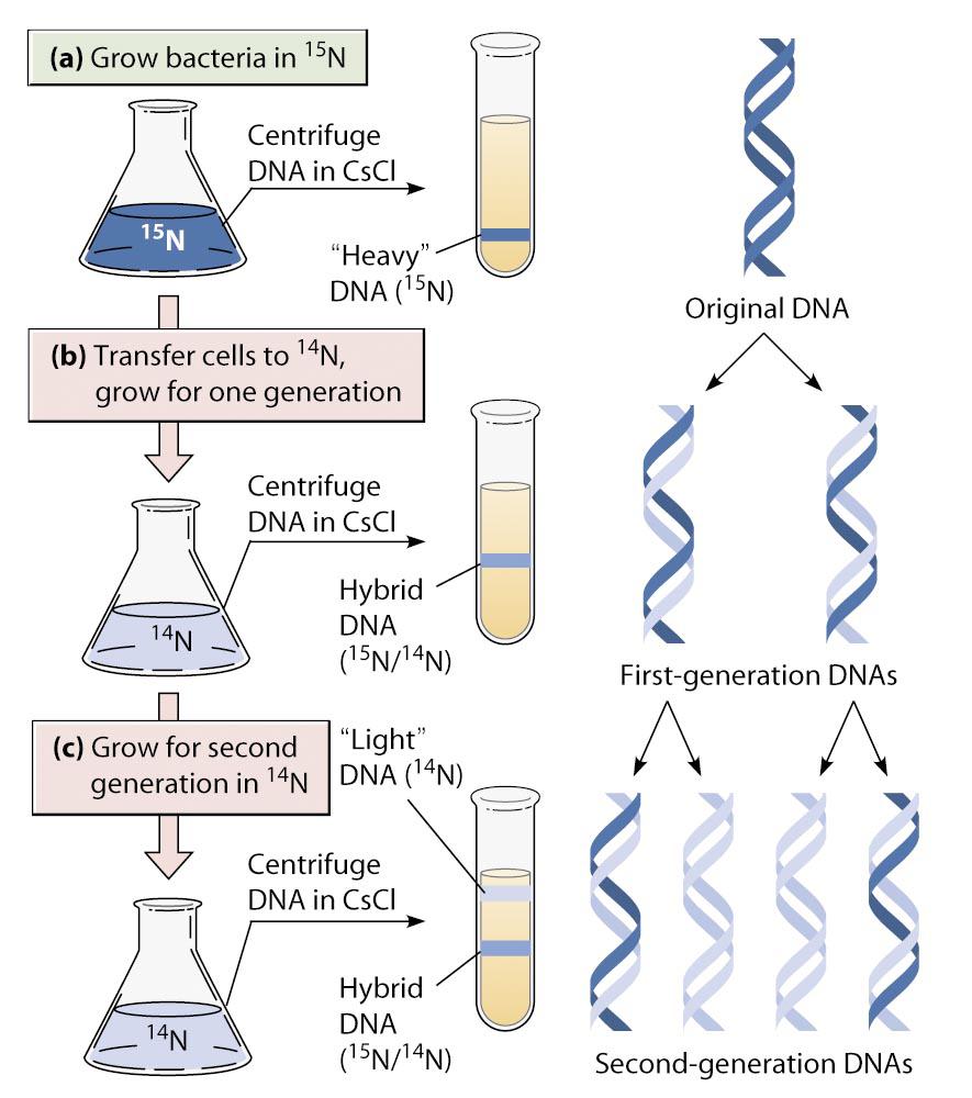
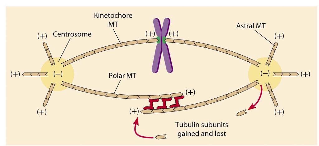
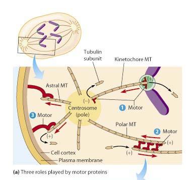
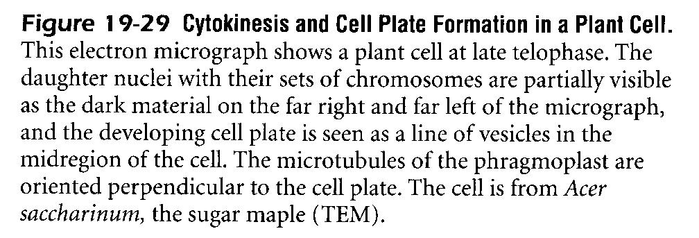
30 Nuclear and Cell Division M phase= Mitosis + Cytokinesis Mitosis= Prophase + Prometaphase + Metaphase + Anaphase + Telophase
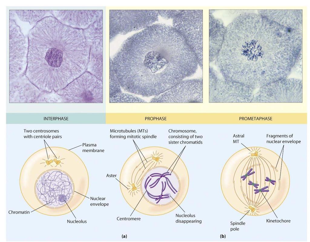
31 染色體出現
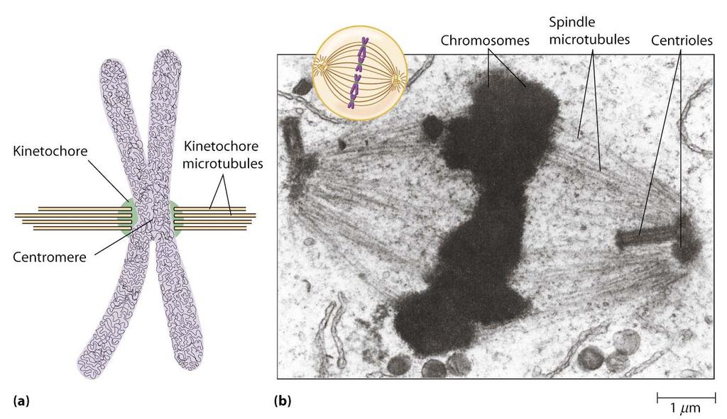
32 著絲點

33 Karyotype 最短時期 AB

34
35 絲變短 兩者離越遠


36

37


38 數目隨物種而有不同 Yeast: 1 Mammalian: 30-40
39 Taxol


40


41

42
43 Chapter 19 The Cell Cycle, DNA Replication, and Mitosis 首頁 OUTLINE Overview of the Cell Cycle DNA Replication DNA Damage and Repair Nuclear and Cell Division Regulation of the Cell Cycle Growth Factors and Cell Proliferation


44

45


46


47


48

49
50

51

52
53

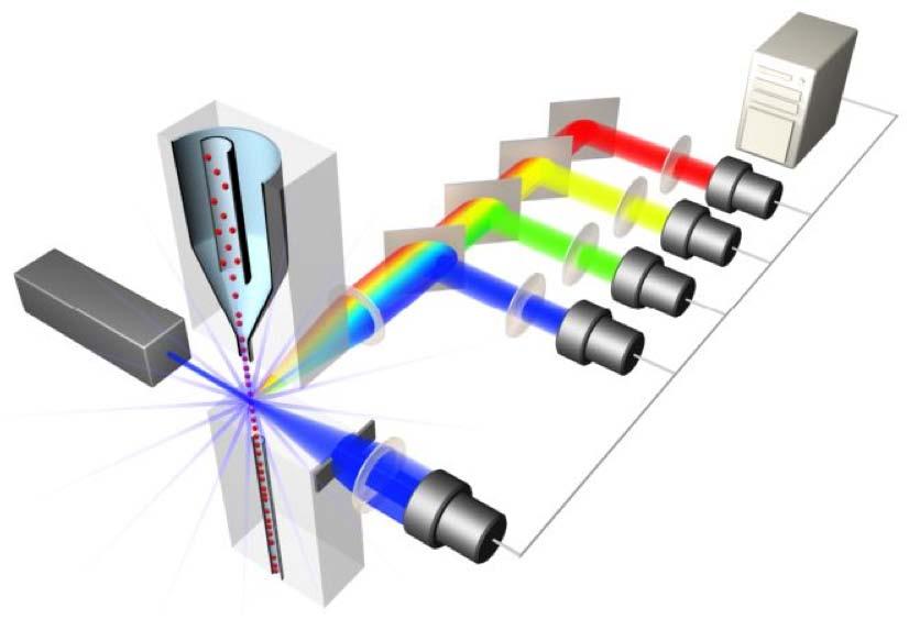
54

55
56 Chapter 19 The Cell Cycle, DNA Replication, and Mitosis 首頁 OUTLINE Overview of the Cell Cycle DNA Replication DNA Damage and Repair Nuclear and Cell Division Regulation of the Cell Cycle Growth Factors and Cell Proliferation


57

58
59 Cell cycle analysis by flow cytometry
60 Basic Principles in Flow Cytometry
61 流式細胞儀原理圖
62 The automated Microscope Detector & Counter Waste Sample This primitive diagram shows the principle: Cells are passing the microscope objective, and an electronic circuit decides whether the cells is fluorescent or not. This is how a flow cytometer works!
63 Z Y X Sample Sheath Cells are presented to the laser using principles of hydrodynamic focusing Flow chamber Y Z Laser optics Laser Beam X
64 Laminar Fluidic Sheath Core Sheath FITC FL PE FL Outer Sheath 488nm Sct
65 Each cell generates a quanta of fluorescence Photomultiplier Tubes (PMT s) PE FL FITC FL 488nm Sct Discriminating Filters Dichroic Lenses Confocal Lens Forward Light Scattering Detector
66 Negative cells are also detected PE FL FITC FL 488nm Sct Dichroic Lenses Confocal Lens Forward Light Scatter

67 Optical Bench Schematic FL2 Sensor 575BP FL3 Sensor 620BP FL4 Sensor 675BP FL1 Sensor 525BP SS Sensor Fluorescence Pickup Lens 600DL 645DL Laser Beam 488BK 550DL 488DL Flow Cell FS Sensor
68 From Fluorescence to Computer Display Individual cell fluorescence quanta is picked up by the various detectors(pmt s). PMT s convert light into electrical pulses. These electrical signals are amplified and digitized using Analog to Digital Converters (ADC s). Each event is designated a channel number (based on the fluorescence intensity as originally detected by the PMT s) on a 1 Parameter Histogram or 2 Parameter Histogram. All events are individually correlated for all the parameters collected.
69 Light Scattering, 2 Parameter Histogram Bigger 90 degree Light Scatter Y Axis Apoptotic Cells Dead Cells Bigger Cells More Granular X Axis Forward Light Scatter (FLS) Live Cells
70 1 Parameter Histogram Negative Positive Count Dimmer Brighter Channel Number Fluorescence picked up from the FITC PMT
71 DNA/RNA Probes Propidium Iodide Ethidium Bromide Hoechst dyes Cyanine dyes eg TO-PRO-3, SYTO/SYTOX dyes Acridine Orange Pyronin Y Styryl Dyes eg LDS-751 Mithramycin, Chromomycin 7 Aminoactinomycin D (7AAD) Diamino-2-phenylindole (DAPI) DRAQ5 Derek Davies, Cancer Research UK
72 Which probe to use? Excitation wavelength available UV: Hoechst, DAPI 488: PI, 7AAD 633: TO-PRO-3 Specificity None: PI A-T: Hoechst G-C: 7AAD, Chromomycin Viability Hoechst DRAQ5 Derek Davies, Cancer Research UK
73 We can use DNA dyes in two ways: 1. As a dead cell discriminator 2. To quantitate cellular DNA content Derek Davies, Cancer Research UK
74 We can use DNA dyes in two ways: 1. As a dead cell discriminator 2. To quantitate cellular DNA content Derek Davies, Cancer Research UK

75 Dead Dead cell discrimination Dead Note log scale! Derek Davies, Cancer Research UK
76 We can use DNA dyes in two ways: 1. As a dead cell discriminator 2. To quantitate cellular DNA content Derek Davies, Cancer Research UK

77 The Cell Cycle G 1 G 0 S G1: Gap 1 S: Synthetic G2: Gap 2 M: Mitosis G0: cells that cease division M G 2 Derek Davies, Cancer Research UK
78 Cell cycle analysis by flow cytometry Cells must be permeable - can use detergent or fixation (ethanol is best) DNA in cells can be stained with a fluorescent dye DNA probes like Propidium Iodide are STOICHIOMETRIC So we can measure how much DNA is in a cell Basic protocol - fix, wash twice, remove RNA and stain with DNA-binding dye Derek Davies, Cancer Research UK
79 In an ideal world. # of Events Derek Davies, Cancer Research UK Increase in Fluorescence Intensity
80 In the real world. # of Events CV: SD/mean x 100. Derek Davies, Cancer Research UK Increase in Fluorescence Intensity
81 DNA stained with propidium iodide S Phase G1 G2/M Note linear scale! Derek Davies, Cancer Research UK DNA Content
82 A pre-requisite for flow cytometry is that cells should be in a single cell suspension. So what about cell clumps affecting quantitation of DNA content? Derek Davies, Cancer Research UK
83 G1 S G2 # of Events M Derek Davies, Cancer Research UK Increase in Fluorescence Intensity

84 How a voltage pulse from the PMT is generated 1. Laser t Voltage 2. Laser t Voltage 3. Laser t Voltage
85 Height, Area, and Width Voltage Pulse Height (H) Pulse area(a) 0 40 Pulse Width (W) Time (µs)
86 Laser Pulse Area Pulse Height Pulse Width Pulse Area G2/M S Doublets The beginners guide to pulse processing G1 Pulse Width Derek Davies, Cancer Research UK
87 Removal of cell clumps Ungated R1 Doublets Gated Derek Davies, Cancer Research UK
88 So, we have our single cells.. G 0 /G 1 S G 2 /M # of Events Derek Davies, Cancer Research UK Fluorescence Intensity
89 How can we use DNA analysis to get useful and relevant scientific information? We can quantitate the percentage of cells in each phase of the cell cycle and monitor the effect of treatments Derek Davies, Cancer Research UK

90 Example 1: Compare cycles Day 1 Day 3 G1 S G2 Derek Davies, Cancer Research UK

91 Example 2: S phase block T0 T24 Derek Davies, Cancer Research UK G1 S G2
92 Example 3: G2 block T0 T24 Derek Davies, Cancer Research UK G1 S G2