Ubiquitin (76 aa) UB genes encode linear fusions of UB either to itself (poly-ub genes) or to other proteins these fusions are cleaved by
|
|
|
- Dominic Freeman
- 5 years ago
- Views:
Transcription
 yielding mature")
1 Ubiquitin (76 aa) UB genes encode linear fusions of UB either to itself (poly-ub genes) or to other proteins these fusions are cleaved by deubiquitylases (DUBs) yielding mature Ub
2 deubiquitylase FLAG 3 -DHFR-UB R48 -X- Gal-FLAG 3
3 deubiquitylase FLAG 3 -DHFR-UB R48 X- Gal-FLAG 3 FLAG 3 -DHFR-UBNH R48 2 -X- Gal-FLAG 3 DUB
4 deubiquitylase FLAG 3 -DHFR-UB R48 Protein-FLAG 3 FLAG 3 -DHFR-UBNH R48 2 -X- Gal-FLAG 3 DUB FLAG IP
5 URT Pulse-Chase Assays and Immunoprecipitation E. coli and V. vulnificus were transfected with URT-based reporter plasmids cells were grown in LB at overnight, culture was diluted 1:200 in fresh LB and grown until A600 = ml of cells were pelleted at 5000g for 5 min at RT, washed 3x with Pulse Medium (PM: M9 medium, ph 7.0, 0.5% glycerol, 0.5% glucose, 0.1 mm CaCl2, 2 mm MgSO4 and Methionine/Cysteine-free Synthetic Complete (SC) Mixture cells were resuspended in 135 µl of PM and incubated at 37 C for 10 min Proteins were pulse-labeled with 15 µl of Express [35S] Protein Labeling Mix (1.175 Ci/mmol, Perkin Elmer) for 3 min at 37 C labeling was quenched by the addition of 0.5 ml of Chase-Medium (CM: PM supplemented with Met and Cys at 0.5 mg/ml each) at 37 C at indicated times of chase cells were mixed with an equal volume of TDS buffer (1% SDS, 5 mm dithiothreitol (DTT), 50 mm Tris-HCl, ph 7.4, containing complete protease-inhibitor mixture (Roche)), followed by immediate freezing in liquid N2 Frozen samples were directly heated at 95 C for 10 min, diluted with 10 volumes of TNN buffer (0.5% NP40, 0.25 M NaCl, 5 mm EDTA, 50 mm Tris-HCl, ph 7.4, containing complete protease-inhibitor mixture (Roche)) lysate was added to 10 µl of magnetic beads linked to anti-flag antibody M2 (Sigma) and incubated at 4 C for 3 hrs IP was washed 4x in TNN buffer, resuspended in 20 µl of SDS-sample buffer, and incubated at 95 C for 5 min samples were fractionated by SDS-PAGE followed by autoradiography
 destabilizing N-terminal residues are recognized by the ClpS N-recognin and are")

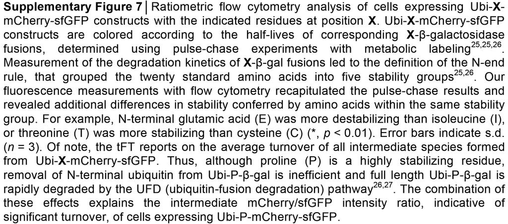
6 The rule N-end pathway in E. coli The Aat L/FR,K-transferase conjugates Leu to N-terminal Arg or Lys. Substrates bearing the indicated primary (bulky hydrophobic) destabilizing N-terminal residues are recognized by the ClpS N-recognin and are delivered to the ClpAP protease The rule N-end pathway V. vulnificus V. vulnificus, which contains both the Aat L/FR,K-transferase and the Bpt LD,Etransferase. As a result, N-terminal Asp and Glu, which are stabilizing(nondestabilizing) residues in E. coli, are secondary destabilizing residues in the V. vulnificus N-end rule pathway
7 analyze protein turnover and trafficking in vivo no pulse-chase metabolic labeling necessary (not possible in living cells) no photoactivation/photobleaching
8

9 UBR1 E3 ubiquitin-protein ligase knockout wt overexpression
10 UBR1 E3 ubiquitin-protein ligase knockout wt overexpression

11 UBR1 E3 ubiquitin-protein ligase knockout wt overexpression
12
13 High-throughput screening for regulators of protein turnover constructs with mcherry-sfgfp fused to different degrons were introduced into the SGA (synthetic genetic array) query strain Y8205 each resulting query strain was crossed in triplicate with a genome-wide library of diploid yeast strains with heterozygous gene deletions The library was sporulated and spores were mated with the query strains using a RoToR pinning robot subsequent library manipulations selection of diploids, sporulation, selection of haploids were done using standard SGA protocols resulting haploid strains each carrying an mcherry-sfgfp fusion and a specific gene deletion were grown in 1,536-colony format for h plates were imaged with a wide field IS4000MM-Pro fluorescence imager equipped with a 4- megapixel CCD camera and filters for sfgfp and mcherry fluorescence imaging (replicate 1) or with a wide field Decon imaging station equipped with an LED-based illumination system, a Retiga4000DC camera and filters for sfgfp and mcherry imaging (replicates 2 and 3) Images of a uniformly fluorescent plate were acquired for flat field correction For comparison between different plates/screens, the corrected measurements were normalized by dividing by the per-plate median absolute deviation (MAD) of corrected measurements normalized B-scores of all constructs were strongly affected in ~250 strains lacking genes mostly related to mitochondrial functions. These strains were identified and omitted from the analysis A gene deletion was considered to stabilize if sfgfp intensity was increased (Δ-score sfgfp > 0) (increased abundance) mcherry/sfgfp intensity ratio increased (Δ-score mcherry/sfgfp ratio > 0) (increased stability)
14
15

16