Electron Microscopy Sciences
|
|
|
- Bernice Cooper
- 5 years ago
- Views:
Transcription


1 Electron Microscopy Sciences INSTRUCTIONAL MANUAL CAT , & Nanopatterned Cell Cultureware P.O. Box 550 s1560 Industry Road s Hatfield PA 19440
2 1 Terms Release of Liability This document is provided to you as is for your information and is to be used at your own risk. assumes no responsibility for any inaccuracies (technical, typographical, or otherwise). reserves the right to change information that is contained in this document at any time. (EMS), without limitation makes no representations, warranties, or agreements (either express or implied). Nor does EMS make warranties or conditions of fitness for a particular reason or for the performance of any product referenced in this guide (either express or implied). EMS does not in any way guarantee or represent that you will obtain satisfactory results from using our products as described herein. You assume all risk in connection with your use of our products. Conditions of Use Nanopatterned Cultureware is for life science research use only. You are responsible for understanding and safely performing the protocols described within this guide. EMS does not guarantee any results you may achieve. These protocols are provided as EMS recommendations to you and does not constitute a guarantee of success. Technical Support or call for any assistance. General Information Handling Your cultureware has been plasma treated and gamma sterilized. EMS recommends rinsing the cultureware in a sterile environment with a sterile solution of water, PBS, or culture medium before use. While the nanopattern is durable, EMS advises against making direct contact with the surface. For best results, avoid touching the surface with items such as pipette tips, tweezers, etc. For single well and 6-well plates, the nanopattern is formed on a #1.5 glass coverslip that is attached to the bottom of a polystyrene (PS) culture dish with a biocompatible adhesive. On 24-well plates and higher, the entire bottom surface of the well is a #1.5 thickness glass with the nanopattern formed on it. Take extra care in handling multi-well plates to avoid undue damage to the thin glass bottom. For coverslips, the patterned side can be seen by reflecting light off of the surface. Alternatively, you can place a droplet of sterile dih 2 O and tilting the surface. The droplet will only run in one direction if it is on the nanopatterned side. Nanopattern Composition and Structure The nanopattern is made using a propriety polymer that is optically transparent and non-cytotoxic. The modulus of the polymer is approximately 7 MPa and has a refractive index of It has excellent transmissivity above 350 nm through to IR (>1.2 μm). The structure of the nanopattern is ridge-groove-ridge. The ridges and grooves are 800 nm wide, while the height of the ridge/depth of the groove is 600nm, as described in Figure 1.


3 Nanopattern Composition and Structure, continued 2 Figure 1. Left: A schematic showing the approximate dimensions of the nanopattern cross-section. Right: A scanning electron micrograph of the nanopattern showing the surface and cross section. Coating with Different ECM Molecules Highly-adherent cells can grow directly on the nanopatterned surfaces as shipped, but for certain experiments and cell types it may be beneficial to modify or coat the surface with a variety of extracellular matrix molecules. Much like standard cell cultureware, the nanopatterns are pre-treated with oxygen plasma to facilitate cell adhesion and functionalization. However, EMS recommends that you use a much lower concentration of ECM molecules. The high concentrations that are commonly used with traditional cultureware can form a thick gel-like material that can obscure the nanopattern and negate its function. EMS also recommends that you allow for longer absorption times when incubating proteins on the culture surface. Below are suggested protocols for some common ECM proteins: 1. Do all of your cell culture work in a sterile environment, and handle all materials using standard aseptic techniques. 2. Open the sterile bag that contains your cultureware and rinse the surface of your cultureware 2-3X in a sterile solution. 3. Prepare your ECM protein: a. Fibronectin: Dilute your fibronectin stock to a working concentration. We recommends using a concentration of 5 μg/ml, though up to 50 μg/ml have been used successfully. We highly recommend diluting your fibronectin in PBS that contains Mg2+ and Ca2+. b. Matrigel: Prepare 1:60 MatrigelTM dilution in culture medium. We recommend DMEM:F12. c. Collagen: Prepare a sterile solution of 50 μg/ml collagen in 0.02 M acetic acid. An HCL solution of 0.01 M can be used as well. d. Gelatin: Prepare a sterile 0.2% gelatin solution in PBS. 4. Cover the bottom of the chamber with the ECM protein solution. You may have to use a significant amount of solution. If the drop is very difficult to spread, use the side of a clean, sterile pipette tip to brush the solution across the surface. You may have to do this several times to get full coverage. For 1- and 6- well plates, use just enough solution to cover the nanopatterned insert. There is no need to treat the flat plastic edges of the dish. 5. Incubate the solution overnight at 37 C/5% CO 2 in sterile conditions (a cell culture incubator works well). 6. After incubation, aspirate the solution and immediately seed your cells at your desired concentration. a. We recommends having 5% FBS in your cell culture medium to improve cell attachment. 7. Continue with your standard cell culture protocol.

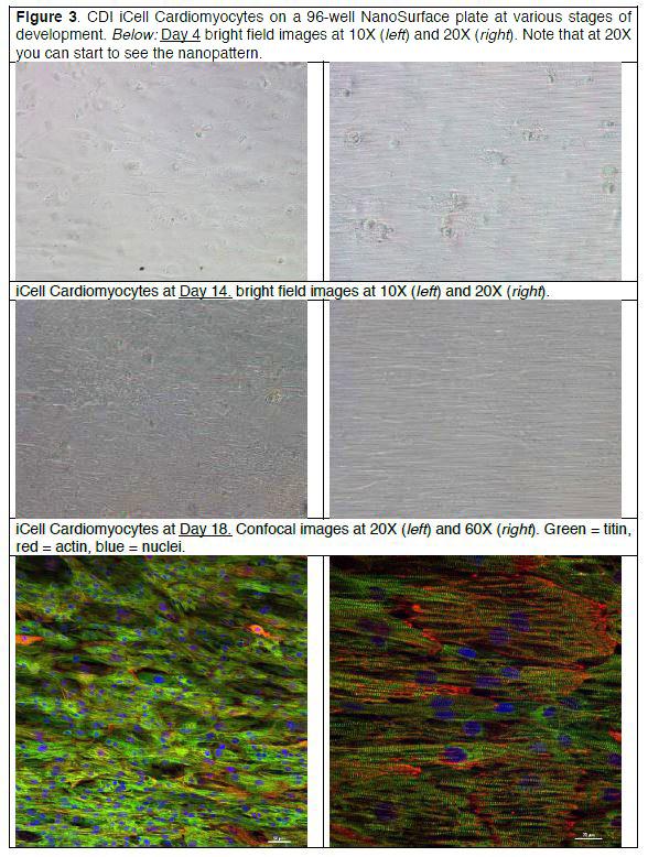
4 3 Cell Seeding Due to the highly variable nature of cell attachment protocols, it is difficult to provide a one-size-fits-all protocol for cell seeding. In general, EMS recommends starting with protocols verified for a given cell type on conventional surfaces and modifying as needed. In some cases, increasing the time of initial seeding can facilitate better attachment to the nanopatterned surfaces. Also, in some cases adding 5% FBS to your medium can help with attachment. Example: ipsc-derived Cardiomyocytes 1. Do all of your cell culture work in a sterile environment, and handle all materials using standard aseptic techniques. 2. This protocol assumes you have already coated the dish with your protein of choice, as outlined in Section Differentiate your ipscs using your protocol of choice for days. 4. Passage the cells and dilute to a concentration of 20 million cells/ml. 5. Seeding will be based on which specific cultureware product you are using: a. For 1- and 6- well plates, use a two-step procedure as shown in Fig. 2: 1. Pipette 0.6 ml of cell solution onto the nanopatterned area of the plate; take care not to spread the drop over onto the polystyrene dish (see figure below). 2. Allow the cells to attach for 6-8 hours (overnight if possible) in the cell culture incubator (37 C, 5% CO2). 3. Fill the dish with a standard working volume (~3 ml) of medium. b. For 24-well and 96-well plates: 1. Pipette ,000 (24-well) or 20-25,000 cells (96-well plate) into the desired wells. 2. Allow the cells to attach overnight in the cell culture incubator (37 C, 5% CO2). 3. Fill the desired wells with a standard working volume (~0.5-1 ml for the 24 well plate; μl for the 96 well plate) of medium. Figure 2. Two-step procedure for coating 35 mm dishes (left) and 6-well plates (not shown). Start with a small droplet (middle image) and allow the cells to attach. After the attachment period, fill the dish or well with a working volume of culture medium (right image). 6. Use standard medium changes using the cell manufacturer s recommended medium (e.g. icell Cardiomyocytes Maintenance Medium or RPMI + B27). 1. NanoSurface recommends replacing only half of the medium during changes. This will allow several soluble growth factors to remain and will also lower the chance of pressing the pipette tip into the sample/cell surface, which can damage the cells. 2) Some representative images of icell Cardiomycoytes plated on NanoSurface 96-well plates are included in Figure 3 (next page).
5 4

 surface topography OR unpatterned control")


 Total Well Volume (μl) Working Volume (μl) 35 mm dish Flat 12.")
6 5 Technical Specifications Nanopatterned Coverslips EMS Catalog , No.1.5 thickness, 25 mm diameter circular coverglass Polymeric surface Either 800:800:600 nm (ridge:groove:depth) surface topography OR unpatterned control surface UV sterile, untreated surface 35mm Petri Dishes EMS Catalog , , , mm Pattern Diameter Cell Culture Treated 6-Well Plates EMS Catalog , mm Glass Diameter 24-Well Plates EMS Catalog , Well Plates EMS Catalog , Specifications Table Item Bottom Shape Approximate Patterned Growth area (cm 2) ) Total Well Volume (μl) Working Volume (μl) 35 mm dish Flat Well Plate Flat Well Plate Flat Well Plate Flat