Table S1. List of antibodies used including isotype controls, biotinylated. secondaries, and fluorophore conjugated tertiary antibodies.
|
|
|
- Morgan Wiggins
- 5 years ago
- Views:
Transcription
1 Table S1. List of antibodies used including isotype controls, biotinylated secondaries, and fluorophore conjugated tertiary antibodies. Antibody Description Distributor Catalogue number Working Concentration Primary Antibodies Rabbit polyclonal to human Annexin II Abcam (Cambridge, UK) ab /2000 Rabbit polyclonal to mouse SDF-1 beta Abcam ab /5 Rabbit polyclonal anti-mouse/rat SDF-1 alpha. ebioscience (San Diego, CA, USA) µg/ml Goat polyclonal anti-human SDF-1. Santa Cruz (CA, USA) SC µg/ml Rabbit polyclonal anti-mouse/rat SDF-1 alpha MBL International (Woburn, MA) JM mg/kg Goat anti-mouse Tie-2 R&D Systems Inc (Minneapolis, MN, USA) AF762 10µg/ml Goat anti-mouse vascular endothelial growth factor R&D Systems Inc receptor 2 (VEGF-R2) BAF644 5µg/ml Biotin anti-mouse intercellular adhesion molecule-1 ebioscience (ICAM-1, CD54). Clone YN1/ µg/ml Purified NA/LE Rat anti-mouse intercellular adhesion molecule-2 (ICAM-2, CD102). Clone 3C4(mIC2/4). In vivo injection. BD Pharmingen (Franklin Lakes, NJ, USA) mg/ml Rabbit polyclonal to human Vascular cell adhesion Santa Cruz molecule-1 (VCAM-1) sc µg/ml Goat polyclonal to the C-terminus of P-selectin of Santa Cruz human origin. Recognises mouse, rat and human. Sc /25 Biotinylated Hyaluronic Acid Binding Protein Seikagaku, (Japan) (HABP) A 20µg/ml Purified goat anti-mouse osteopontin R&D Systems Inc AF808 2µg/ml Isotype Controls Normal Goat IgG control R&D Systems Inc AB-108-C 10µg/ml ChromPure Rabbit IgG control Jackson ImmunoResearch, (West Grove, PA, USA) µg/ml Rat IgG2a NA/LE isotype. Clone R In vivo BD Pharmingen injection mg/ml Secondary Antibodies Biotin-SP-conjugated AffiniPure Goat anti-rabbit IgG Jackson ImmunoResearch, (H+L) µg/ml Biotin Goat anti-rat IgG (H+L) KPL Protein Research Products (Gaithersburg, MD, USA) 1/200 Biotin-SP-conjugated AffiniPure Donkey anti-goat Jackson ImmunoResearch, IgG (H+L) /200 Tertiary antibodies streptavidin, Alexa Fluor 568 conjugate Molecular Probes S (Eugene, OR, USA) 10µg/ml streptavidin, Alexa Fluor 488 conjugate Molecular Probes S /200

2 Table S2. Example of the calculations involved in estimating the distance separating blood vessels in the metaphysis Perfusion-fixed BM followed by post-fixation in osmium both cleared the BVs, making them easier to segment for analysis, and fixed adipocytes, rendering these cells optically opaque, so they could be easily distinguished from BVs. Calculations involved in estimating the distance separating blood vessels in the cellular marrow. All calculations were carried out using Metamorph software. Metaphysis example. A: BVs were segmented using Metamorph software and the total number of BVs in either the metaphyseal or diaphyseal region was recorded. 322 B: Using the same method as described in step A, the average area of each BV was calculated. 202µm C: Using the same method as described in step A, the total area occupied by BVs was calculated µm 2 D: Trabeculae were manually outlined using the drawing tool and the total marrow area was calculated using region analysis in Metamorph. The total extravascular marrow was then calculated by subtracting the total BV area obtained in step C from the total marrow area µm 2 E: To calculate the average area of extravascular marrow occupied by each BV, the total extravascular marrow was divided by the number of BVs as calculated in step A. This calculation assumes an even distribution of BVs. 3621µm 2 F: Assuming each blood vessel is round, the average radius of each blood vessel in µm is the square root of the average BV area (step B) divided by π. See below diagram. 8µm G: The average radius of the area occupied by each blood vessel is the square root of E divided by 2. See below diagram. 30µm H: The average distance separating two blood vessels is 2 x ( G F ). See below diagram. 44µm
3
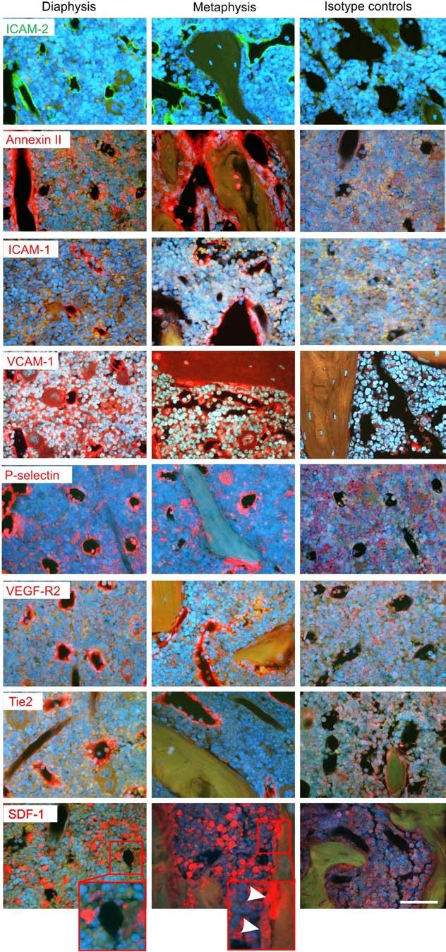
4 Figure S1. Cell adhesion molecules (CAMs) are ubiquitously expressed on the BV endothelium throughout the femur The chemokine, SDF-1, was not observed on the BM endothelium with IHC. Micrographs illustrating the expression of several CAMs and the chemokine, SDF-1, which are important to the homing of HSPC to the BM. Two of the CAMs, VEGF-R2, and Tie2, are included as positive controls. Ubiquitous expression of ICAM-2, Annexin II, ICAM-1, VCAM-1, and P- selectin was observed throughout the femur. SDF-1, whilst present in the cellular marrow and on bone lining cells (arrow heads, middle column inset), was not expressed on BV endothelium (first column inset). The first column depicts micrographs of the diaphysis, the second column metaphysis and the third column reveals immunolabelling of either diaphysis or metaphysis by the relevant isotype control. Scale bar = 50 µm.
5 Figure S2. BV separation, number, and width are similar in WT and Has3 mice To determine if the homing defect observed during very early stages post-transplant in Has3 recipients is due to factors other than a lack of Has3-synthesised HA, BVs within the metaphysis were examined for any abnormalities. Histomorphometry was performed on 1ìm resin sections of perfusion-fixed femurs using Metamorph analytical software. No significant differences were observed between the distances separating individual BVs, the width of BVs or the number of BVs between WT and Has3 mice. Each symbol represents an individual animal. Data is mean + S.E.M.

6
7 Figure S3. Cell adhesion molecules (CAMs) are ubiquitously expressed on the BV endothelium throughout the femur of Has3 mice ICAM-2, Annexin II, ICAM-1, VCAM-1, and P-selectin are ubiquitously expressed on sinusoidal vessels throughout the femur. Two CAMs, VEGF-R2, and Tie2, are included as positive controls. The first column are sections of the diaphysis, the second column sections of the metaphysic, and the third column reveals immunolabelling of either diaphysis or metaphysis by the relevant isotype control. Scale bar = 50 µm.