Freire et al. - Supplemental Material for Trypanosoma brucei translation-initiation factor homolog EIF4E6 is linked to posttranscriptional regulation
|
|
|
- Hilary Hoover
- 5 years ago
- Views:
Transcription
1 Freire et al. - Supplemental Material for Trypanosoma brucei translation-initiation factor homolog EIF4E6 is linked to posttranscriptional regulation Table S1 Oligonucleotides used in this study Table S2 Conserved core residues in T. brucei eif4es FIG S1 Schematic representation of genetically-modified T. brucei cell lines used in this study and validation of fusion proteins FIG S2 Conservation of protein sequence among related kinetoplastid protozoa FIG S3 Protein-level controls for RNAi knockdown of TbE6 FIG S4 Effect of RNAi against TbE6 on social motility FIG S5 Yeast two hybrid assay performed with TbE6 against all Tb4G homologs Supplemental References 1
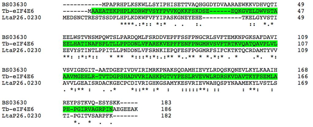
2 Table S1. Oligonucleotides used in this study. Vector Fragment Cloning site Oligonucleotide TbE6 PKO cptp GAD/GBK p2t7-177 p2171 5' UTR 3' UTR partial A partial B HindIII EcoRI XbaI SacI ApaI SalI SalI NotI NdeI BamHI HindIII SpeI HindIII BamHI tttttaagcttcagcgcataaagggtctgctagaag tttttgaattcgtgcgccaggaacatccaag ttttttctagaggtgaagcacaccatacacacaac tttttgagctccccaaatgtccttcgcatcatcg tttttgggcccgaggagcagaaaggtgtagagttgg tttttgtcgactgagtagcttgaactttcgtgcg tttttgtcgacggctgtaccgctcgtgttag tttttcagcggccgcgtcttcgcctcctccccttccg catatggccgctgaggctact ggatcccttcgcctcctccccttcc tttttaagcttatggccgctgaggctact tttttactagtcttcgcctcctccccttcc aagcttatggccgctgaggctact ggatcccttcgcctcctccccttcc TbG5 PKO cptp GAD/GBK 5' UTR 3' UTR partial HindIII EcoRI XbaI SacI ApaI NotI NdeI BamHI tttttaagcttgagacgagggtaagtgaggga tttttgaattctccagtgatgcagttgtagc ttttttctagatgtaataagagtgggtggtacg tttttgagctcttgcgatcaggtaagaacaggag tttttgggccctaagcctgttccgtcccgg ttttcagcggccgcgttttagatttctcaaacgcagcaaaggc catatggagcaccgcgccg ggatcctttagatttctcaaacgcagcaaaggc TbG5-IP PKO Tb (previously known as : cptp Tb ) GAD GBK 5' UTR 3' UTR partial XhoI EcoRI XbaI NotI ApaI NotI SmaI SacI SmaI PstI ctcgagaacaagatggacctttcggaatgc gaattcaacaagaaaggtcccttccctttga tctagagggaagggagagaaggaagaaagg gcggccgcttacacacacacacacacactcac gggcccttcatattcgaggccacttttgcc CAgcggccgcAACCCAGAAGCTTCATTCTGCACT tttttcccgggtatgtcactaaaccctaacgctcct ttttgagctccccagaagcttcattctgcacttc tttttcccgggtatgtcactaaaccctaacgctcct tttttctgcagcccagaagcttcattctgcacttc Table S2. Conserved core residues in T. brucei eif4es human W W W WE W eif4e a 73 b 102 a /3 166 c _ TbE1 W W F WE W TbE2 W29 W W WE W TbE3 W Y W WE W TbE4 W Y W WE W TbE5 W W W YE W TbE6 W F29 H FE W a = W56 & W102 are involved in pi-pi cap-binding interactions b = W73 interacts with eif4g c = W166 recognizes the methyl group of the m 7 G cap. 2

3 A TbE6 /PTP PHLEO R TbE6-PTP E6-PTP NEO R pc-ptp-e B TbE6 +/PTP RNAi (29-13) TbE6 E6-PTP PUR R C pc-ptp-e6 177 bp E6 PHLEO R 177 bp p2t7-177-e6 TbG5 /PTP PHLEO R TbG5-PTP G5-PTP NEO R pc-ptp-g D TbG5-IPkDa /PTP PHLEO R TbG5-IP-PTP G5-IP-PTP NEO R pc-ptp-g5-ip FIG S1 Schematic representation of genetically-modified T. brucei cell lines used in this study and validation of fusion proteins. The integrated plasmids locations are indicated by the thick lines under the genes, not drawn to scale. Clear boxes represent the target gene or selectable marker as indicated; grey boxes represent genomic fusion targets; black boxes represent the PTP tag; stippled boxes represent the 177-bp repeats that are the insertion site for the RNAi vector. Arrowheads represent T7 promoters. Alongside each integration schematic are blots of protein extracts from the specified cell lines. Polyacrylamide concentration used in western blots: E5-PTP 12.5%; G5-PTP 8%; G5-IP- PTP 10%. 3
4 A B 4
 residues; dashes represent gaps introduced to maximize the alignment.")
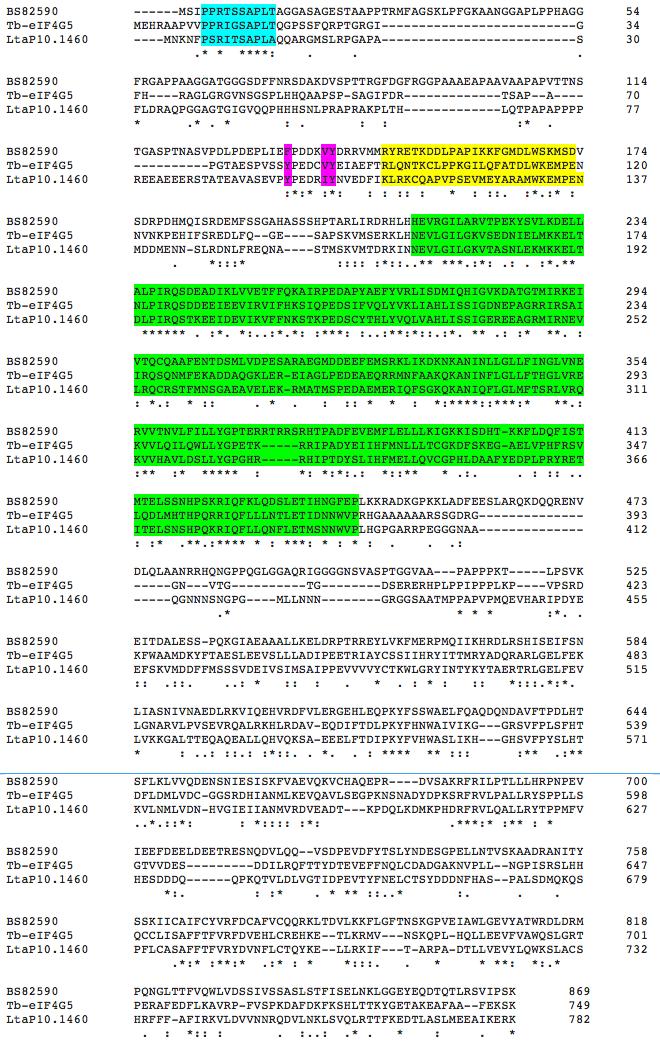
5 C FIG S2 Conservation of protein sequence among related kinetoplastid protozoa. Asterisks = identical amino acids; colons = conserved amino acids; dots = semiconserved (shape) residues; dashes represent gaps introduced to maximize the alignment. Predicted protein sequences from were aligned using Clustal Omega. A. TbE6. The eif4e core domain is shown by green shading; T. brucei (Tb), L. tarentolae (LtaP ), and B. saltans (BS03630). B. TbG5, The MIF4G domain is indicated by green shading. Pink shading represents a Ynnnnϕϕ motif associated with eif4e interaction (Mader et al.,1995). Yellow shading represents identified similarity with the HMG box, and blue shading represents a well-conserved sequence at the amino terminus; T. brucei (Tb), L. tarentolae (LtaP ), and B. saltans (BS82590). C. TbG5-IP. The NTPase domain is indicated by yellow shading and the N 7 G methyltransferase domain by green shading. NH-terminal sequences, which show no substantial conservation, are not shown T. brucei (Tb), L. tarentolae (LtaP ), and B. saltans (BS54325).. 5
Tet Tet (+)Tet + Tet FIG S3 Protein-level controls for RNAi knockdown of TbE6.")
 was induced for 2, 3 or 4 days. Serial two-fold dilutions of the uninduced culture (- Tet) were run in parallel to indicate the relative protein levels.")
6 A Tet, day 2 + Tet day TbE6 (α-prota) 1/8 1/4 1/ kda TbEIF4A1 40 kda B days + Tet (- )Tet Tet (+)Tet + Tet FIG S3 Protein-level controls for RNAi knockdown of TbE6. (A) A western blot of RNAi-induced cultures of TbE6 shown in Fig. 4A were probed with the anti-pap reagent that detects the protein A component of the PTP tag. RNAi (+ Tet) was induced for 2, 3 or 4 days. Serial two-fold dilutions of the uninduced culture (- Tet) were run in parallel to indicate the relative protein levels. The membranes were reprobed with antibody against the T. brucei eif4a1 (Dhalia et al., 2006) to control for loading. (B) RNAi-revertants do not appear after two weeks under Tet induction. The growth characteristics of triplicate cultures induced for RNAi against TbE6. The included western blot shows levels of knockdown of the cognate tagged protein similar to that observed at earlier times (panel A) in three different cultures. 6


7 Tet n=17 + Tet n=18 FIG S4 Effect of RNAi against TbE6 on social motility. All replicates from two experiments are shown. Plates were either uninduced (-Tet) or induced for RNAi (+Tet). 7
8 1.5 mm 3AT 2.5mM 3AT 5.0 mm 3AT control + control TbE6:TbG1 TbE6:TbG2 TbE6:TbG3 TbE6:TbG4 TbE6:TbG5 FIG S5 Yeast two hybrid assay performed with TbE6 against all Tb4G homologs. Data shown represent eif4e homologs in the bait plasmid (pgbk) and eif4g homologs in the prey plasmid (pgad). Similar results were obtained with the reciprocal combination of bait and prey (not shown). SUPPLEMENTAL REFERENCES 1. Dhalia, R., N. Marinsek, C. R. Reis, R. Katz, J. R. Muniz, N. Standart, M. Carrington, and O. P. de Melo Neto The two eif4a helicases in Trypanosoma brucei are functionally distinct. Nucleic Acids Res 34: Mader, S., H. Lee, A. Pause, and N. Sonenberg The translation initiation factor eif-4e binds to a common motif shared by the translation factor eif-4 gamma and the translational repressors 4E-binding proteins. Mol Cell Biol 15: