Protein Data Bank and Structure Display with PyMOL
|
|
|
- Elvin Patrick
- 5 years ago
- Views:
Transcription
1 Protein Data Bank and Structure Display with PyMOL Ching-Shu Suen ( 孫慶姝 ) Supervisor: Dr. Ming-Jing Hwang ( 黃明經 ) Bioinformatics Service Support BioIT, IBMS ( 生醫所 ) Academia Sinica
2 Outline About the RCSB PDB and the PDB Archive Searching for your structures Looking at structures Advanced applications in PDB Structure visualization NGL & JSmol viewers Structure comparison -- jfatcat-rigid Drug target mapping --DrugBank Protein-ligand interaction -- Ligand Explorer PyMOL Basic Applications PyMOL Advanced Applications
3 Worldwide Protein Data Bank (wwpdb) RCSB PDB (USA) (Nucleic Acids Res :D271-D281) Established in 1971 at Brookhaven National Laboratory (BNL) Taken over in 1998 by Research Collaboratory for Structural Bioinformatics PDBe (Europe) PDBj (Japan) (Nucleic Acids Res :D486-D492) (Nucleic Acids Res :D282-D288) BMRB (USA) Biological Magnetic Resonance Bank (BioMagResBank)


4 Molecular Structure for Analysis of Protein Function and Active Sites Site-Directed Mutagenesis Structure-Based Drug Design Protein Docking Protein-Protein Interaction Protein Evolution Structure Comparison Structure Movements
5 RCSB PDB Statistics (1) Y early G rowth of T otal S tructures Yearly Total Numbe r of Entrie s Ye a r
6 RCSB PDB Statistics (2) Statistics Exp. Method Proteins Nucleic Acids Protein / NA complexes Other Total X-ray 120,564 1,954 6, ,750 NMR 10,826 1, ,338 Electron Microscope 1, ,364 Other Multi Method Total 133,464 3,248 7, ,840
 Top bar menu Education PDB-101 Query area")
7 RCSB PDB ( Top bar menu Education PDB-101 Query area Enter what you want to know

8 Education portal - PDB-101
 Interactive")
9 Take an Interactive Tour of the PDB (1) Interactive Animations
")
10 Take an Interactive Tour of the PDB (2)

11 Searching for your structure (1)
12 Searching for your structure (2)
 Query result")
13 Searching for your structure (3) Query result browser

14 Retrieving your structure 1CQP
15 PDB format (1) 1CQP Header, title and author records HEADER IMMUNE SYSTEM 10-AUG-99 1CQP TITLE CRYSTAL STRUCTURE ANALYSIS OF THE COMPLEX LFA-1 (CD11A) I-DOMAIN / TITLE 2 LOVASTATIN AT 2.6 A RESOLUTION COMPND MOL_ID: 1; COMPND 2 MOLECULE: ANTIGEN CD11A (P180); COMPND 3 CHAIN: A, B; COMPND 4 FRAGMENT: I-DOMAIN, RESIDUES ; COMPND 5 SYNONYM: INTEGRIN ALPHA L, LYMPHOCYTE FUNCTION-ASSOCIATED ANTIGEN 1; COMPND 6 ALPHA POLYPEPTIDE; COMPND 7 ENGINEERED: YES; COMPND 8 OTHER_DETAILS: COMPLEXED WITH LOVASTATIN WHICH OCCURS NATURALLY IN COMPND 9 FUNGI SOURCE MOL_ID: 1; SOURCE 2 ORGANISM_SCIENTIFIC: HOMO SAPIENS; SOURCE 3 ORGANISM_COMMON: HUMAN; SOURCE 4 ORGANISM_TAXID: 9606 KEYWDS ROSSMANN FOLD, STRUCTURAL BASIS FOR LFA-1 INHIBITION, IMMUNE SYSTEM EXPDTA X-RAY DIFFRACTION AUTHOR J.KALLEN,K.WELZENBACH,P.RAMAGE,D.GEYL,R.KRIWACKI,G.LEGGE,S.COTTENS, AUTHOR 2 G.WEITZ-SCHMIDT,U.HOMMEL REVDAT 3 12-NOV-14 1CQP 1 KEYWDS REVDAT 2 24-FEB-09 1CQP 1 VERSN REVDAT 1 07-AUG-00 1CQP 0 JRNL AUTH J.KALLEN,K.WELZENBACH,P.RAMAGE,D.GEYL,R.KRIWACKI,G.LEGGE, JRNL AUTH 2 S.COTTENS,G.WEITZ-SCHMIDT,U.HOMMEL JRNL TITL STRUCTURAL BASIS FOR LFA-1 INHIBITION UPON LOVASTATIN JRNL TITL 2 BINDING TO THE CD11A I-DOMAIN. JRNL REF J.MOL.BIOL. V
16 PDB format (2) 1CQP Remark records Heteroatom records (non-standard residues e.g. ligands, ions and water) REMARK 2 RESOLUTION ANGSTROMS. REMARK 290 CRYSTALLOGRAPHIC SYMMETRY REMARK 290 SYMMETRY OPERATORS FOR SPACE GROUP: P REMARK 290 CRYSTALLOGRAPHIC SYMMETRY TRANSFORMATIONS REMARK 290 THE FOLLOWING TRANSFORMATIONS OPERATE ON THE ATOM/HETATM REMARK 290 RECORDS IN THIS ENTRY TO PRODUCE CRYSTALLOGRAPHICALLY REMARK 290 RELATED MOLECULES. REMARK 290 SMTRY REMARK 290 SMTRY REMARK 290 SMTRY REMARK 290 SMTRY REMARK 290 SMTRY REMARK 290 SMTRY HET MG A HET MG B HET 803 A HET 803 B HETNAM MG MAGNESIUM ION HETNAM 803 LOVASTATIN HETSYN 803 MK-803; LOVALIP; MEVACOR FORMUL 3 MG 2(MG 2+) FORMUL (C24 H36 O5) FORMUL 7 HOH *86(H2 O)
17 PDB format (3) 1CQP Atom records ATOM 1 N GLY A N ATOM 2 CA GLY A C ATOM 3 C GLY A C ATOM 4 O GLY A O ATOM 1462 CA ILE A C ATOM 1463 C ILE A C ATOM 1464 O ILE A O ATOM 1465 CB ILE A C ATOM 1466 CG1 ILE A C ATOM 1467 CG2 ILE A C ATOM 1468 CD1 ILE A C ATOM 1469 OXT ILE A O TER 1470 ILE A 309 ATOM 1471 N GLY B N ATOM 1472 CA GLY B C ATOM 1473 C GLY B C ATOM 1474 O GLY B O ATOM 2931 N ILE B N ATOM 2932 CA ILE B C ATOM 2933 C ILE B C ATOM 2934 O ILE B O ATOM 2935 CB ILE B C ATOM 2936 CG1 ILE B C ATOM 2937 CG2 ILE B C ATOM 2938 CD1 ILE B C ATOM 2939 OXT ILE B O TER 2940 ILE B 309 occupancy B factor element
18 PDB format (4) 1CQP HETAM records HETATM 2941 MG MG A MG HETATM 2942 C1 803 A C HETATM 2943 C2 803 A C HETATM 2944 C3 803 A C HETATM 2945 C A C HETATM 2971 MG MG B MG HETATM 2972 C1 803 B C HETATM 2973 C2 803 B C HETATM 2974 C3 803 B C HETATM 2975 C B C HETATM 3001 O HOH A O HETATM 3002 O HOH A O HETATM 3003 O HOH A O HETATM 3004 O HOH A O CONECT CONECT CONECT CONECT CONECT CONECT MASTER END

19 Looking at your structure (1) 1CQP Click it to launch Ligand Explorer

20 Looking at your structure (2) 1CQP Actual protein in full-length Goes to protein feature view Metal-binding regions Structure determined region -helix -strand

21 Protein feature view 1CQP Viewer: NGL Predicted possible disorder region hydrophobic
")
22 Looking at your structure (3) 1CQP
23 Browsing your ligand Related Data Resources for 803 eg. PubChem, DrugBank, Chemical information
24 Looking at your structure (4) 1CQP
25 Searching for ligand Chemical sketch click on the Submit Query

26 Structure visualization NGL Blue to red Download as PNG image Opening JSmol applet

27 Structure visualization JSmol (1) Press on the right bottom of mouse to show the drop-down menu

28 Structure visualization JSmol (2) Select Hetero By HETATM 803 Style Scheme Ball and Stick Surface Dot surface Select Hetero By HETATM MG Color Atoms Cyan Measurements Click for distance measurement on the screen: Click on MG and [803]311:A C21 #1474 Distance: 19.8Å File Export Export PNG image
")
29 Structure comparison -- jfatcat-rigid (1)
")
30 Structure comparison -- jfatcat-rigid (2)
")
31 Structure comparison -- jfatcat-rigid (3) Sequence-Structure alignment

32 Structure comparison -- jfatcat-rigid (5) This structure alignment also can be viewed using the stand-alone Java Web Start application. Click on Launch Web Start Structure alignment Sequence alignment RMSD = 1.72Å ; SeqID = 98%

33 Drug and Drug Target Mapping
 Save the")
34 Protein-ligand interaction (1) Save the image Y257

35 Protein-ligand interaction (2)


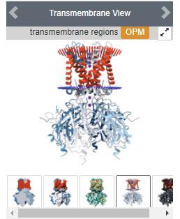
36 Other features Human Gene View Pathway View Transmembrane Proteins

37 Multiple file downloading
38
39 What PyMOL Can Do Open source Support multiple file formats including pdb, mol2, sdf,... Manipulate multiple molecules High quality rendering Read in density maps in CCP4 or X-PLOR format Van der Waals surface rendering Extensive animation generation Written in C and Python languages Get the free source code or purchase licenses ( and

40 Visualize 1CQP using PyMOL External GUI window Name panel A: Active S: Show H: Hide L: Label C: Colour Command lines Viewer window Mouse Mode 3-Button Viewing 3-Button Editing Frame indicator
41 The command language The majority of simple functions, such as open, save, are available via the external GUI menu. Commands can also be used to interact with PyMOL. Commonly-used commands: load <$PYMOL_PATH/filename> select </object_name/segiment_identifier/chain_identifier/residue_identifier/name_identifier> color <color> show <representation type> hide < representation type > set <parameter> zoom <select or object> distance <two atoms> Arguments are separated by one or more commas. show cartoon, chain A
 A rename selection chain_a 1cqp H everything")
 A remove atoms Display")
42 PyMOL Basic Application (1) File Open 1cqp.pdb Display Sequence PyMOL>select /1cqp//A (sele) A rename selection chain_a 1cqp H everything (chain_a) S cartoon (chain_a) C by ss click on 803 (sele) S sticks (sele) C by element File Export Image As PNG [or]draw/ray Save Image to File PyMOL>select /1cqp//B (sele) A remove atoms Display Sequence click on 803 and Y257 (sele) S sticks PyMOL>distance 257/OH, 311/O1
 PyMOL>select /1cqp//A (select /3e2m//A) (sele) A rename selection 1cqp_a (3e2m_a) (3e2m_a) C yellows; A align to selection 1cqp_a (RMS = 0.")
 all H everything 3m6f (1cqp_a & 3e2m_a) S cartoon Click on 803 (E2M & BJZ) S sticks; C by element Click on 1cqp_a Y257 (sele) S lines; C by element; S label")
43 PyMOL Basic Application (2) Display Background white File Open 1cqp.pdb (3e2m.pdb & 3m6f.pdb) PyMOL>select /1cqp//A (select /3e2m//A) (sele) A rename selection 1cqp_a (3e2m_a) (3e2m_a) C yellows; A align to selection 1cqp_a (RMS = 0.317Å) 3m6f A align to selection 1cqp_a (RMS = 0.37Å) all H everything 3m6f (1cqp_a & 3e2m_a) S cartoon Click on 803 (E2M & BJZ) S sticks; C by element Click on 1cqp_a Y257 (sele) S lines; C by element; S label Setting Transparency Cartoon 50% Wizard Measurement Distances; click on oxygen atoms of Y257 & 803 measure01 C skyblue; H labels measure02 C skyblue; H labels File Export Image As PNG File Save Session As *.pse

44 PyMOL Advanced Applications Electrostatic surface Plugin Molecular movement Animation Script
 File Open 1cqpa.pdb File Open 15209457729-pot-PE0.")
45 Electrostatic surface and plugin An input file of APBS (Adaptive Poisson-Boltzmann Solver) is required to generate the electrostatic surface for the structure. This file can be obtained from PDB2PQR web server. ( File Open 1cqpa.pdb File Open pot-PE0.dx Plugin Initialize Plugin System Plugin APBS Electrostatics run File Open 1cqp.pdb 1cqp C by chain; H waters Mouse Mode Residues click on 803 of chain B (sele) C by element carbon atoms in yellow click on MG C magentas purple Setting Transparency Sphere 50%

46 Molecular movement File Open 1cqp.pdb select /1cqp//B (sele) A remove atoms File Open 3m6f.pdb Plugin Alignment/Superposition one by one 1cqp A center 1cqp A generate morph wizard Morph Wizard sele2: 3m6f; method: linear run Display Sequence click on Y257 of morph01 (sele) S sticks; C by element carbon atoms in yellow Morph Wizard run

47 Movie and animation File Open 1cqp.pdb 1cqp A present ligand sites solid surface 1cqp L chains Movie 30 FPS; Program Camera Loop X-Roll 4 seconds File Export Movie As Save Movie as File Export Movie As PNG Images At this step, 120 PNG images are saved. An external software is used to convert these images into a movie or an animation.

48 A Simple PyMOL Script Make a text file of commands: BasicScript.pml load C:\Users\USERX\Desktop\1cqp.pdb bg_color white select chb, chain B remove chb hide everything show cartoon set cartoon_color, marine set cartoon_transparency, 0.5 select ligand, /1cqp//A/311 show sticks,ligand set_bond stick_radius, 0.5 label (311/C21),"%s" % "Lovastatin" alter /,ss="L" alter /,ss="L" rebuild center all draw Run this script file by typing in: run FILEPATH\BasicScript.pml

49 Thank You