Super Resolution Microscopy - Breaking the Diffraction Limit Radiological Research Accelerator Facility
|
|
|
- Jane Greene
- 5 years ago
- Views:
Transcription


1 Super Resolution Microscopy - Breaking the Diffraction Limit Radiological Research Accelerator Facility Sabrina Campelo, Dr. Andrew Harken
2 Outline Motivation Fluorescence Microscopy -Multiphoton Imaging -Microbeam II Super resolution Microscopy -Abbe Diffraction Limit -STED Imaging -gsted Imaging Conclusions & Future Work


3 Motivation Biomolecules that require imaging are typically 1-50nm in size Most super resolution imaging techniques cause damage to biological cells Microtubule 25nm
4 Multiphoton Imaging Multiphoton Imaging Two photons at the same time and at the same place with doubled wavelength Two lower energy photons can combine to have the same effect as one higher energy photon. Yamada, M., Lin, L. L., and T. W. Prow. (2014). Multiphoton microscopy applications in biology. Fluorescence Microscopy: Super Resolution and Other Novel Techniques.
5 Multiphoton Microscopy Advantages Limitations Improved axial resolution Higher depth of light penetration Reduced out of focus bleaching Broader excitation spectra Higher bleaching in the focus More costly Broader excitation spectra
 Multiphoton Imaging (900")
6 Multiphoton Imaging: Spatially Localized Single Photon Imaging (488 nm) Multiphoton Imaging (900 nm)

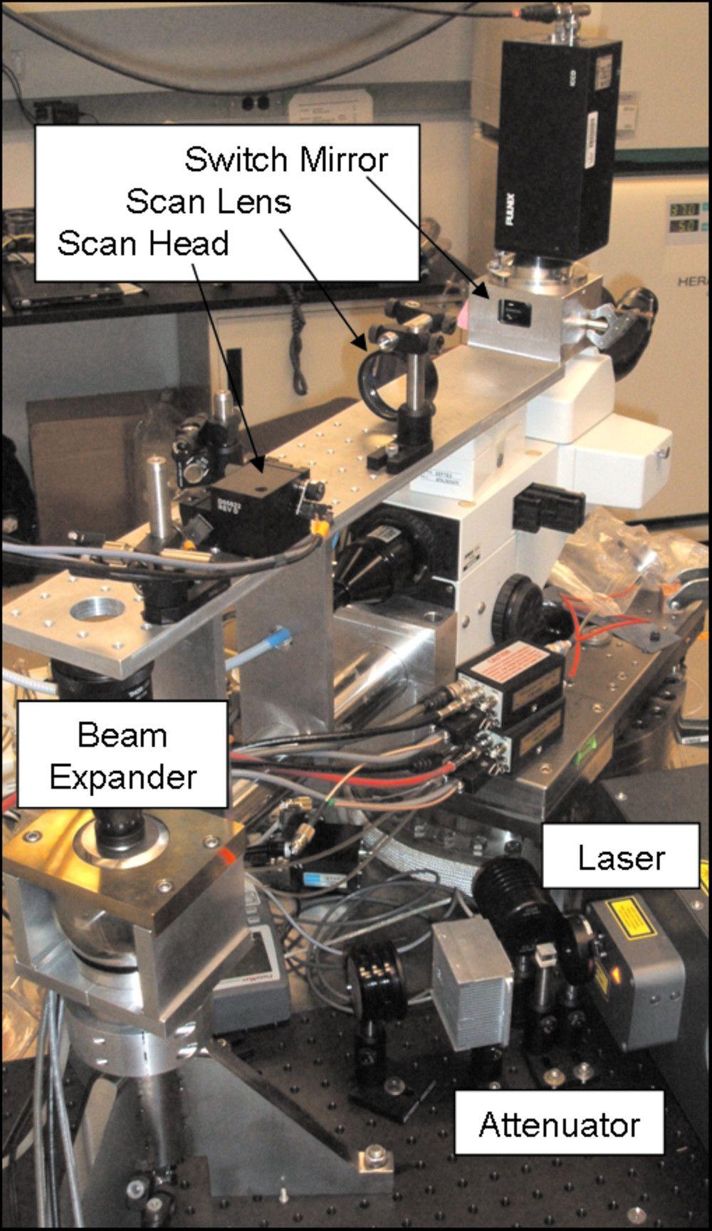
7 Microbeam II at RARAF

8 Multiphoton Imaging Standard Cell Image 10 μm Diffraction limited image of sub-diffraction beads 250 nm 350 nm illumination => 253 nm We are at the diffraction limit!
9 Multiphoton Imaging Reset at Microbeam II
10 Multiphoton Imaging Reset 10µm green fluorescent bead 1010nm wavelength 10µm
11 Multiphoton Imaging Reset 10µm green fluorescent bead 1010nm wavelength 10µm 250nm
12 Outline Motivation Fluorescence Microscopy -Multiphoton Imaging -Microbeam II Super resolution Microscopy -Abbe Diffraction Limit -STED Imaging -gsted Imaging Conclusions & Future Work

13 Abbe Diffraction Limit Resolution is diffraction limited The microscope image is the interference effect of a diffraction phenomenon. Diffraction from different samples and interference between the diffractive beams gives rise to the image of the sample. Ernst Abbe
14 Abbe Diffraction Limit The smallest resolvable distance (d) between two points may never be smaller than half the wavelength of the imaging light

15 Stimulated Emission Depletion (STED) Super resolution technique Image: PubMed.gov Pulsed laser excite fluorophores Depletion beam force surroundings to ground state Resulting in smaller point spread function Greater Resolution!
16 Stimulated Emission Depletion (STED) Not limited by the wavelength of light used Inhibiting the fluorescence at its outer part allows sharper image of focal point. Image: Max Planck Institute for Biophysical Chemistry
17 Time Gated Stimulated Emission Depletion (G-STED) Confocal STED G-STED Image: Optics Express

18 Fluorescence Lifetime Imaging (FLIM) Building images through analysis of fluorescence lifetimes -Separate fluorophores with similar spectra -Minimize effect of photon scattering Image: PicoQuant

19 Fluorescence Lifetime Imaging (FLIM) Ti:Sa Chameleon Ultra II Laser Average Power 3 W Chameleon Tuning Range 680nm-1080nm
 PMT Amplifier Rise Time.")
20 Fluorescence Lifetime Imaging (FLIM) PMT Amplifier Rise Time.23ns

21 Fluorescence Lifetime Imaging (FLIM) TimeHarp 260 Nano TCSPC Unit 250ps resolution Histogramming software
22 Fluorescence Lifetime Imaging (FLIM) 1) Data acquisition 2) Exponential fit of decay curves in each pixel 3) Lifetimes color code
23 Conclusion Breaking the 250nm diffraction limit Multiphoton Imaging ~250nm STED ~ < 20nm living cells g-sted FLIM


24 Research Team & Acknowledgements Research Advisor Dr. Andrew Harken Microbeam technologies Radiation detector physics Special Thanks to: Amy Garwood Georgia Karagiorgi John Parsons Columbia University & Nevis Labs Members at RARAF National Science Foundation REU Programs