In-Room Treatment Verification Using Film and CBCT
|
|
|
- Theodora Simmons
- 5 years ago
- Views:
Transcription


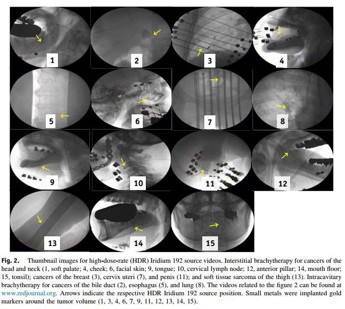
1 SAM: HDR Brachytherapy: Treatment Verification Methods In-Room Treatment Verification Using Film and CBCT Oana Craciunescu, PhD, DABR Department of Radiation Oncology Duke University Medical Center
2 Role of imaging in modern brachytherapy Application insertion Planning Image-guided brachytherapy (IGBT) Treatment Verification Applicator design Facilitate real-time dosimetry Dose summation Functional imaging
3 Objectives To describe current applications of Film/Fluoro and CBCT(kV) in brachytherapy, especially as it relates to inroom treatment verification We have time for the With What and For What, but not for the How
 Radiochromic film")
4 FILM/Fluoro: With what? Standard simulator Flat panel detector (just film) Radiochromic film OR-based X-ray unit AccuBoost (mammographic equipment) C-arm
5 FILM/Fluoro: For What? LDR/HDR GYN: Applicator insertion Planning Orthogonal Semi-orthogonal planning with C-arm with/without jig Pre-TX verification LDR prostate: Needle loading by 3 rd party Post implant film verification HDR breast IGBT with AccuBoost More recent reported uses: Real time HDR TX verification with modified C-arm fluoroscopy (2016) Pre-TX Catheter verification for HDR prostate (2017) Guidance for free-hand needle placement in HDR GYN (2017)

6 FILM: 2D Planning GYN + (Potential refilming for TX verification)

7 FILM: LDR Prostate TX verification of Implant I-125 AgX100 Seed Prostate Implant Radiograph Usually using x-ray unit in OR


8 FILM: LDR prostate 3 rd party loaded sterile needles + autoradiograph
 From J Hiatt,")

9 FILM: HDR Breast Boost (AccuBoost ) From J Hiatt, Non-invasive Image-guided Breast Brachytherapy, AAPM 2015


10 FILM: HDR prostate

11 FILM: HDR GYN Free-hand Interstitial Needles Olsen, Craciunescu, Chino, under review, IJ Contemporary Brachytherapy, 2017 Duke University Medical Center
12 Fluoro: Prostate HDR needle checks


13 Fluoro: HDR Applications


14 Fluoro: HDR GYN, C-arm, VIR-method


15 Fluoro: Real-time HDR TX Verification
16 kv-cbct: With what? CBCT enabled simulators C-arms Note: reports of MV-CBCT use in brachytherapy: Since MV CBCT images are less affected by high atomic number materials, such as metal objects, they can complement the information provided by kv CT(or CBCT) in images with metalinduced streak artifacts. Deschovich et al., Brachytherapy,vol. 5, Issue 2, 85-86, 2006


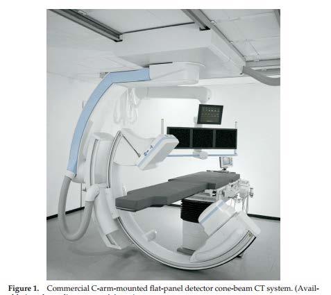

17 kv-cbct Commercially available systems: Varian, Acuity - C-arm CBCT Nucletron, Simulinx
18 CBCT: For what? GYN (G) & Prostate (P) Planning (G, P): alone or with MRI/US Applicator/Needle reconstruction and position (G, P) Free-hand needle placement (G) Gold marker displacement (P) Post-plan/seed localization LDR (P) C-arm CBCT for QA of LDR (P) Breast: Multi catheter HDR interstitial Balloon-based
19 CBCT Stand alone for planning As in-room imaging for applicator placement and pre-tx verification As part of a hybrid method when access to MRI is limited Use of MRI at least at 1 st FX and identify HRCTV/IRCTV Continue subsequent fractions with CT or CBCT


20 Duke Brachy Suite BrachySuite Console CBCT Console CBCT Console + Access to 3.0 T MRI in Rad Onc on same hallway
21 Advantages of a CBCT in Brachy Suite Intra-operative imaging Large mechanical clearance (scan in stirrups, make adjustments) Can be easily combined with other imaging modalities Primary, secondary (US, MRI) Minimize applicator/needles motion Limiting the patient s motion is expected to limit post insertion applicator motion, which in return leads to more accurate planning. Good for applicator delineation Ability to image and verify before treatment Can scan, plan and treat under anesthesia


22 Image Quality vs. Patient Size AP = 17 cm AP = 25 cm AP = 31 cm AP = 35 cm Technique: 150 SID, kvp = 120, ma = 80, ms = 13 Large patients attenuate more, resulting in detection of fewer photons (increased noise, reduced signal, increased HU discrepancy, ie computer mistakes a thick absorber for high density material)

23 CBCT vs CT (Female pelvis, small size)

24 CT vs. CB contours
")

25 CBCT vs. CT (breast) Courtesy of Dorin Todor, VCU

26 CBCT Prostate
27 GYN


28 GYN: CBCT-based planning Opt imag protocol

29 GYN Interstitial

30 GYN: CBCT-based planning


31 GYN: CBCT-based Planning

32 OAR: Good agreement with MRI (small patient size, good quality CBCT)

33 OAR: Poor agreement with MRI Craciunescu et. Al, Brachytherapy, vol. 15, S , 2016


34 CBCT: GYN, Free-hand Needle Placement Guidance Duke University Brachytherapy

35 CBCT: GYN, Free-hand Needle, Planning CBCT: Needles reconstruction MRI: HRCTV + normal tissue
36 Prostate

37


38 VCU setup, HDR Prostate Courtesy of Dorin Todor, VCU


39 C-arm CBCT for LDR prostate on-line verification


40 Post-Operative Seed Localization Intra-Operative Seed Localization
41 Conclusions X-ray films/fluoro and kv-cbct (Simulators, C-arms) have a role in in-room pre/post TX verification for several brachytherapy applications Thorough understanding of advantages and limitations is needed before using as sole imaging procedure for inroom treatment verification and/or planning
42 Indeed, we often mark our progress in science by improvements in imaging. Martin Chalfie Next CT and MRI in room TX Verification

43 Thank you!