Percutaneous Spinal Injections problem presentation for HW1
|
|
|
- Pauline Gibson
- 5 years ago
- Views:
Transcription
1 Percutaneous Spinal Injections problem presentation for HW1 Gabor Fichtinger, PhD Director of Engineering, Associate Research Professor of Computer Science and Radiology NSF-Funded Funded Engineering Research Center for Computer- Integrated Surgical Systems and Technology
2 Why Needles? Potentially significant impact on medical practice Minimally invasive (compared to open surgery) Faster recovery Less morbidity Fewer complications Lower cost Repeatable in many indications Sharply increasing number of procedures Challenging but doable Constrained process formally describable Major challenges (in addition to open/lap surgery): no visibility no access no room to maneuver no room to recover
3 Why Spine? Why pain management? In US alone 70% of population affected in lifetime Single most expensive disease Pain management: alleviate pain caused by stressed/pressured/pinched spinal nerve by suppressing sensory input at nerve root Numb with lidocaine/novocaine etc.
4 Key technical issues Accuracy Longevity of pain relief Collateral damage Pain during procedure Acceptable ~ 1mm Time Volume/throughput = money Good surgeons < 10 min Mediocre/poor surgeons ~ min Toxic radiation primarily to physician!!! Good surgeons < total 5.0 sec beam time Mediocre/poor surgeons ~ 30 sec beam time

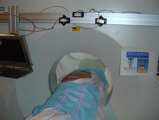
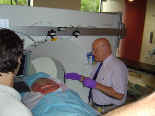
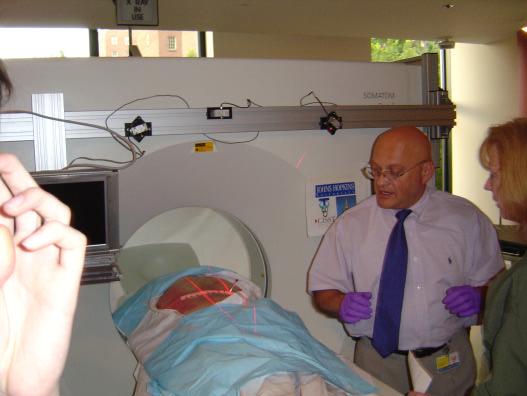
5 Current standard 1: CT guidance
6 Affix IZI Biopsy Strip Pick Entry and Target
7 Current workflow for CT-guided procedure 1. Put patient in prone to scanner 2. Palpate vertebra 3. Affix IZI Biopsy Strip fiducials 4. Take thin volume scan 5. Select slice of interest 6. Pick target and entry 7. Determine angle and depth 8. Pull out patient to outer laser plane 9. Identify entry on skin 10. Touch needle to entry point 11. Enter needle manually 22G beveled 12. Maintain insertion angle by sight 13. Keep needle in laser plane 14. Judge current insertion depth by feeling 15. Insert contrast (optional) 16. Push patient back to scan plane 17. Take confirmation CT 18. Pull out patient 19. Inject therapeutic agent
8 The challenge Transfer entry, angle and depth over patient Control all 3-DOF simultaneously during insertion


9 Current standard 2: C-arm Fluoroscopy Mobile Real-time Inexpensive Broad coverage Courtesy of Siemens Siemens Limited rotation X-ray dose Low soft tissue contract Need for calibration Need for tracking

10 Current workflow for Fluoro-guided procedure 1. Put patient in prone on table 2. Palpate vertebra 3. Set C-arm in anticipated needle direction 4. Turn on beam 5. Adjust C-arm angle to optimal 6. Reach into beam with needle 7. Touch entry with needle tip 8. Keep needle perpendicular to beam 9. Optimize entry location 10. Fulcrum with needle till barrel-view 11. Turn off the beam 12. Rotate C-arm by 90 degrees 13. Turn beam on, keep beam on 14. Insert needle 22G beveled 15. Monitor insertion depth/deflection in image 16. Stop needle at target position 17. Insert contrast (optional) 18. Take confirmation C-arm image 19. Inject therapeutic agent

11 Handheld Needle Guide
12 CT-Mounted Laser Guide


13 CT-Mounted Image Overlay Goal : Simple and radiation-free X-ray vision during surgery Approach : Merging data model with physical space through half-mirror Future work : Cadaver studies in June/July 2003 Industry partner: Siemens Funded by Siemens, PI: Fichtinger

14 Robot assistance: 3-DOF RCM-PAKY Robot 7-DOF passive arm Needle Joysticks and safety switches Locking arm 2-DOF Remote Center of Motion robot 1-DOF needle injector w/ mounted stereotactic fiducials Amplifier box Table side robot mount Robot: D. Stoianovici


15 Robot-CT Registration: Stereotactic Adapter Robot registered to CT from a single image using stereotactic frame on the end-effector Credit: D. Stoianovici, L. Kavoussi, A. Patriciu, S. Solomon (JHU Bayview)

16 CT-Robot Registration: Stereotactic Adapter

17 Surgical planning


18 Robot-CT Registration: Scanner s Laser Robot registered to CT using the scanner s alignment laser Credit: D. Stoianovici, L. Kavoussi, A. Patriciu, S. Solomon, JHU Bayview and G. Fichtinger, ERC

19 The URobotics 6-DOF 6 Accubot robot 7-DOF passive arm 3-DOF Cartesian motion 2-DOF rotation motion stage 1-DOF Needle insertion stage Mounting bridge Robot: D. Stoianovici

20 Robot-CT Registration: Optical Tracking
21 Robot-CT Registration: Optical Tracking


22 Robot-CT Registration: Fiducial carrier attached to spinous process


23 Robot-C-arm registration: Simple fiducial-based registration in biplane fluoroscope

24 Corkscrew fiducials in monoplane fluoroscope


25 Joystick Controlled Robot Under Fluoroscopy