Lung Cell Apoptosis. Man Yi, Rosetta Belcastro, Samuel Shek, Daochun Luo, Martin Post, A Keith Tanswell ON-LINE DATA SUPPLEMENT
|
|
|
- Angela Davis
- 5 years ago
- Views:
Transcription
1 Fibroblast Growth Factor-2 and Receptor-1α(ΙΙΙc) Regulate Postnatal Rat Lung Cell Apoptosis Man Yi, Rosetta Belcastro, Samuel Shek, Daochun Luo, Martin Post, A Keith Tanswell ON-LINE DATA SUPPLEMENT
2 SUPPLEMENT METHODS Lung tissue fixation and section preparation. Lungs were initially cleared of blood by flushing the pulmonary circulation with phosphatebuffered saline (PBS) containing 1 U/mL heparin, fixed over 12 hours by perfusion with 4% (wt/vol) freshly-dissolved paraformaldehyde. A constant airway pressure of 20 cm water was maintained, via a tracheal catheter, throughout the fixation process in order to prevent lung recoil. The lungs were briefly washed three times in PBS then, after using a graded ethanol series, embedded in paraffin at 60 C. The lungs were cut into 5-µm sections for staining with hematoxylin and eosin, or for bromodeoxyuridine (BrdU) immunohistochemistry. For the preparation of frozen sections, the fixed lungs were cryopreserved overnight in 30% (wt/vol) sucrose in 4% (wt/vol) freshly-dissolved paraformaldehyde/0.1 M PBS, ph 7.4, at 4 C, then placed in Tissue-Tek OCT compound (Sakura Finetek USA, Torrance, CA), frozen under liquid nitrogen, and stored at -70 C in sealed plastic bags. Before sectioning, the blocks were equilibrated at -15 to -19 C for 30 minutes. Sections (10 µm) were cut at -22 C using a CM-300 cryostat (Leica Microsystems, Deerfield, IL) and applied to microscope slides for storage at -20 C for a minimum of 3 days. The sections were used within 2 weeks. Western Blot Analysis. Freshly-excised lungs were homogenized and sonicated (40 Watts for 30 seconds) in RIPA cell lysis buffer (10 mm monosodium phosphate, 0.3 M sodium chloride, 0.1% (wt/vol) sodium
3 dodecyl sulphate, 1% (vol/vol) Nonidet P-40, 1% (vol/vol) sodium deoxycholate, 2 mm disodium phosphate, ph 7.2) with the protease inhibitors leupeptin, aprotinin (1 µg/ml each) and 1 mm phenylmethylsulfonyl fluoride. The homogenate was allowed to stand on ice for 10 minutes, centrifuged at 7,000 x g for 10 minutes. Samples were stored at -80 C until analysis. For cytoplasmic protein analysis, the lung tissue was removed and homogenized in buffer containing 1 mm EDTA, 10 mm Hepes (ph 7.6), 42 mm KCl, 5 mm MgCl 2, 1 mm phenylmethylsulfonyl fluoride, and protease inhibitors. After a15-minute incubation on ice, samples were centrifuged at 21,000 rpm at 4 C for 30 minutes. The supernatant, representing the cytoplasmic fraction, was stored at -80 C. Lung tissue lysates containing 80 µg total protein were boiled for 5 minutes in SDS sample buffer [60 mm Tris-HCl, 10% (wt/vol) SDS, 5% (vol/vol) glycerol, 2 mm β- mercaptoethanol, ph 6.8], then fractionated under reducing conditions by SDS polyacrylamide gel electrophoresis for 2 hours at 120 Volts. The proteins were transferred to polyvinylidene fluoride membranes (Pall Corporation, Pensacola, FL) and blocked in 0.1% (vol/vol) Tween in PBS with 5% (wt/vol) non-fat dry milk for 1 hour at room temperature. Membranes were incubated overnight at 4 C with 1:1000 dilution of rabbit polyclonal antibodies to cleaved caspase-3, -7, Bax (Cell Signaling Technology, Beverly, MA) or 1:200 dilution goat polyclonal antibody to cytochrome c (Santa Cruz Biotechnology, Santa Cruz, CA) in the same blocking solution. A mouse monoclonal antibody to Bcl-x L and a rabbit polyclonal antibody to Fas (Santa Cruz Biotechnology, Santa Cruz, CA) were diluted 1:200 with 0.1% (vol/vol) Tween in PBS with 5% (wt/vol) non-fat dry milk. After a thorough wash, the membranes were
4 incubated with secondary horseradish peroxidase-conjugated anti-mouse (Calbiochem- Novabiochem Corporation, San Diego, CA), diluted 1:10,000, or anti-rabbit (Cell Signaling Technology, Beverly, MA), diluted 1:1000, or anti-goat (Calbiochem-Novabiochem Corporation, San Diego, CA), diluted 1:10,000, for 90 minutes. As an internal control, glyceraldehyde 3-phosphate dehydrogenase (GAPDH) was detected using a 1:500 dilution of a rabbit polyclonal antibody to GAPDH (Santa Cruz Biotechnology), and a 1:1000 dilution of an horseradish peroxidase-conjugated secondary anti-rabbit antibody (Cell Signaling Technology). Blots were imaged and bands quantified by enhanced chemiluminescence detection as previously described (E 1). Immunoprecipitation Lung tissue was homogenized in lysis buffer containing 50 mm Hepes (ph 7.5), 100 mm NaCl, 1 mm EGTA, 1 mm dithiothreitol, 10 mm MgCl 2, 1% (vol/vol) Triton X-100, 10 µg/ml aprotinin, 10 µg/ml leupeptin, 1 mm sodium orthovanadate and 25 mm sodium fluoride for 10 minutes on ice. Cellular debris was removed by centrifugation at 10,000 rpm for 10 minutes at 4 C, and the supernatant was retained for immunoprecipitation. Protein content of extracts was determined by using the Bio-Rad protein assay kit. Rabbit anti-human flg antibody (Santa Cruz Biotechnology) 4 ng/mg protein, or rabbit anti-rat Erk1/2 antibody (QED Bioscience Inc. Hornby, ON, Canada) 2 ng/mg protein, and protein G agarose (Sigma, Saint Louis, Missouri) 80 µl/500 mg supernatant protein, were added to supernatant and incubated at 4 C for 3 hours.
5 Beads were isolated by centrifugation and washed three times with lysis buffer and analyzed by immunoblotting using the p-tyr antibody (Santa Cruz Biotechnology). Immunohistochemistry. A BrdU In-Situ Detection Kit (BD Biosciences Pharmingen, San Diego, CA) was used for immunohistochemical staining of BrdU in paraformaldehyde-fixed paraffin-embedded lung tissue. Slides were deparaffinized in xylene (2 x 5 minutes), then exposed to 100% alcohol (2 x 3 minutes) then 95% (vol/vol) alcohol (1 x 3 minutes). Endogenous peroxidase activity was blocked by incubation with 3% (vol/vol) hydrogen peroxide in PBS for 10 minutes. The slides were placed in the provided antigen retrieval solution, heated in a microwave oven at 89 C for 10 minutes then allowed to cool at room temperature for 20 minutes. The biotinylated anti- BrdU antibody was applied at a 1:10 dilution, with the provided diluent, to the sections for a 1- hour incubation at room temperature. After the slides were rinsed 3 times, the supplied streptavidin-horse radish peroxidase mixture was applied for 30 minutes. Tissue sections were covered with the 3, 3 diaminobenzidine (DAB) substrate solution for up to 5 minutes to obtain the desired color intensity. The slides were counterstained with hematoxylin, then dehydrated in an alcohol series, then cleared in xylene. Frozen sections were used for cleaved caspase-3 immunohistochemistry. Slides were rinsed 3 times in PBS at room temperature and sections were then permeabilized with 1% (vol/vol) Triton X-100 and 1% (wt/vol) bovine serum album in PBS for 4 minutes at room temperature. Incubations were with a 1:100 dilution of rabbit anti-cleaved caspase-3 antibody
6 (Cell Signaling Technology, Beverly, MA) in 5% (vol/vol) normal goat serum and 1% (wt/vol) bovine serum album in PBS at 4 C overnight, followed by a 1:300 dilution of the secondary flurorescein-conjugated goat anti-rabbit antibody (Calbiochem-Novabiochem Corporation, San Diego, CA) in 1% (wt/vol) bovine serum album in PBS for 1 hour at room temperature. The slices were mounted with Vectashield mounting medium with 4, 6-diamidino-2-phenylindole (DAPI: Vector Laboratories, Burlingame, CA). For quantification of BrdU-positive and cleaved caspase-3-positive cells, values were averaged from 20 random images per slide, and then averaged from three slides per animal. Mean values from each of four animals per group were expressed per mm 2. TUNEL Assay Paraffin-embedded tissue sections were dewaxed, rehydrated, and treated with proteinase K, The TUNEL assay was performed using an In Situ Cell Death Detection Kit (Roche Applied Science, Penzberg, Germany), according to the protocol provided by the manufacturer. Slides were then examined by fluorescence microscopy.
7 SUPPLEMENT REFERENCE E1. Jankov RP, Luo X, Belcastro R, Copland I, Frndova H, Lye SJ, Hoidal JR, Post M, Tanswell AK. Gadolinium chloride inhibits pulmonary macrophage influx and prevents O 2 -induced pulmonary hypertension in the neonatal rat. Pediatr Res 2001;50:
8 SUPPLEMENT FIGURE LEGENDS Figure E1. TUNEL assay. Vehicle-treated lungs from day-7 pups were perfusion-fixed and paraffin-embedded. TUNEL reaction-positive cells were identified by their red color under fluorescence microscopy. DAPI stains nuclei blue under fluorescent light. In the merged images (insert a, c) it was apparent that TUNEL-positive cells included red blood cells that had not been cleared, even after a thorough perfusion of the pulmonary circulation prior to fixation. Figure E2. Analysis of Erk1/2 phosphorylation. Erk1/2 was immunoprecipated from lung homogenates of three of four average-sized day-7 pups in each group, using a rabbit polyclonal antibody, then run on Western blots for immunodetection of phosphotyrosine. Erk1/2 phosphorylation was reduced in pups that had been injected with either sfgf-r1 or an anti- FGF-2 neutralizing antibody, but not after injection of an anti-fgf-1 neutralizing antibody. Figure E3. BrdU incorporation in neonatal rat lung as a marker of cell proliferation. Lung tissue from rat pups at 7 days of age after receiving intraperitoneal injections of neutralizing antibody to FGF-1 or FGF-2, or their respective isotype IgGs, on days 3, 4, 5 and 6 of life. There was no obvious effect of antibody injections on cell proliferation. Bar length = 100 µm.
9 Figure E4. Quantification of BrdU-positive cell numbers in lung tissue from day-7 rat pups. The numbers of BrdU-positive cells in lung tissue from 7-day-old pups that had received neutralizing antibody to FGF-1 (FGF-1Ab) or FGF-2 (FGF-2Ab) were not significantly different from values for 7-day-old pups that had received their respective rabbit (NR-IgG) or mouse (NM-IgG) isotype IgGs. Values are means ± SEM for three animals in each group.
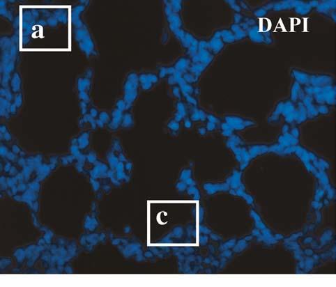


10 Figure E1.

11 Figure E2.


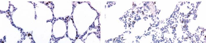

12 Figure E3.
13 Figure E4.