Sample collection & laboratory methods for identification bacteria. Lab. 2 A.T. Samira
|
|
|
- Cuthbert Baker
- 5 years ago
- Views:
Transcription
1 Sample collection & laboratory methods for identification bacteria Lab. 2 A.T. Samira


2 Learning objectives At the end of the presentation, participants should understand the: Procedures, preparation, processing and transport of specimens
3 Successful laboratory investigations Advance planning Collection of adequate and appropriate specimens Sufficient documentation Biosafety and decontamination Correct packaging Rapid transport Choice of a laboratory that can accurately perform the tests Timely communication of results
4 Specimen collection: key issues Consider differential diagnoses Decide on test(s) to be conducted Decide on clinical samples to be collected to conduct these tests consultation between microbiologist, clinicians and epidemiologist
5 Transport medium Allows organisms (pathogens and contaminants) to survive Non-nutritive - does not allow organisms to proliferate For bacteria i.e., Cary Blair For viruses - virus transport media (VTM)
6 Blood for smears Collection Capillary blood from finger prick make smear fix with methanol or other fixative Handling and transport Transport slides within 24 hours Do not refrigerate (can alter cell morphology

7 Blood for cultures Collection Venous blood infants: ml children: 2 5 ml adults: 5 10 ml Requires aseptic technique Collect within 10 minutes of fever if suspect bacterial endocarditis: 3 sets of blood culture
8 Blood for cultures Handling and Transport Collect into bottles with infusion broth change needle to inoculate the broth Transport upright with cushion prevents hemolysis Wrap tubes with absorbent cotton Travel at ambient Store at 4oC if can t reach laboratory in 24h
9 Serum Collection Venous blood in sterile test tube let clot for 30 minutes at ambient temperature glass better than plastic Handling Place at 4-8oC for clot retraction for at least 1-2 hours Centrifuge at RPM for 5-10 min separates serum from the clot
10 Cerebrospinal fluid (CSF) Collection Lumbar puncture Sterile tubes Aseptic conditions Trained person

11 CSF Handling and transportation Bacteria preferably in trans-isolate medium, pre-warmed to C before inoculation transport at ambient temperature (relevant pathogens do not survive at low temperatures)
12 Stool samples Collection: Freshly passed stool samples avoid specimens from a bed pan Use sterile or clean container do not clean with disinfectant During an outbreak - collect from patients
; dry ice for Ag, PCR")
13 Stool samples for bacteria Timing during active phase Sample amount and size fresh sample and two swabs from patients, controls and carriers (if indicated) Method Cary-Blair medium For Ag detection/pcr no transport medium Storage refrigerate at 4oC if testing within 48 hours, -70oC if longer; store at -15oC for Ag detection and PCR Transport 4oC (do not freeze); dry ice for Ag, PCR detection
14 Throat swab (posterior pharyngeal swab) Hold tongue away with tongue depressor Locate areas of inflammation and exudate in posterior pharynx, tonsillar region of throat behind uvula Avoid swabbing soft palate; do not touch tongue Rub area back and forth with cotton or Dacron swab
15 Nasopharyngeal swab Tilt head backwards Insert flexible fine-shafted polyester swab into nostril and back to nasopharynx Leave in place a few seconds Withdraw slowly; rotating motion
16 Sputum Collection Instruct patient to take a deep breath and cough up sputum directly into a wide-mouth sterile container avoid saliva or postnasal discharge 1 ml minimum volume
17 Respiratory samples Handling and Transport All respiratory specimens except sputum are transported in appropriate media bacteria: Amie s or Stuart s transport medium Transport as quickly as possible to the laboratory to reduce overgrowth by oral flora For transit periods up to 24 hours ambient temperature for bacteria
18 Glass slides for microscopy Label slides individually use glass marking pencil ensure markings don t interfere with staining process Each slide should bear: patient name unique identification number date of collection
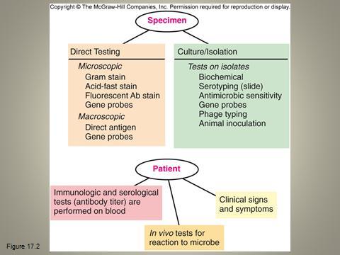
19 Preparation for the Survey of Microbial Diseases Methods used to identify bacteria to the level of genus and species Phenotypic methods Morphology Physiology or biochemistry Immunologic method Serological analysis Genotypic techniques More and more often used as a sole resource for identifying bacteria.
20 Phenotypic Methods Microscopic morphology Macroscopic morphology Physiological/Biochemical characteristics Chemical analysis
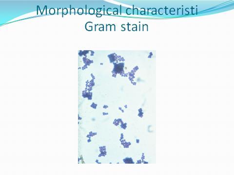
21 Microscopic Morphology Cell shape and size Gram stain reaction Acid fast reaction Special structures
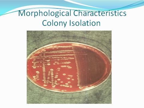
22 Macroscopic Morphology Colony appearance Speed of growth Patterns of growth
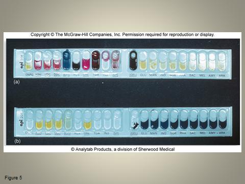
23 Physiological/Biochemical Characteristics Traditional mainstay of bacterial identification Diagnostic tests for determining the presence of specific enzymes and assessing nutritional and metabolic activities Examples Fermentation of sugars Capacity to metabolize complex polymers Production of gas Presence of enzymes Sensitivity to antimicrobic drugs
24
25 Cultivation of Specimen Isolation media Biochemical testing Carbohydrate fermentation (acid and/or gas) Hydrolysis of gelatin, startch, and other polymers Enzyme actions such as catalase, oxidase, and coagulase By-products of metabolism
26
27
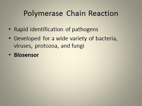
28 Polymerase Chain Reaction Rapid identification of pathogens Developed for a wide variety of bacteria, viruses, protozoa, and fungi
29
30