Local vasoconstriction. is due to local spasm of the smooth muscle (symp. reflex) can be maintained by platelet vasoconstrictors
|
|
|
- Alberta Holland
- 5 years ago
- Views:
Transcription
1 Hemostasis


2 Hemostasis ( hemo =blood; sta= remain ) is the stoppage of bleeding, which is vitally important when blood vessels are damaged. Following an injury to blood vessels several actions may help prevent blood loss, including: Formation of a clot
3 Hemostasis STAGE I

4 Local vasoconstriction is due to local spasm of the smooth muscle (symp. reflex) can be maintained by platelet vasoconstrictors
5 Hemostasis STAGE II

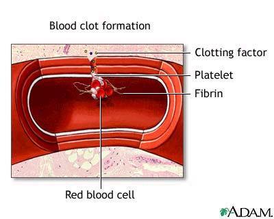
6 Formation of platelet aggregate Injured blood vessel releases ADP, which attracts platelets (PLT) PLT comming in contact with exposed collagen release: serotonin, ADP, TXA2, which accelerate vasoconstriction and causes PLT to swell and become more sticky
7 Platelets
8 Platelets (thrombocytes) thrombocytes, are not true cells, but rather cytoplasmic fragments of a large cell in the bone marrow, the megakaryocyte blood normally contains 150,000 to 400,000 per microliter (µl) of platelets


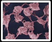
9 The image shows a number of platelets stained purple associated with some RBC's.
10 Platelets At any one time, about two-thirds of the body's platelets are circulating in the blood and one-third are pooled in the spleen. the life span of platelets is between 1 and 2 weeks if not consumed in the process of blood clotting, they are destroyed by macrophages in the liver and spleen

11 The micrograph shows activated platelets adhering to some damaged cells
12 Hemostasis STAGE III
13
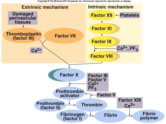
14 Calcium ions Are required for promotion and acceleration of almost all blood clotting reactions Except: activation of XII and XI (intrinsic mechanism) Ca 2+
15 Vitamin K the "K" in Vitamin K came from the Danish word "koagulation" Vitamin K is a cofactor needed for the synthesis (in the liver) of: - factor II (prothrombin), VII, IX, and X - proteins C and S deficiency of Vitamin K predisposes to bleeding. Conversely, blocking the action of vitamin K helps to prevent inappropriate clotting (eg. by Warfarin )
16 Fibrinolysis
17 Clot Dissolution 1. Plasmin is formed from plasminogen - enzyme called activator (e.g. enzymes from urine, tears, saliva or bacterial enzyme streptokinase) 2. Plasmin as an enzyme is involved in breaking down fibrin into soluble fragments (fibrinolysis) Plasminogen Activator (e.g. t-pa) Plasmin Fibrin soluble fragments Plasminogen may be produced by eosinophils
18

19 Anticoagulants Hirudo medicinalis produce Hirudin that inhibits Thrombin
20 Anticoagulants Although tissue breakdown and platelets destruction are normal events in the absence of trauma, intravascular clotting does not usually occur because: - the amounts of procoagulants released are very small - natural anticoagulants are present (Antithrombin III, Heparin, Antithromboplastin, Protein C and S, fibrin fibers)
21 Natural anticoagulants Antithrombin III inhibits factor X and thrombin Heparin from basophils and mast cells potentiates effects of antithrombin III (together they inhibit IX, X, XI, XII and thrombin) Antithromboplastin (inhibits tissue factors tissue thromboplastins) Protein C and S activated by thrombin; degrade factor Va and VIIIa

22 Abnormalities of hemostasis
 - increased destruction (autoimmune processes) - increased PLTs consumption (DIC) Thrombocytopenia Hemorrhagic spots")
23 Severe reduction in the number of PLTs - thrombocytopenia this causes spontaneous bleeding as a reaction to minor trauma in the skin - reddish-purple blotchy rash it may result from: - decreased production (toxins, radiation, infection, leukemias) - increased destruction (autoimmune processes) - increased PLTs consumption (DIC) Thrombocytopenia Hemorrhagic spots (petechiae)


24 Thrombocytopenia Lethal when PLTs<10G/L Bleeding occurs when PLTs<50G/L Norm: G/L

25 Hepatic failure Most of the clotting factors are formed in the liver Subconjunctival hemorrhage


26 Hemophilia A (lack of F VIII) and B (lack of F IX) are transmitted genetically and affect only males. Females carry the gen but do not show symptoms. Von Willebrand s disease loss of large component of fviii


27 Hemophilia A (lack of F VIII; 85%) Spontaneous or traumatic subcutaneous bleeding Blood in the urine Bleeding in the mouth, lips, tongue Bleeding to the joints, CNS, gastrointestinal tract Mild hemophilia after injection in buttock
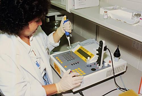
28 Tests of coagulation
29 Selected causes of abnormal coagulation tests Partial Thromboplastin Time (aptt) Prothrombin Time (PT) Thrombin Time (TT) Bleeding Time (BT) Factor deficiency (except VII) VII, X, V, II, fibrinogen deficiency Low or absent fibrinogen Thrombocytopenia Antibodies to clotting factors Antibodies Dysfibrinogenemia, hypofibrinogenemia Von Willebrand s disease Heparin Warfarin; Vit K defficiency (mild to severe) Heparin Drugs (Aspirin, NSAIDs, high dose penicillins, etc.) Excessive Warfarin Excessive Heparin Cirrhosis, Uremia, PLTs dysfunction
30 International Normalised Ratio (INR) The result for the PT is expressed as a ratio (prothrombin clotting time for patient plasma divided by time for control plasma); Correction factor (International Sensitivity Index) is applied to the prothrombin ratio and the result issued as INR. Therapeutic interval: Therapeutic interval for oral anticoagulant therapy: Application: Monitoring oral anticoagulant therapy (eg. Warfarin); note that heparin will not prolong INR (heparinase is included within the INR reagent)!!!!!!!!!!!!! For heparin therapy we monitor aptt and/or aptt ratio
31 INR oral anticoagulants Norm:INR about 1.0. For patients on anticoagulants, the INR typically should be between 2.0 and 3.0 for patients with atrial fibrillation, or between 3.0 for patients with mechanical heart valves = 4.0 be individualized for each patient. An INR can be too high; a number greater than blood is clotting too slowly (a risk of uncontrolled bleeding) INR less than 2.0 may not provide adequate protection from clotting.

32 Bleeding time procedure: Clean the earlobe with an alcohol Prick the earlobe with an automatic lancet Note the time when blood first appears on the skin After half a minute (30sec) place the edge of the filter paper on the top of the drop of blood. Perform the operation at half minute (30 sec) interval The end point or bleeding time is the first half minute when no blood is seen on the filter paper.