Simple, intuitive and accessible MRI solution for preclinical research. M-Series Compact MRI Systems
|
|
|
- Beatrice Alexina Armstrong
- 5 years ago
- Views:
Transcription


1 Simple, intuitive and accessible MRI solution for preclinical research M-Series Compact MRI Systems


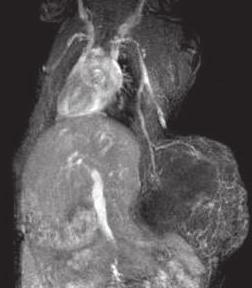
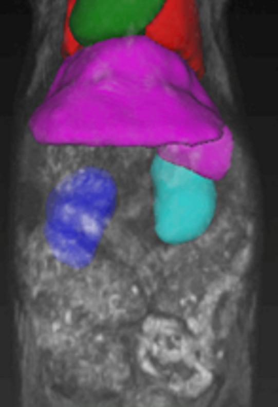
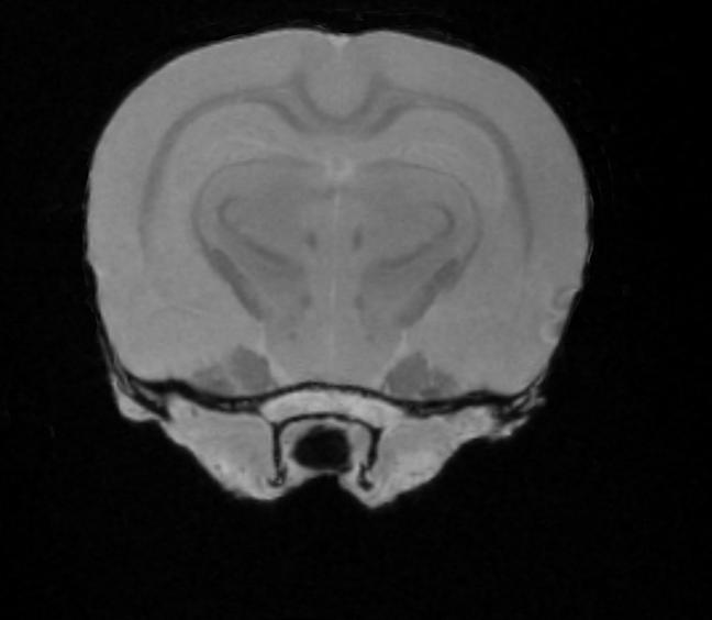
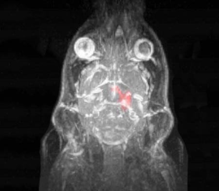
2 Application Oriented Imaging Molecular Imaging Using Contrast Agents Detection and quantification of cellular activity targeted and enhanced with contrast agents Anatomy and Morphology In vivo soft tissue imaging for morphological characterization. 2D and 3D imaging can be performed quickly and easily for preclinical model assessment. Neurobiology In vivo anatomical imaging of the brain, spine and spinal cord for assessment and follow-up of neurologically-based diseases Cancer Research Detection, follow-up, and quantification of tumor development and progression. Multi-modality Imaging Ex vivo Imaging Easy registration with other modalities such as Optical, PET, SPECT and CT to enable powerful multi-modality pheno-typing. High-resolution, high throughput, 3D MR-based histology imaging of fixed samples and embryos for toxicological and developmental studies. 2 3


3 M-Series : Compact MR Imaging Non-invasive 3D anatomical, functional and molecular results in mice and rats Simple to operate Intuitive software interface and analysis tools require no prior experience in MR imaging to fully execute the workflow and imaging. No additional infrastructure necessary to maintain the magnetic field Aspect's permanent magnet technology removes the need for cryogens, plumbing, chemicals and supplemental power supplies or coolers. Pain-free installation, ready for imaging from day one Simply wheeled into position and moved around based on the needs of the working lab, with imaging possible just a few hours after installation. Simplified and Optimized Preclinical Research Aspect Imaging is the world's leader in compact, high-performance MRI systems: Powerful results without the cost, complexity and technical burden of conventional MRI systems. With the M-SeriesTM platform, academic researchers and pharmaceutical companies can harness the power and insights of MRI, deriving quantified answers to their biological questions - quickly, easily and cost effectively. No running cost of upkeep Negligible running cost with a maintenance free magnet, no moving parts or cooling. Standard warranty is 12 months with options for extended warranty for up to 5 years. Power fluctuations and outages have no impact on the permanent magnet. No dedicated housing facilities Requires no dedicated power supply, no shielding, isolation from other metal objects or other magnetic field restrictions. Height 1320 mm Height 1080 mm 734 mm Depth 950 mm 4 Width 790 mm 5


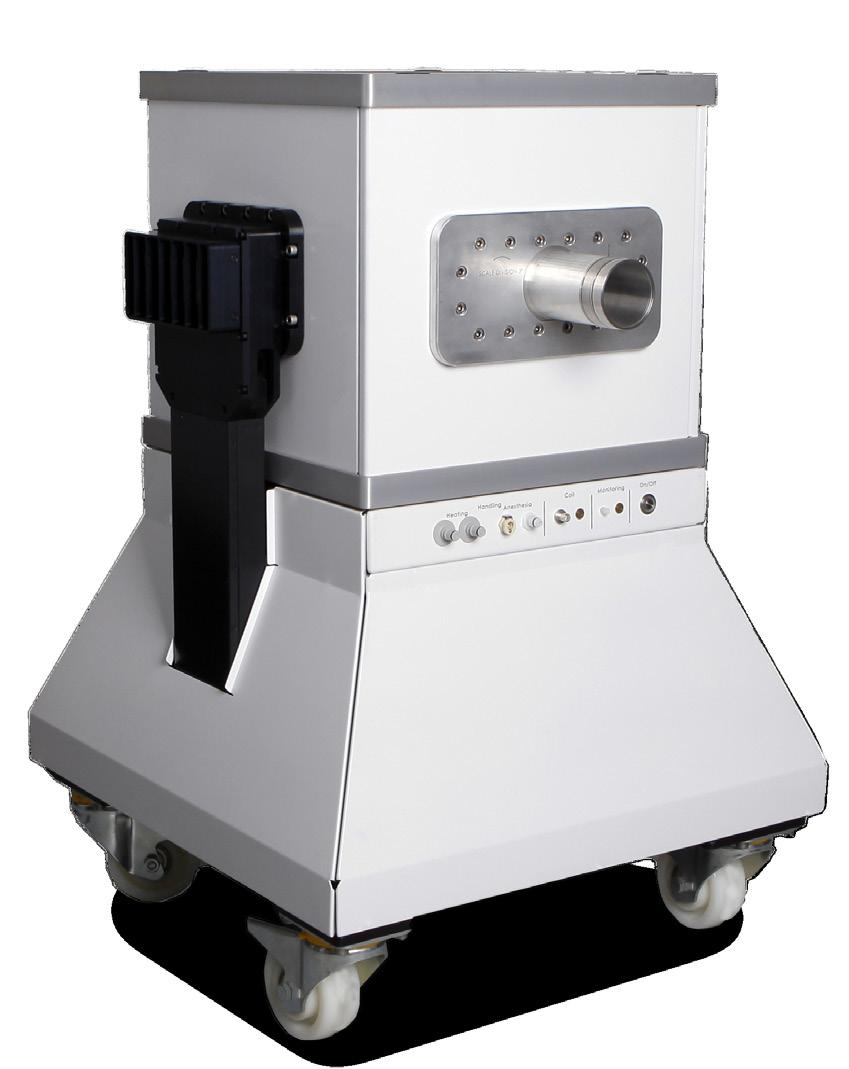
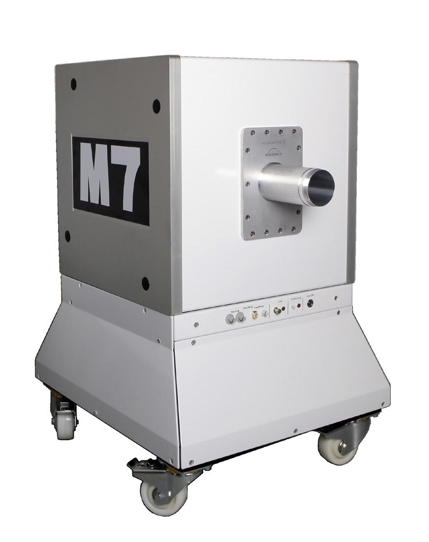
4 M3 : For Mice and Small Animal Imaging M7 : From Small Mice to Large Rats Aspect's MRI systems offer a comprehensive preclinical solution to quantify the expression of disease, monitor disease progression and assess therapeutic efficacy and response in lab rodents. The smaller sized M3 makes the power of MRI systems available to the significant portion of academic researchers requiring only mice imaging. The larger-scale M7 delivers imaging for both mice and rats. High performance, compact, permanent magnet on a portable cart Integrated mini electronics cabinet PC workstation with simple and intuitive operating software Scalable with easy magnet only upgrade. Workstation, software and electronics are the same for all M-Series compact MRI systems Flexibility and customization available for more advanced MRI users A complete solution including animal handling, physiology monitoring and anesthesia delivery Best-in-class post processing, analysis and data management solution M-Series Imaging Software Platform Acquisition software for preclinical MR imaging, integrating a coherent suite of sequences: Spin echo with the following options: Respiration/cardiac triggering Preceding inversion recovery pulse Diffusion weighted imaging Gradient echo with the following options: 2D and 3D Respiration/cardiac triggering Dynamic acquisition i.e. Dynamic Contrast Enhanced (DCE) IR Snap for T1 map generation Fast spin echo 2D and 3D Respiration/cardiac triggering Variable echo train length Multi-point fat/water separation Extensive post-processing tool - VivoQuant Image Analysis and Co-registration Software - includes: 3D ROI segmentation tools with automatic, semi-automatic and manual segmentation Co-registration tools to generate multi-modality MR images (automated, manual and fiducially-aided), e.g. PET/MR Supports multiple input data formats, and multiple output formats including video loop generation Modeling tools to generate T1, T2, and ADC maps Co-registration capabilities with optical and PET imaging 6 7


5 Optimized Animal Handling System Fully Integrated Animal Handling System A full suite of application-specific RF coils and animal handling beds and accessories: Type Dimensions Inner Diameter Length Application Facilitating a complete setup for preclinical imaging with designated coil for different imaging applications Mouse head 23 mm 25 mm Neurological imaging in mice Motorized calibration mechanism enabling automatic coil-tuning Mouse body 30 mm 50 mm Extremity, abdominal and thoracic cavity imaging in mice Water heated animal bed maintaining hydrated body temperature Mouse whole body 30 mm 80 mm Whole body imaging in mice LumiQuant - mouse body 38 mm 50 mm Multi-modal imaging in mice Obesity studies in mice Rat head 35 mm 40 mm Neurological imaging in rats Rat body 50/60 ellipsoid 90 mm Large rat body 71 mm 90 mm MR-based Histology Module Automated multi-sample ex vivo imaging Accommodate long scan times (>1 hr) for high spatial resolution Easy to use multi-sample ID and data management system Extremity, abdominal and thoracic cavity imaging in rats Extremity, abdominal and thoracic cavity imaging in large rats Physiological monitoring system (respiration, ECG and temperature) Delivery and evacuation of isoflurane-based anesthesia Small animal physiological monitoring Respiration, ECG and temperature monitoring Respiration and ECG output triggering to MRI spectrometer Additional readouts monitor Isoflurane-based anesthesia Vaporizer with temperature and flow-rate compensation Scavenging cube for waste gas Breathing circuit with 3 access points Multi-nuclear Capabilities Optional imaging coils for advanced multi-nuclear imaging Supports imaging and detection of 13 C, Xe and F Multi-modality Capabilities Simultaneous PET/MRI with SimPET (MR compatible PET insert from Brightonix Imaging). MIM (Multimodal Imaging Module) for MRI and optical co-registration (3D Bioluminescence by IVIS SpectrumCT) 8 9
6 Taking notes 10


7 DOC Rev.3 Product specification and descriptions in this document are subject to change without notice. Aspect Imaging Made with recycled paper.