Kinesin Walks on Microtubules
|
|
|
- Stuart Patterson
- 5 years ago
- Views:
Transcription

1 Kinesin Walks on Microtubules

2 Other Motor Proteins

3 Question: Alone or in Groups?
4
5 Our live-cell imaging: DIC (differential interference contrast)
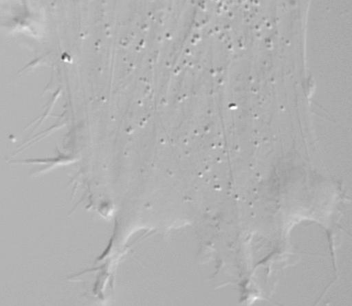
6 Normal DIC Microscopy Image 50x50 m Orca ER camera 125 ms exposure 60X water immersion NA 1.0 Nikon Eclipse Chick motoneurons glass-bottom dish


7 Measuring Vesicle Diameter A. B µm 0.35 µm intensity pixel diagonals 50x50 m, Orca ER camera, 125 ms exposure, 60X water immersion NA 1.0, Nikon Eclipse, NT2 cell, glass-bottom dish

8 Vesicle Volume Measurements Works below diffraction limit! Shtridelman et al. 2009
9 How does MEDIC work? (motion-enhanced DIC) Camera Code: 12 bits 8 bits A B Select front buffer 8-image Queue + Subtract - Average background 12 8 bits Look-up table monitor 400-image Queue key press HDD Hill, Macosko, Holzwarth 2008

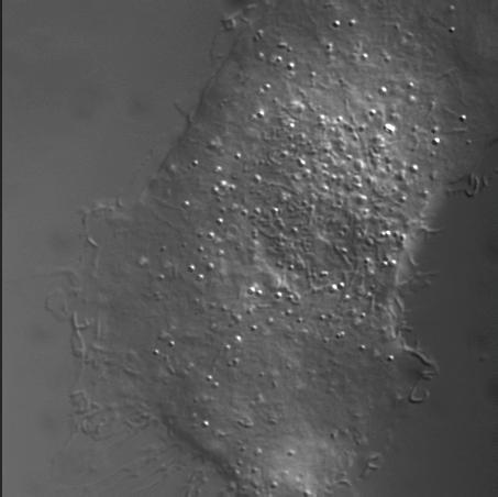
10 Normal DIC Microscopy Movie 50x50 m Orca ER camera 8 frames/s 60X water immersion NA 1.0 Nikon Eclipse Chick motoneurons glass-bottom dish

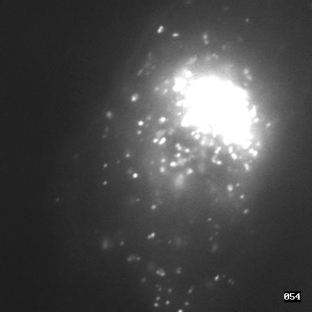
11 MEDIC movie of a Motoneuron 50x50 m Orca ER camera 8 frames/s 60X water immersion NA 1.0 Nikon Eclipse Chick motoneurons glass-bottom dish


12 Simultaneous MEDIC+fluorescence 50x50 m Orca 125 ms 60X oil NA 1.4 Nikon Ti-E Mouse fibroblast on glass Stained with LysoTracker Hirokawa, Science 1998

13 Computerized Vesicle Tracking 50x50 m Orca ER camera 8 frames/s 60X water immersion NA 1.0 Nikon Eclipse Chick motoneurons glass-bottom dish Movie -130 X pixel number Y pixel number


14 Obtaining trajectories (x-y y -t) A. t= 0s t= 2s t= 4s t= 6s B. y ( m) X 2 red = 1.09 = X 2 red Number of of Segments t (s) Orca ER camera, 125 ms exposure, 60X water immersion NA 1.0, Nikon Eclipse, NT2 cell, glass-bottom dish, scale bar 5 m Shtridelman et al. 2008
15 Slope of Distance vs Time is Velocity Very fast! Hill et al (PC12 cells)
?")

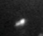
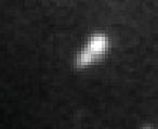
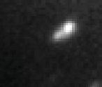
16 How does kinesin move so fast in a cell (although it s highly viscous)? Kinesin 300 nm Vesicle DIC image Microtubule -100 nm- DIC image Electron microscopy image Hirokawa, Science 279, 1998
 Shtridelman et al.")
17 Larger Cargo Moves Slower Velocity ( m/s) Diameter( m) Shtridelman et al. 2009


18 Larger Cargo Often Moves Retro Macosko et al. 2008


19 Cargo Slows 1 3 Days in Culture Macosko et al. 2008


20 Velocity histograms give insight Chick neuron Macosko et al. 2008
21 Same Pattern as in PC12 cells motor Hill et al Number of Occurances motors 3 motors 4 motors 0 v/v
22 And in NT2 Cells Shtridelman et al. 2009


23 Other Organisms As Well Rat striatal neurons Zahn, 2004 Hill, 2003 Levi, 2006 Frog Kural, 2006 Chick Breuer, 1975! ?
24 Hypothesis: more motors=more speed get slopes Recap: B. 6 X 2 red = 1.09 = 0.05 plot histogram y ( m) X 2 red Number of Segments t (s) B. 6 X 2 red = 1.09 = 0.05 y ( m) 4 2 X 2 red Number of Segments t (s). Explain peaks by number of motors (from many files)
25 Model that may explain hypothesis Velocity ( m/s) Diameter( m) Stokes Law: F and v linearly related (get viscosity from Stokes-Einstein)
26 Measuring Intracellular Viscosity

27 Model that may explain hypothesis
28 Model that may explain hypothesis (adding curves for 2 and 3 motors) 1 motor 3 motors 2 motors Shtridelman et al. 2008
29 Latest data from NT2 cells Shtridelman et al. 2009


30 Vesicle Transport: Physicists View V ~nv F Drag F k F ' Drag nf k
31 Summary of single molecule studies Live-cell imaging: MEDIC, sub-diffraction sizing Single molecule extended to groups: force-velocity Insights from polymer physics: Stokes, viscocity How could this help whole cell studies? Example: Vesicle transport in rat cortical neurons was found increase with increasing plating density. I.e. MEDIC and vesicle tracking reveals unnoticed differences in cell cultures. Bauer et al (Neuroscience Lett)
32
33
34 In Vitro single molecule experiments Viscous drag


35 Magnetic drag magnet Magnetic tweezers Changing gears: drug discovery
36 2 nd project: discovering cancer drugs Holy grail in cancer therapy: Targeted cancer therapeutics Need to find molecules that only target cancer cells Two key insights: 1. Using single molecule techniques we can, for the first time, observe individual binding events. 2. Then: using an AFM* we pick the single high affinity binding molecules from a large pool of uninteresting molecules. *It may turn out to be cheaper or easier to use other methods However, we have a problem: 1. Single molecule binders are hard to identify, even with PCR. 2. An additional insight: Put multiple copies of the same molecule on each bead to amplify the identification process.