Received for publication April 7, 1955
|
|
|
- Elvin Leonard
- 5 years ago
- Views:
Transcription
1 FLUORESCENCE MICROSCOPY FOR MEASURING FIBRIL ANGLES IN PINE TRACHEIDS RALPH O. MARTS, Forest Products Laboratory, 1 Forest Service U. S. Department of Agriculture, Madison, Wisconsin Received for publication April 7, 1955 ABSTRACT. Observation and measurement of fibril angles in increment cores or similar small samples from living pine trees was facilitated by the use of fluorescence microscopy. Although some autofluorescence was present, brighter images could be obtained by staining the specimens with a 0.1% aqueous solution of a fluorochrome (Calcozine flavine TG extra concentrated, Calcozine red 6G extra, rhodamine 6G, rhodamine 6GD extra, or a succession of flavine and rhodamine). Staining for 2-5 min followed by a 5-10 sec washing in distilled water and drying 15 min at C prepared radially split surf aces of specimens for microscopic observation with ultraviolet light. Measurement of fibril angles, important for the determination of wood strength and the properties of its pulp, was made with a protractor eyepiece. Photomicrography was feasible also, and the need of preparing microtome sections was obviated. INTRODUCTION The strength and longitudinal shrinkage of wood and the properties of pulp are greatly influenced by the angle at which fibrils lie with respect to the longitudinal axis of wood fibers. In summerwood fibers, these fibril angles have been found to vary widely, from near parallelism to 45 or more (Pillow, et al, 1953). The smaller angles, less than 10, have been found to be associated with the favorable strength and dimensional stability of such wood as that of the southern yellow pines and the good tearing strength of kraft paper made from these woods. On the other hand, larger fibril angles, 20-45, contributed particularly to poor stiffness and excessive longitudinal warping in lumber and to inferior tearing strength of kraft paper. For these reasons, the sizes of fibril angles in summerwood of yellow pines are recognized as an important index of wood and paper properties. Such an index has a particular value in forest products research, since it provides an estimate of the intrinsic properties of sound and clear wood on the basis of fiber structure. Through the determination of fibril angles (together with wood density), it is possible to obtain a reasonable evaluation of those properties for superior or elite phenotypes and for hybrids. The 1Maintained at Madison, Wis., in cooperation with the University of Wisconsin. STAIN TECHNOLOGY, VOL. 30, No. 5, SEPTEMBER
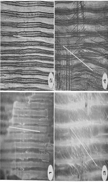
2 244 STAIN TECHNOLOGY applications of the method extend also to the effects of silvicultural treatment on wood structure. The technics for direct evaluation of fibril angles were developed from the discovery that the microscopical checking of certain softwood tracheids was clearly shown by fluorescence microscopy. Such checking in the secondary walls of compression wood fibers lies among the cellulose strands or the fibrils, thereby indicating that their arrangement tends to be at larger angles than usual in nomal summerwood fibers (Pillow and Luxford, 1937). For some normal summerwood, however, the fibril angles were found to be as large as, or even larger than, those in most compression wood although typical checking was absent. PURPOSE The research on improved microscopy of fiber structure at the Forest Products Laboratory had the immediate objective of simplifying the observations and measurements of fibril angles, particularly for increment cores or ocher small samples from living trees. Such methods are needed to avoid damage to the living trees and also to permit observation of structural. characteristics without the cutting of sections and the attendant complicated histological preparations. EQUIPMENT AND PROCEDURES Fluorescence is a characteristic of certain materials whereby absorption of invisible ultraviolet rays excites emission of visible light that lasts only during the period of excitation. Although wood is antofluorescent, color contrasts and brighter images usually result after treatment with certain fluorescent chemicals called fluorochromes. Essential for fluorescence of small pieces of wood to evaluate fibril angles is a microscope designed particularly for surface examination of the specimen. The present examinations were found to require a FIG. 1. A radial surface view of summerwood fibers of slow-growth loblolly pine photograped by incident fluorescence microscopy Fibril angle indicated by white line. FIG. 2. A photomicrograph for comparison made by transmitted light from a section of the same summerwood zone Fibril angle indicated by white line. FIG. 3. A radial surface view of compression wood fibers of fast-growth pine photograped by incident fluorescence microscopy. ZOO. Fibril angle indicated by white line. Checks in the secondary walls are typical of cornpression wood. FIG. 4. A photomicrograph for comparison made by transmitted light from a section of the same growth zone as Fig Fibril angle indicated by white line.
3
4 246 STAIN TECHNOLOGY microscope with a stage that served as a coarse focusing devise for the specimen and an optical system that transmitted invisible ultraviolet rays in the region from mp to the opaque specimen on the stage. Such optical elements require quartz or special glass that transmits ultraviolet; that is, material that will not absorb those wavelengths. The illumination of the object is by means of a ring condenser system with the circular path of the illuminating rays outside the optical rays of the objectives and, thus, the object becomes self-luminous. Such illumination gives improved visibility to structural features that are not easily distinguishable at high magnifications with visible light. The ultraviolet source was a directcurrent carbon arc, a liquid filter of 10% copper sulfate with a few drops of sulfuric acid, and suitable ultraviolet transmitting filters that allowed no visible light to pass to the object. Radial splits of dry wood of loblolly pine (Pinus taeda L.), which had excellent color contrast, were examined at magnifications from 30-50diameters. When desired, certain detailed features of fibers could be magnified up to 500 diameters. A protractor eyepiece was used to measure fibril angles. A blue fluorescence was given by southern yellow pine wood in which the various structural features were differentiated by the use of fluorochromes. The fluorochromes acted best on bright, dry wood. Also, splitting of radial surfaces was more satisfactory with dry material. The fluorescent image was sufficiently bright to use binocular vision with 6, 7, or 10 paired Huyghens eyepieces for observations and measurements with a protractor eyepiece. Treatments on loblolly pine specimens with dry heat at temperatures of C for periods ranging from 15-20min to several hours were found to induce some checking in normal fibers. Although such checking was limited in normal summerwood to the ray crossing, it occurred more or less independently of the size of the fibril angles. Specimens of loblolly pine wood about the size of increment cores and representing different growth rates were split along the fibers as nearly as possible on the radius with a sharp knife, such as a cartilage knife, a microtome knife, or a single-edged safety razor blade. The type of knife used depended somewhat on the size and shape of the sample to be split. A holder was found necessary to facilitate leveling the specimen surface next to the objective lens, particularly if top and bottom surfaces of the wood specimen were not parallel. After the split was made, the surface could be examined immediately by fluorescence microscopy for the detection of compression wood. For other structural features, however, the treatments of the specimens with fluorochromes gave improved contrast and brighter images Details of the color contrasts achieved with the use of fluoro-
5 TABLE 1. FLUOROCHROMES USEFUL FOR EXAMINATION OF DRY WOOD STRUCTURE AND FOR AIDING IN THE MEASURING OF FIBRIL ORIENTATION IN SUMMERWOOD FIBERS OF PINE BY INCREASING COLOR CONTRAST Structure of pine wood Autofluorecence Color of fluorescence Fluorescence obtained with fluorochromes * Flavine Rhodamine 6 G Flavine followed by rhodamine 6 G Walls of summerwood tracheids.... Blue Blue Blue Blue Walls of springwood tracheids..... Blue Yellowish green Blue Blue green Pit apertures Light blue Luminous yellow Luminous yellow Luminous yellow Bordered pit annulus Light blue Luminous yellow Luminous yellow Luminous yellow Torn cell edges and cell-wall checks Light blue Luminous yellow Luminous yellow Luminous yellow Torus Red Red Dentate ray tracheids Projections light blue Yellowish green Projections edged luminous Projections luminous yellow yellow or blue green Ray parenchyma Blue Pale orange yellow Red Red Secretory parenchyma of resin ducts Pale yellow Light blue Bright yellow Sometimes bright yellow Crassulae Red Red Starch grains Transparent blue Transparent blue Bright golden yellow Bright golden yellow *Aqueous solutions of fluorochrome 1:1000. Period required to take effect, 2-5 min., then washed in distilled water, blotted, and dried in an oven at about 195 C, for 15 min. or longer. Calcozine flavine TG extra concentrated, lot furnished by American Cyanamid Company, Calm Dyestuff Department, Bound Brook, N. J. The following were found to be satisfactory: Calcozine red 6 G extra, lot furnished by American Cyanamid Company, Calco Dyestuff Department, Bound Brook, N. J.; rhodamine 6 G, C.I. No. 752, lot furnished by National Aniline Division, Allied Chemical & Dye Corporation, New York, N. Y.; rhodamin 6 GD extra, K. Hollborn & Sohne, Leipzig; rhodamine 6 GD (extra), lot 05156, George T. Gurr Ltd., London S.W. 6., England.
6 248 STAIN TECHNOLOGY chromes on loblolly pine are given in Table 1. The procedure for treatment with a single fluorochrome was as follows: 1. Treat the dry wood specimen in a 0.1% aqueous fluorochrome solution 2-5min in a low-pressure vacuum container in order to increase penetration of the solution. 2. Wash the specimen in distilled water, 5-10sec then blot it with clean falter paper. 3. Dry in an oven at C.for 15 min or more. Treatment with two fluorochromes (for example, using Calcozine flavine TG extra concentrated as the first fluorochrome) has the advantage of giving a highly increased overall brightness as a background for the color contrasts given with the second fluorochrome (either Calcozine red 6G extra, rhodamine 6G, or rhodamine 6GD extra). Structural features and the fluorescence colors obtained are given in Table 1. The stated time limits for treatment are not rigid and may require modification according to the nature of the material under examination. In our laboratory, improved results were obtained when specimens were dried after treatment with the first fluorochrome before being treated with the second. Fig, 1-4 show results that were obtained by fluorescence microscopy as compared with the more complicated preparations needed for transmitted-light microscopy. Although this technic with fluorescence microscopy lends itself to simple preparations for direct examinations, it is not suitable for photomicrographs of large fields because the split surfaces have irregular contours. Only features visible in one focal plane of the camera are in sharp detail on the photographic film or plate. Fluorescence microscopy aided by fluorochromes is superior to other methods not only in the saving of time and in labor, but because the fluorescence colors give excellent visual differentiation of structural features within the wood. In addition, certain fluorochromes readily differentiate starch grains in the wood elements. REFERENCES PILLOW, MAXON Y., TERRELL, BESSIE Z., and HILLER, CHARLOTTE H Patterns of variation in fibril angles of loblolly pine. U. S. Dept. of Agric., Forest Service, Forest Products Lab. Report No. D1935., and LUXFORD, R. F Structure, occurrence, and properties of compression wood. U. S. Dept. of Agric. Tech. Bull. 546.