Topochemical and TEM/FESEM-studies on tension wood fibres of Acer spp., Fagus sylvatica L. and Quercus robur L.
|
|
|
- Barbara Stone
- 5 years ago
- Views:
Transcription
1 Topochemical and TEM/FESEM-studies on tension wood fibres of Acer spp., Fagus sylvatica L. and Quercus robur L. Uwe Schmitt Johann Heinrich von Thunen-Institute (vti) Federal Research Institute for Rural Areas, Forestry and Fisheries Institute of Wood Technology and Wood Biology Leuschnerstrasse Hamburg/Germany
2 Co-Authors Christian Lehringer University of Hamburg Geoffrey Daniel Swedish University of Agricultural Sciences Burgi Gierlinger Max Planck Institute - Potsdam Gerald Koch von Thunen-Institute Hamburg
3 Objectives To apply various microscopic techniques for obtaining more detailed knowledge on tension wood fibres Special emphasis on: Methods applied: - the fine structure of the cell wall of tension wood fibres - the chemical composition of tension wood fibres - the occurrence of aromatic compounds in the G-layer - light microscopy, scanning and transmission electron microscopy - cellular UV-spectroscopy -Ramanspectroscopy


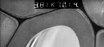
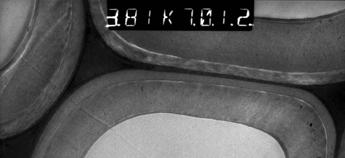
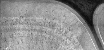
4 Material - maple fibers - beech fibers - oak fibers All with distinct tension wood Stem section of Fagus sylvatica with pronounced tension wood. Individual fibers with G-layers for topochemical analysis (SEM)


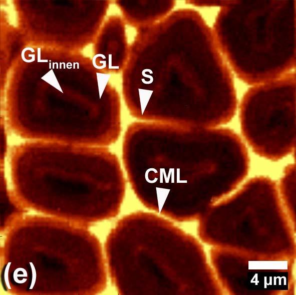
5 General Tension wood fibres of hardwoods develop a special wall layer, the so-called gelatinous layer or G-layer The G-layer consists of concentric lamellae of cellulose microfibrils parallel aligned to the fibre axis The cellulose is highly crystalline, and the content of hemicelluloses amounts to only a few percent The occurrence of lignin in the G-layers is controversially discussed
6 Light and electron microscopy - conventional light microscopy different staining techniques (safranin, astrablue) - field emission scanning electron microscopy - transmission electron microscopy staining with potassium permanganate

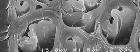


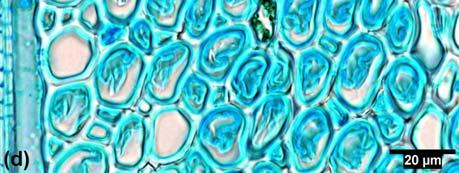
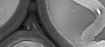
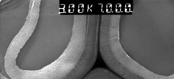
7 Acer Beech Oak


8 For tension wood fibers of oak FESEM-technique sometimes revealed a multilayered G-layer structure.


9 Beech Oak
 ZEISS for the topochemical analyses of lignin within individual cell wall")
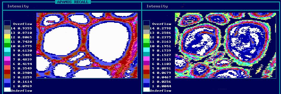
10 Cellular UV-spectroscopy - recording absorbance values of cell wall constituents by using UV light of different wavelengths Universal-Microscope-Spectrophotometer (UMSP 80) ZEISS for the topochemical analyses of lignin within individual cell wall layers

11 Point analyses - with varying wavelengths (resolution 0.25 µm 2 ) Vessel 0.6 Fiber Absorbance Fibre S2 Fibre CML Extractives Parenchyma Wavelength [nm] Typical UV absorbance spectra of individual cell wall layers and cell lumen deposited phenolic compounds in the woody tissue of Fagus sylvatica (wavelength range nm) [Koch et al., Holzforschung 2003]


12 Scanning analyses - with a constant wavelength (resolution 0.25 µm 2 ) Wavelength = 278 nm


13
14 0,4 Absorbance maximum for hardwood lignin around 276 nm Oak/Control Fibre CML Absorbance 0,3 0,2 0,1 Fibre S2-outer layer Fibre S2-middle Fibre S2-inner layer 0, Wavelength [nm]
15 First absorbance maximum for hardwood lignin shifted to ~ 268 nm 0,4 -indicates higher syringyl content 0,3 Oak/G-fibre Fibre-GL outer layer (oak) Fibre-GL middle (oak) Absorbance 0,2 0,1 Second absorbance maximum at ~ 300 nm -indicates more conjugated double bonds = evidence for aromatic compounds Fibre-GL inner layer (oak) 0, Wavelength [nm]
16 0,20 -typical lignin absorbance Oak and beech Fibre S2-inner layer (beech) 0,15 Fibre GL-inner layer (beech) Absorbance 0,10 0,05 Fibre GL-inner layer (oak) -evidence for aromatic compounds 0, Wavelength [nm]
17 Raman spectroscopy - recording topochemical differences between wall layers by using inelastic scattering of monochromatic laser light Wave numbers for the detection of aromatic compounds 1,508 aryl ring 1,601 aryl ring 1,620 conjugated ring; C=C bonds of coniferyl alcohol 1,660 conjugated ring; C=C bonds of coniferylaldehyde Evidence for the occurrence of aromatic compounds in the G-layer of tension wood fibres


18 Lignin Carbohydrates Tension wood fibres of Acer Tension wood fibres of Fagus

19

20 Beech tension wood, RAMAN - similar for Acer cm -1 CML S2 GL
21 Wave numbers for the detection of aromatic compounds aryl ring 1,601 aryl ring conjugated ring; C=C bonds of coniferyl alcohol conjugated ring; C=C bonds of coniferylaldehyde Evidence for the occurrence of aromatic compounds in the G-layer of tension wood fibres

22 Oak tension wood, RAMAN CML S2 GL middle GL inner cm -1
23 Oak enlargement cm-1 CML S2 GL middle GL inner
24 CONCLUSIONS Electron microscopy sometimes revelaed a concentric layering in the G-layer of tension wood fibres TEM demonstrated some dark staining constituents in the G-layer when using potassium permanganate as staining agent Cellular UV-spectroscopy indicated a certain amount of aromatic compounds in the G-layer Raman spectroscopy confirmed the results obtained with UV-spectroscopy.