ISOLATION AND CHARACTERISATION OF AQUATIC FUNGI FROM OPEN DRAINS IN PORT HARCOURT MUNICIPALITY, SOUTHERN NIGERIA.
|
|
|
- Bernice Ferguson
- 5 years ago
- Views:
Transcription
1 ISOLATION AND CHARACTERISATION OF AQUATIC FUNGI FROM OPEN DRAINS IN PORT HARCOURT MUNICIPALITY, SOUTHERN NIGERIA. *Ogbonna D. N. and Okorie S. Department of Microbiology Rivers state University of Science and Technology Nkpolu-Oroworukwo, P.M.B 5080, Port Harcourt, Nigeria. ogbonna. edu. ng* *Correspondencing author Abstract Gari slide culture media was used to isolate fungi from open drains in Port Harcourt Municipality. Fungi grown onto Sabouraud Dextrose Agar (SDA) were purified by repeated sub-culturing unto gari slide culture media. Microcultures of the fungi isolated on SDA were made by transferring spore suspensions of each isolate onto a 1 cm disc of the same medium placed on a microscope slide in a Petri dish. All inoculated slides were incubated at 30±2 0 C for 5 days, and then examined under the microscope. Fungi isolated on SDA and gari medium were Aspergillus niger, Aspergillus tamarii, Aspergillus versicolor, Cryptococcus neoformans, Mucor spp, Penicillium chrysogenum, Penicillium marneffei, Phoma spp, Rhizopus spp, Scopulariopsis spp and Torulopsis glabrata. Also a micrograph of Torulopsis spp and Cryptococcus spp as yeasts on Gari slide culture medium are presented. However, the patterns and rate of growth of the fungal species were more in sediments than in water samples which could be responsible for the degradation of receiving water bodies. Keywords: Aquatic fungi, open drains, Gari slide culture media INTRODUCTION Microorganisms are present in all natural habitats, as well as in humanmade environments such as open drainage channels. Such reservoirs have features such as darkness, long retention times and stagnation zones that favour the proliferation of microorganisms NigerJ.mycol Vol.9, (especially fungi) and biofilm formation, as well as, toxin production (Hageskal et al., 2009). Apart from their significant ecological role in helping nutrient turnover, water can become contaminated or highly impacted by their activities (Hageskal et al., 2006; Besner et al., 2011; Douterelo et al., Nigerian Journal of Mycology Vol. 9 (2017) 40
2 Ogbonna & Okorie 2014, EPA, 2016 ) causing bad taste and odour. Through the inhalation of spores after aerosolisation of water, especially when water passes through taps and showers water loses its potability (Warris et al., 2001; Hageskal et al., 2009; Anaissie et al., 2011; Richardson and Richardson, 2015). Studies of the structural characteristics of moulds could be simplified by placing on microscope slides (about 1 cm square) blocks of agar cut from appropriate culture media; the upper edges of the agar blocks are inoculated with the desired moulds; then after adequate incubation of inoculated blocks, the slides along with the agar block are examined under the microscope (Sokari et al., 1996). This is because moulds can be identified to the generic level often without special staining or biochemical techniques since their structural details could be readily visible microscopically. However, the structural differences can be readily observed only if the mould colony is not disturbed during preparation or mounting for microscopic examination (Sokari et al., 1996), and this is achieved by means of monocultures on microscopes slides. The aim of the present study, therefore, was to identify fungi isolated from open drainage channels in Port Harcourt using gari media as an inexpensive, but effective alternative culture medium to conventional dehydrated mycological media for isolating and identifying moulds. MATERIALS AND METHODS Collection of sample Wastewater samples were collected from open drains along the Ntanwogba creek with sterilized plastic bottles. Each sample bottle was rinsed with the appropriate sample before the final collection according to the standard methods (APHA, 2012). To collect the water sample, base of the sterilized sample bottle was held with one hand, plunged about 30cm below the water surface with the mouth of the sample container positioned in an opposite direction to water flow (APHA, 2012). The container was filled with wastewater samples from different locations starting from the upstream (Afam /Kaduna street behind the Winners chapel) to the downstream (at Abacha road, all sites in Port Harcourt Rivers State Nigeria ) leaving a gap of about 2cm and then covered. Sediment samples for analysis were also collected along the same water course. To collect the sediment samples, the bottles were opened and held with the left hand while using the right hand with a plastic scooper to scoop the sediment sample. The sample bottles were filled with sediment sample and covered immediately. Thereafter, each Nigerian Journal of Mycology Vol. 9 (2017) 41

3 Isolation and Characterisation of Aquatic Fungi Isolated from Open Drains in Port Harcourt Municipality, Southern Nigeria sample was immediately labelled and transported in a cooler packed with ice blocks for analysis. Sample collection was carried out from February to August Fig 1. Map of Port Harcourt showing sampling stations Mycological Study Serial dilution Ten-fold serial dilutions of the samples were made according to Oliveira et al. (2016). Inoculation and incubation One milliliter of appropriate ten - fold serial dilution of the sample was inoculated onto appropriate Sabauraud dextrose agar in triplicates using pour plate method of Hageskal et al. (2009) and spread plate method of Oliveira et al. (2016). Inoculated plates were incubated at 28±2 0 C for hours. Visible discrete colonies on incubated plates were counted and expressed as colony forming unit per gram (cfu/g) of sediment samples and colony forming units per milliliter (sfu/ml) of waste water samples. Nigerian Journal of Mycology Vol. 9 (2017) 42
4 Ogbonna & Okorie Maintenance of pure culture Fungi grown onto SDA were purified by repeated sub-culturing unto gari slide culturing media. Pure cultures were preserved on gari slide culture media at ambient temperature (28±2 0 C) for further tests. Garri slide culture method According to the method of Sokari et al. (1996), individual, fairly large grains of gari were placed in a glass Petri dish and autoclaved at C. Using a flamed forceps, one granule of the gari was transferred from the glass Petri dish unto a glass slide. Thereafter, fungal growth was touched with a wire inoculating needle and then placed on gari granule on glass slide. Inoculated granule was transferred into a sterile Petri dish layered with moistened cotton wool, and incubated at 30 0 C for 3-5days. After incubation, structures of fungal growth were enhanced by touching edge of coverslip with cotton blue in lactophenol. Identification of fungal species was done phenotypically based on macroscopic and microscopic morphological features of cultivation in gari slide culture medium. Characterization and identification of fungal isolates. Pure cultures of fungal isolates were identified based on cultural parameters, microscopic technique and biochemical tests including carbohydrate utilization as described by Cruickshank et al. (1975). Characterization and identification of fungal isolates was done according to Kelly et al. (2003) and Samson et al. (2004) RESULTS AND DISCUSSION Predominant fungal isolates from the open drains were characterized and identified as Aspergillus niger, Penicillium chrysogenum, Aspergillus tamarii, Cryptococcus neoformans, Aspergillus versicolor, Torulopsis glabrata, Rhizopus spp, Mucor spp, Scopulariopsis spp Penicillium marneffei and Phoma spp (Table 1; Figs 1-9 a and b). Species of Torulopsis and Cryptococcus (Table 2; Figs a and b) were the only yeast isolates from the open drainage system. Fungi are ubiquitous in soils and wastewater preferring cool and moderate climate, especially with the abundance of organic materials (Nicoletti et al., 2009). However, Cryptococcus neoformans and Rhizopus species were isolated more frequently from both water and sediment samples in the open drains. The patterns and rate of growth of the fungal species were more in sediments than in water samples. Doggett (2000) had earlier reported densities of filamentous fungi and yeasts in similar systems. Their presence in wastewater indicates that there is possible contamination by fungal Nigerian Journal of Mycology Vol. 9 (2017) 43
5 Isolation and Characterisation of Aquatic Fungi Isolated from Open Drains in Port Harcourt Municipality, Southern Nigeria pathogen (Prescott et al., 2005; Oliveira et al., 2016) which may be responsible for the degradation of receiving water bodies. Their presence was most likely through the runoff of fertilizers or sewage which contain excess nutrient that plants, algae and fungi can utilize for growth. Their abundance and activities in drainage systems have profound effect on the physicochemical characteristics of the wastewater especially at the banks of the creek. This is as a result of drastic narrowing of the creek channels induced by the declining riparian vegetation along the system particularly at the degrading wastewater open drains. Table 1. Morphological Characteristics and identity of fungal Isolates Nigerian Journal of Mycology Vol. 9 (2017) 44

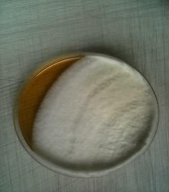
6 Ogbonna & Okorie Plate 1(a) Morphological features of Penicilium chrysogenum as seen on gari medium Nigerian Journal of Mycology Vol. 9 (2017) 45


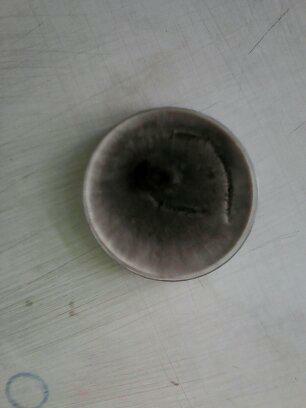
7 Isolation and Characterisation of Aquatic Fungi Isolated from Open Drains in Port Harcourt Municipality, Southern Nigeria Plate 2 Plate 3 Plate 4 Nigerian Journal of Mycology Vol. 9 (2017) 46


8 Ogbonna & Okorie Plate 5 Plate 6 Plate 7 Nigerian Journal of Mycology Vol. 9 (2017) 47


9 Isolation and Characterisation of Aquatic Fungi Isolated from Open Drains in Port Harcourt Municipality, Southern Nigeria Plate 8 Plate 9 Plate 10 Nigerian Journal of Mycology Vol. 9 (2017) 48


10 Ogbonna & Okorie Plate 11 CONCLUSION Fungi isolated from all the open drains examined are considered to produce mycotoxins and/or are opportunistic human pathogens. Water storage in drainage systems generates stagnation, stratification, particle accumulation, dead zones, depletion of residual disinfectant, and biofilm formation. These parameters combined with chemical, physical characteristics of the water system (like high turbidity and temperature, ph, total organic carbon and dissolved oxygen), are favorable to microbial growth making reservoirs a potential high risk of water quality degradation by fungi. In this current work, all sampling sites presented Aspergillus and other species, and these species are recognized producers of aflatoxins. Paterson et al [1997) and Oliveira et al (2016) demonstrated production of aflatoxins in a cold water storage tank. Water subjected to storage (e. g., reservoirs, cisterns and bottles) for long periods, may contain increased mycotoxin concentrations. In the long term, daily consumption of large amounts of water containing low levels of mycotoxin, may pose a health hazard (Paterson et al., 1997; Skaar and Hageskal, 2015; DEFRA, 2016, Oliveira et al., 2016). Therefore, frequent surveillance for fungi and the setting of limits are required to improve the mycological quality of drinking water. Presence of microorganisms in such environments affects human health, causing various diseases from allergic reaction to more serious systemic infections. The choice of Gari to Sabouraud media was not meant to be selective in separating clearly fungal taxa. This could be explained by the fungi not being under stress conditions due to the low or zero residual chlorine and the presence of considerable amounts of dissolved oxygen and Nigerian Journal of Mycology Vol. 9 (2017) 49
11 Isolation and Characterisation of Aquatic Fungi Isolated from Open Drains in Port Harcourt Municipality, Southern Nigeria organic matter in water samples. Furthermore, Sabouraud, as a richer medium, gave better conditions for the water fungi (conidia and propagules) to grow, which is reflected by the number of isolates obtained in this study. REFERENCES Anaissie, E. J., Kuchar, R. T., Rex, J. H., Francesconi, A., Kasai, M., Muller, F. M. and Walsh, T. J ( ). Fusariosis associated with pathogenic Fusarium species colonization of a hospital water system: A new paradigm for the epidemiology of opportunistic mold infections. Clinical Infectious Diseases 33: American Public Health Association (APHA); American Water Works Association (AWWA); W a t e r Environmental Federation (WEF) (2012). Standard Methods for the Examination of Water and Wastewater, 22nd ed. ; American Public Health Association; American Water Works Association; Water Environmental Federation: Washington, DC, USA, Besner, M. -C., Prévost, M. and Regli, S. (2011). Assessing the public health risk of microbial intrusion events in distribution systems: Conceptual model, available data, and challenges. Water Resources 45: Cruickshank, R., Duguid, J. P., Mamion, R. P. and Swain, R. H. A., (1975). Medical Microbiology II, Churchill Press, London, p. 32 Demain, A. L. and Davies, J. E. (1999). Manual of Industrial Microbiology and Biotechnology, 2 nd Ed, American Society for Microbiology Press, Washington D. C. DEFRA (2016). Review of Fungi in Drinking Water and the Implications for Human Health. Available online: defra. gov. uk/research/completed-research/re ports/dwi pdf (accessed on 9 February 2016). Doggett, M. S. (2000). Characterisation of fungal biofilms within a municipal water distribution system. Applied and Environmental Microbiology, 66 (3): Douterelo, I., Boxall, J. B., Deines, P., Sekar, R., Fish, K. E. and Biggs, C. A. (2014). M e t h o d o l o g i c a l approaches for studying the microbial ecology of drinking water distribution systems. Water Resources. 65: Environmental Protection Agency (EPA) (2016). Health Risk from Microbial Growth and Biofilms in Drinking Water Distribution Systems. Available online: Nigerian Journal of Mycology Vol. 9 (2017) 50
12 Ogbonna & Okorie 2007_05_18_disinfection_tcr_whitep aper_tcr_biofilms. pdf Hageskal, G., Knutsen, A. K., Gaustad, P., de Hoog, G. S and Skaar, I. (2006). Diversity and significance of mold species in Norwegian drinking water. Applied Environmental Microbiology 72: Hageskal, G., Lima, N. and Skaar, I. (2009). The study of fungi in drinking water. Mycological Research. 113: Kelley, J.. Kinsey, G., Paterson, R. and Brayford, D. (2003). Identification and Control of Fungi in Distribution Systems; AWWA Research Foundation and American Water Works Association: Denver, CO, USA Nicoletti, R. Manzo, E. and Ciovatta, M. L. (2009). Occurrence and bioactivities of funicone related compound. Oliveira, H. M. B; Santos, C; Russell, R; Paterson, M; Gusmão, N. B and Lima, N (2016). Fungi from a Groundwater- Fed Drinking Water Supply System in Brazil. International Journal of Environmental Research and Public Health 13: Paterson, R. R. M., Kelley, J. and Gallagher, M. (1997). Natural occurrence of aflatoxin and Aspergillus flavus (Link) in water. Letters of Applied Microbiology 25: Prescott, L. M., Harley J. P. and Klein D. A (2005). Microbiology. 6 th ed. McGraw Hill, London. Pp Richardson, M. D. and Richardson, R. (2015). Aspergillus and aspergillosis. In Molecular Biology of Food and Water Borne Mycotoxigenic and Mycotic Fungi; Paterson, R. R. M., Lima, N., Eds. ; Food Microbiology Series; CRC Press: Boca Rotan, FL, USA, pp Skaar, I. and Hageskal, G. (2015). Fungi in drinking water. In: Molecular Biology of Food and Water Borne Mycotoxigenic and Mycotic Fungi; Paterson, R. R. M., Lima, N., Eds. ; FooMicrobiology Series; CRC Press: Boca Rotan, FL, USA, pp Samson, R. A. ; Hoekstra, E. S. and Frisvad, J. C. (2004) Introduction to Food and Airborne Fungi, 7th ed. ; Centraalbureau voor Schimmelcultures: Utrecht, The Netherlands. Sokari, T. G., Yubedee, A. G., and Lugbe P. B. (1996). Garri as an effective medium for certain Mycological studies. Niger Delta Biologia. 1(1): Warris, A., Gaustad, P; Meis, J. F. G. M. Voss, A., Verweij, P. E. and Abrahamsen, T. G. (2001). Recovery of filamentous fungi from water in a paediatric bone marrow transplantation unit. Journal of Hospital Infections 47: Nigerian Journal of Mycology Vol. 9 (2017) 51