Serial dilution and colony count (Viable count) Pour plate. Spread plate Membrane filtration. Turbidity. Microscopic cell count
|
|
|
- Charles Francis Cannon
- 5 years ago
- Views:
Transcription


1 Aljawharah Alabbad 2016
2
3 Serial dilution and colony count (Viable count) Pour plate Spread plate Membrane filtration Turbidity Microscopic cell count
4 Many studies require the quantitative determination of bacterial populations. The two most widely used methods for determining bacterial numbers are : 1.The standard plate count method. 2.Spectrophotometer (turbid metric) analysis.
5 An indirect measurement of cell density (live bacteria). Based on turbidity and indirectly measures all bacteria (cell biomass), dead and alive.

6 The bacteriological tests used most often are the Standard Plate Count (SPC)
7 If the sample is serially diluted and then plated out on an agar surface in such a manner that single isolated bacteria form visible isolated colonies, the number of colonies can be used as a measure of the number of viable (living) cells in that known dilution.


8 We are determining the number of Colony- Forming Units (CFUs) in that known dilution.
9 Bacteria may be clumped together. Therefore, when doing the plate count technique, we generally say we are determining the number of Colony- Forming Units (CFUs) in that known dilution.
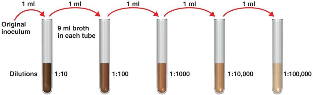
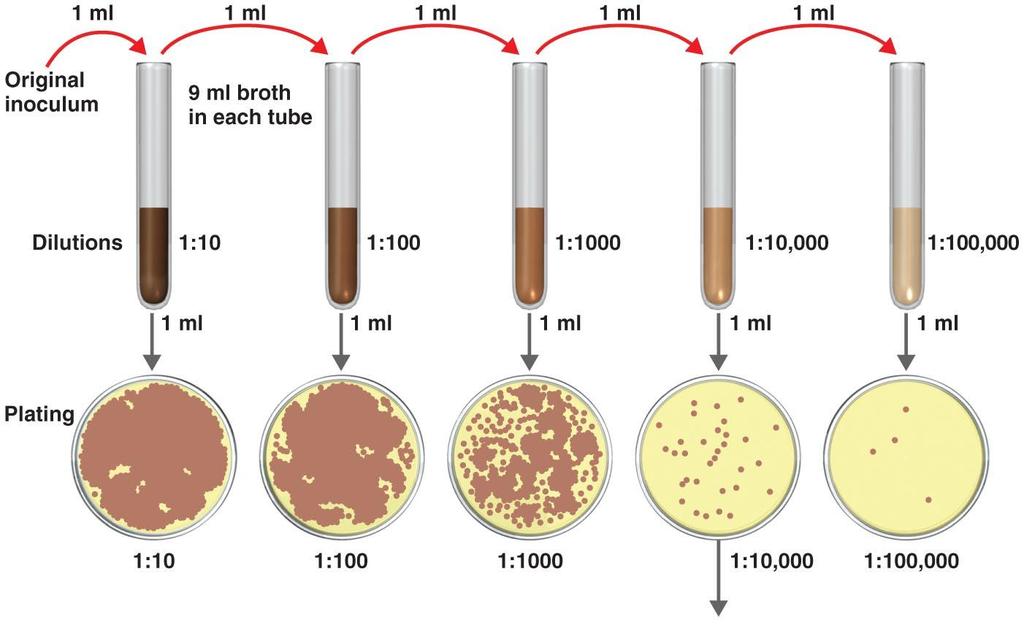
10 The bacterial sample is diluted by factors of 10 and plated on agar. After incubation, the number of colonies on a dilution plate showing between 30 and 300 colonies is determined.
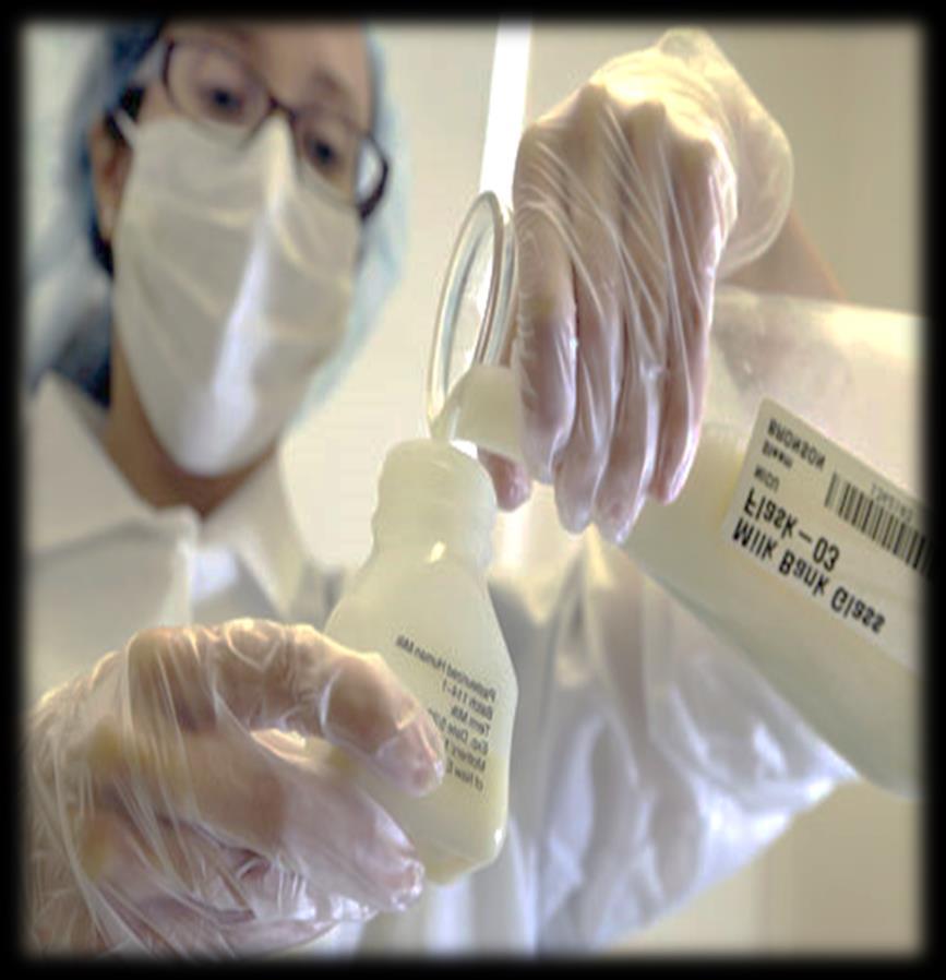
11 Bacterial contamination of raw milk can generally occur from three main sources: 1.Within the udder. 2.Outside the udder. 3.From the surface of equipment used for milk handling and storage.


12


13 Test tubes. Pipettes (1 ml, graduated). Nutrient agar. Bent glass rod. Alcohol 70%. Raw milk


14 1. Take six test tubes and add 9 ml of distilled sterilized water (DSW) in each tube and label them as 1,2,3,4,5,6 2. Transfer 1ml of the sample (unsterilized milk) to tube no.1 contained 9ml DSW and Reflame and cap the sample. 3. Mix the tube thoroughly, this will makes the first dilution. 4. Transfer 1 ml from the 1 st dilution to test tube no.2. And this makes the second dilution. 5. Repeat the same pattern with other tubes as shown in the diagram.


15 Using a Pipette to Remove Bacteria from a Tube Using a Vortex Mixer to Mix Bacteria Throughout a Tube

16 For a more accurate count it is advisable to plate each dilution in duplicate or triplicate and then find an average count.

17

18 6. From the last three dilutions, transfer 1ml to the nutrient agar plate. 7. Using a turntable and sterile bent glass rod, immediately spread the solution over the surface of the plates. 8. Replace the lid and re-sterilize the glass rod with alcohol and flaming, and repeat for each plate. 9. Incubate the plates converted for 24 hrs. at 37 C. 10. Count the colonies of bacteria after incubation.

19 Using a Bent Glass Rod and a Turntable to Spread a Bacterial Sample.
20

21 Count the colonies on each plate. Select all of the Petri plates containing between 30 and 300 colonies.
22 At the end of the incubation period, count by looking at the bottom of the plate (while keeping the Petri plate closed). Agar is translucent you should not have to open the plate.

23 Select all of the Petri plates containing between 30 and 300 colonies. Count the colonies on each plate.
24 Calculate the number of bacteria (CFU) per milliliter or gram of sample by dividing the number of colonies by the dilution factor multiplied by the amount of specimen added to agar plate.
25 A simple calculation is given below: CFU ml in original sample # colonies counted (dilutionfactor)(volume plated,inml) CFU = C (D V) While : CFU = Number of bacteria cell in ml. C = Number of colonies in plate. D = dilution of sample. V = volume plated in ml.
26 Example 1 : If 32 colonies in plate of 1/ dilution and volume plated 0.5 then the count is : 32 (1/10,000x0.5) = 640,000 cell /ml
27 Example 2 : You count 46 colonies on your plate You put 1 ml of bacterial culture into 99 ml of saline and plated 0.1 ml Dilution 1/100 CFU = 46 (1/ ) = = CFU/ml
28 A plate having colonies is chosen because this range is considered statistically significant. This plate has between 30 and 300 colonies and is a suitable plate for counting.
29 If are less than 30 colonies on the plate, small errors in dilution technique or the presence of a few contaminants will have a drastic effect on the final count. too few to count (TFTC). This plate less than 30 colonies and is unsuitable plate for counting.
. This plate has over 300 colonies and cannot be used for counting.")
30 Likewise, if there are more than 300 colonies on the plate, there will be poor isolation and colonies will have grown together. too numerous to count (TNTC). This plate has over 300 colonies and cannot be used for counting.

31
32 Activity Calculate CFU If you count 75 colony in plat of 1/1000 dilution and volume plated 0.5 ml.