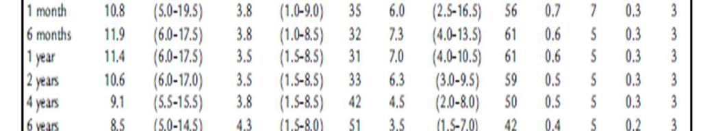
MCV:HCT/RBC,fl MCH:Hb/RBC,pg MCHC:HCT/Hb,gd RDW:anisocytosis
|
|
|
- Brandon Lloyd
- 5 years ago
- Views:
Transcription

1 Dr Goudarzipour
2 MCV:HCT/RBC,fl MCH:Hb/RBC,pg MCHC:HCT/Hb,gd RDW:anisocytosis


3


4

5


6
7 Neutropenia? Anemia? Physiologic anemia?
8 NL PLT? WHATS MPV?


9


10


11


12
13
14 examples 2 Y/O male WBC:6700:PMN:60,L:40 RBC: Hb:11.7 MCV:76 PLT:135000
15 Case -2 3 month years,female WBC:6500,L:30,P:70 Hb:8.7 MCV:90 PLT:
16 Case-3 7 y/o,female WBC:5390,P:63,L:37 RBC: Hb:10.5 MCV:64 MCH:23 PLT:
17 CASE 4 2 y/o male WBC:14500,P;78,l;22 RBC:5400 Hb;8 HCT:13 MCV:99 MCH:32 MCHC:36 PLT:78000
18
19 Background Iron deficiency is the most common nutritional deficiency in children and is worldwide in distribuiotion. The incidence of iron-deficiency anemia is high in infancy % of children under 5 years of age in developing countries.
20 Babies are born with iron stored in their bodies. Because they grow rapidly, infants and children need to absorb an average of 1 mg of iron per day. Since children only absorb about 10% of the iron they eat, most children need to receive 8-10 mg of iron per day. Breastfed babies need less, because iron is absorbed 3 times better when it is in breast milk
21

22
23 Tissue Effects of Iron Deficiency I. Gastrointestinal tract Anorexia-common and an early symptom Pica-pagophagia (ice) geophagia Atrophic glossitis II. Central nervous system Irritability Fatigue and decreased activity
24 Tissue Effects of Iron Deficiency III. Cardiovascular system Cardiac hypertrophy IV. Musculoskeletal system Deficiency of myoglobin and cytochrome C Adverse effect on fracture healing V. Immunologic system Evidence of increased propensity for infection
25


26 Absorbtion of Iron


27
28 PREVALENCE Peak prevalence occurs during late infancy and early childhood Rapid growth with exhaustion of gestational iron Low levels of dietary iron Complicating effect of cow s milk-induced exudative enteropathy
29 PREVALENCE A second peak is seen during adolescence due to rapid growth and suboptimal iron intake. This is amplified in females due to menstrual blood loss
30 Causes of Iron-Deficiency Anemia I. Deficient intake Dietary (milk, 0.75 mg iron/l) II. Inadequate absorption Poor bioavailability: absorption of heme Fe.Fe2.Fe3; breast milk iron.cow s milk Antacid therapy or high gastric ph (gastric acid assists in increasing solubility of inorganic iron) Cobalt, lead ingestion
31 Causes of Iron-Deficiency Anemia III. Increased demand Growth pregnancy IV. Blood loss


32
33 The best sources of iron include: Baby formula with iron Breast milk (the iron is very easily used by the child) Infant cereals and other iron-fortified cereals Liver

34


35


36

37


38


39
40 symptoms Blue-tinged or very pale whites of eyes Blood in the stools Brittle nails Decreased appetite (especially in children) Fatigue Headache Irritability Pale skin color (pallor) Shortness of breath Sore tongue Unusual food cravings (called pica) Weakness Note: There may be no symptoms if anemia is mild.
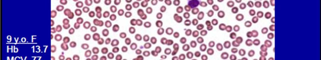
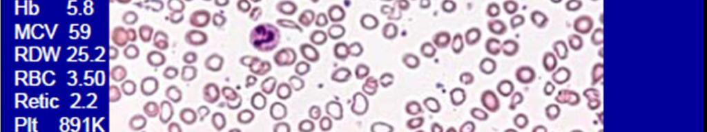
41 P/E and lab 1. Hemoglobin: Hemoglobin is below the acceptable level for age 2. Red cell indices: Lower than normal MCV, MCH and MCHC for age. 3.Increase RDW Blood smear: Red cells are hypochromic and microcytic with anisocytosis hemoglobin level falls below 10 g/dl.
42 P/E and lab Basophilic stippling can also be present but not as frequently The RDW is high (.14.5%) Reticulocyte count: The reticulocyte count is usually increase in bleeding).(normal Platelet count: The platelet count varies from thrombocytopenia to thrombocytosis. free erythrocyte protoporphyrin (FEP) levels. increase
43 P/E and lab The normal FEP level is mg/dl. The upper limit of normal is 40 mg/dl Serum ferritin: The level of serum ferritin reflects the level of body iron stores (below than 12). Normal ferritin levels, however, can exist in iron deficiency when bacterial or parasitic infection, malignancy or chronic inflammatory conditions co-exist.because ferritin is an acute-phase reactant
44 P/E and lab Serum iron and iron saturation percentage: limitations: Wide normal variations (age, sex, laboratory methodology) Time consuming Subject to error from iron ingestion Diurnal variation Falls in mild or transient infection.
45 Stages of Iron Depletion 1. Iron depletion: tissue stores are decreased without a change in hematocrit or serum iron level 2. Iron-deficient erythropoiesis Iron decrease reticuloendothelial macrophage
46 TIBC increase With out change in HCT 3. Iron-deficiency anemia: Anemia Increase RDW Increase FEP
47 Differential Diagnosis
48 Differential Diagnosis
49 TREATMENT Oral Iron Medication Dose: mg/kg elemental iron three times daily. In children with gastrointestinal side effects, iron once every other day Duration: 6 8 weeks after hemoglobin level and the red cell indices return to normal.
50 Response: Peak reticulocyte count on days 5 10 following initiation of iron therapy. Following peak reticulocyte level, hemoglobin rises on average by g/dl/ hematocrit rises 1%/day during first 7 10 days. Thereafter, hemoglobin rises slower: g/dl/day.
51 Failure to respond to oral iron: Poor compliance failure or irregular administration of oral iron; Inadequate iron dose Ineffective iron preparation Insufficient duration Persistent or unrecognized blood loss Incorrect diagnosis thalassemia, sideroblastic anemia Coexistent disease that interferes with absorption or utilization of iron Impaired gastrointestinal absorption due to high gastric ph
52 Parenteral Therapy Noncompliance or poor tolerance of oral iron. Severe bowel disease (e.g., inflammatory bowel disease) Chronic hemorrhage Rapid replacement of iron stores is needed. Erythropoietin therapy is necessary, e.g. renal dialysis.
53 Blood Transfusion In children with sever infection specially with cardiac dysfunction or Hb less than 4 g/dl.


54


55


56


57


58
59 References Glader B. Iron-deficiency anemia. In: Kliegman RM, Behrman RE, Jenson HB, Stanton BF, eds. Nelson Textbook of Pediatrics. 18th ed. Philadelphia, Pa: Saunders Elsevier; 2007: chap 455. Heird WC. The feeding of infants and children. In: Kliegman RM, Behrman RE, Jenson HB, Stanton BF, eds. Nelson Textbook of Pediatrics. 18th Ed. Philadelphia, Pa: Saunders Elsevier; 2007: chap 42. O'Connor NR. Infant formula. Am Fam Physician. 2009;79:
60