Acoustic Characterization of Materials
|
|
|
- Denis Matthews
- 5 years ago
- Views:
Transcription
1 Acoustic Characterization of Materials Bernhard R. Tittmann Group Leader CAV
2
3 Introduction Graduate Students 4 Ph.D. Candidates 4 U.S. 1 Non-US 3 M.S Candidates 3 U.S. 1 Non U.S. Visiting Scholars Dr. Atsushi Baba-Hitachi Jao Sakamoto Univ. of Brazil

4 Matt Guers
 and potentially commercial reactors as well.")
5 ATR Sensors The goal of this work is to develop an insitu ultrasonic technique for in-core material characterization of specimens in the Advanced Test Reactor (ATR) at Idaho National Labs (INL) and potentially commercial reactors as well. Testing of materials for new reactor constructions Health monitoring of existing commercial reactors 5
6 Existing Setup Waveguide Remote transducer 6

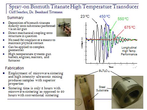
7 Cliff Searfoss

8 Kate Boudreau
9 Patch Transducer

10 Atsushi Baba, Ph.D.
![Amplitude [mv] Laser-Based Ultrasonics for](/docs-images/90/103447004/images/11-0.jpg "Non-destructive Evaluation Summary Non-contact material")


11 Amplitude [mv] Laser-Based Ultrasonics for Non-destructive Evaluation Summary Non-contact material evaluation technology by Laser UT Application Detection of flaw, delamination inclusion in the materials Theory Generation: Pulsed Laser Detection: Laser interferometer Thermoelastic mode Experimental Setup Generation of ultrasound Generation Laser Detection Laser Propagation of ultrasound (1) No Flaw (2) Flaw Delamination Pulsed Laser L-wave Abrasion mode L wave No wave Time [ s] Time [ s]

12 Hyeoung-Sick Ju
 velocity : sensitive to adhesion -Theory Anisotropic layered media: dispersion / angular dependence 1.")
13 Film Bond strength via Acoustic Microscopy Motivation - Needs for acoustic evaluation of thin film adhesion - Actively functioning films: MEMS and semiconductors - Anisotropic substrates (Si or GaAs) for active function films Application - Surface acoustic wave (SAW) velocity : sensitive to adhesion -Theory Anisotropic layered media: dispersion / angular dependence 1. Wave matrix method T - V(z) technique of scanning P( x ) acoustic u u u microscopy Wave-field vector (SAM) U - Displacement :Oscillation in vertically P( xmeasured signal (Δz) 3) T( x3) U - Matrix coupling SAW velocity measurement 2. Force-like weakness model - Displacement continuity at the interface L S - discontinuity stresses 3i 3i / 3i : stress degradation factor 3. SAW velocity in V(z) measurement (1) (2) (3) (4) (5) (6) U U U U U U c R c w c f w z 2 T SAW dispersion by interfacial weakness V(z) for anisotropic substrate Δz UH-3 SAM system for V(z) measurement

14 João M. Salvi Sakamoto

 laminated composites was")

 showing")

15 TRANSVERSE IMPACT OF CFRP LAMINATES UNDER STATIC LOAD OF AXIAL COMPRESSION WITH ACOUSTIC MICROSCOPY Objective: The effect of an initial compressive load on the impact-induced damage of Carbon Fiber Reinforced Plastic (CFRP) laminated composites was experimentally studied. An Air-gun Type of Impact Apparatus A loading fixture for static load of axial compression Schematic diagram showing locations of delaminations Superimposed C-scan images (pulse-wave-mode) showing delaminations at interfaces. A and B. Frequency: 30 MHz. SAM images of the specimen for applying an initial load up to the prescribed strain of 400μ through the upper and lower clamped edges.

16 David Parks
17 Modulus Variation with Temperature
18 Attenuation Variation with Temperature

19 Brian Rheinhardt

20 Leave-in-place High Temperature Transducer

21 Xiaoning Xi

22

23 Sahar Louyeh


 Poly (Styrene-")

24 Atomic Force Microscopy / Ultrasonic Atomic Force Microscopy Surface characterization tool: - Many interactions such as magnetic, electronic, thermal, conductive, topographical and elastic properties can be detected High resolution: nm-sub Å Record nn forces Image in ambient environment Fixation and staining not required Local determination of mechanical properties (Application of ultrasonic atomic force microscopy) Poly (Styrene- methymethacrylate) Image size: nm E-beam evaporated Ir thin film coating Image size: nm SEM AFM Surface roughness = 108 Angstrom
25 Conclusion Atomic Test Reactor Sensors High Temperature Spray-on Transducer Water Level Detector NDE of Composites Photo-Acoustic System Thin Film Bond Strength Detector Impact Detection of Composite Structures High Temperature Ultrasonics Leave-in-place contact transducer Atomic Force Microscopy of materials