In Vitro and In Vivo Comparison of New Biodegradable Magnesium-Based Implants for Orthopedic Uses.
|
|
|
- Felicia Sherman
- 5 years ago
- Views:
Transcription
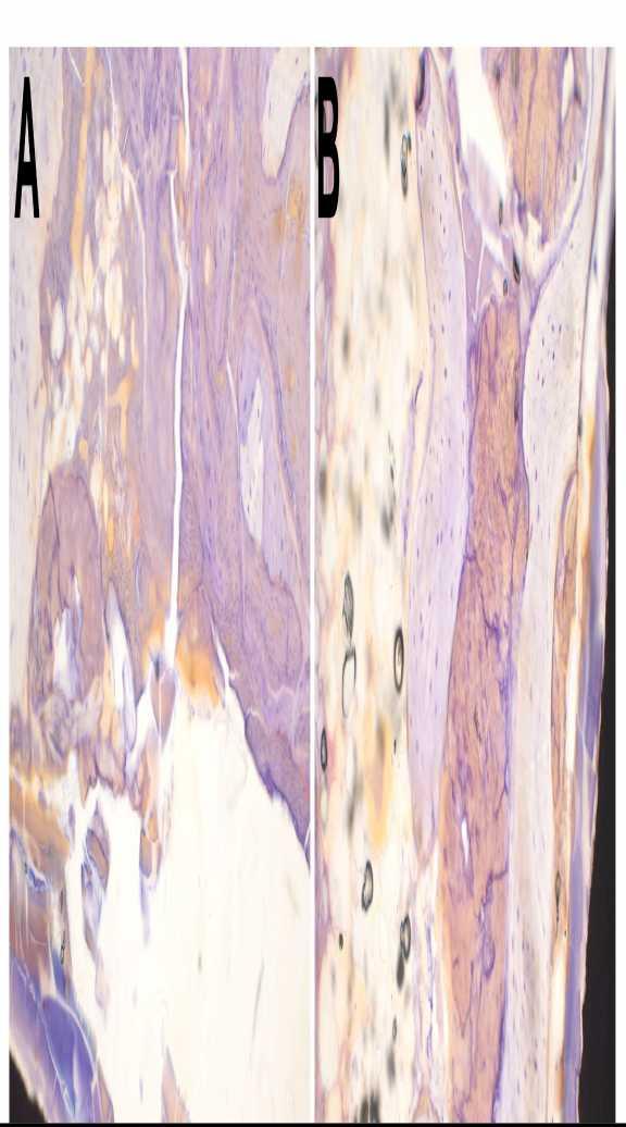
1 In Vitro and In Vivo Comparison of New Biodegradable Magnesium-Based Implants for Orthopedic Uses. Olga Charyeva, DDS 1, Ulrich Thormann, Dr. 2, Sven Schmidt, Dr. 2, Lydia Heimann 3, Ursula Sommer, Dr. 4, Katrin Lips, Prof. 4, Reiner Schnettler, Prof AAP Biomaterials, Justus-Liebig University, Dieburg, Germany, 2 Clinic and Polyclinic for Trauma Surgery, Giessen, Germany, 3 AAP Biomaterials, Dieburg, Germany, 4 Experimantal Trauma Surgery, Justus-Liebig University, Giessen, Germany. Disclosures: O. Charyeva: None. U. Thormann: None. S. Schmidt: None. L. Heimann: 3A; AAP Biomaterials & Implantate GmbH. U. Sommer: None. K. Lips: None. R. Schnettler: None. Introduction: Clinical problems like risk of postoperative infection (1) and increased incidence of pediatric trauma requiring surgical intervention (2) raised the need for temporary orthopedic implants that would resorb after the bone healing is complete. This would decrease high costs associated with repeated surgeries, minimize recovery times, decrease the risk of postoperative infections, and thus promote higher quality of life to the patients. The specific requirement for orthopedic implants, aside from being bioresorbable, is the ability to bear high loads. Magnesium was suggested as a suitable material for these purposes because it is biocompatible; has excellent mechanical properties; is natural for human body, and seems to stimulate new bone formation (3). However, an important problem with magnesium is high corrosion rate with consistent hydrogen gas formation on contact with fluids. The specific aim of the study was to investigate how alloying and combining magnesium with other metals and established biomaterials might improve cyto- and biocompatibility as well as minimize gas production. Methods: Seven types of degradable magnesium-based materials were tested for cyto- and biocompatibility in this study: I) 99.99% magnesium; II) magnesium and silver alloy (Mg2Ag); III) magnesium and gadolinium alloy (Mg10Gd); IV) magnesium and yttrium alloy (W4); V) magnesium, yttrium and earth metals alloy (WE43); VI) magnesium and hydroxyapatite composite; VII) magnesium and calcium phosphate cement paste. For cytocompatibility, viability, proliferation and differentiation were investigated on primary bone marrow derived mesenchymal cells. In vivo biocompatibility was assessed after intraosseous implantation of samples into the femora of 33 male New Zealand white rabbits under general anesthesia. Animals were sacrificed after 6 and 12 weeks and the bone samples were analysed histomorphometrically. Results: The most favorable in vitro cell reaction was observed for the magnesium silver with viable cells directly contacting the alloy. The lowest cell viability was seen for the pure magnesium group followed by magnesium-gadolinium alloy. The pure magnesium's gas emission was highest of all groups in vitro. In vivo, pure magnesium implants were surrounded by a corrosion layer; new bone formation was observed primarily around gas voids. The highest amount of new bone formation was for magnesium-calcium phosphate cement; this material was also surrounded by highest amount of multinucleated cells of all groups. Magnesium-hydroxyapatite composite had extremely fast degradation rate already at 6 weeks alongside with the least amount of new bone formation, and the lowest amount of multinucleated cells at the implantation site. Discussion: Carefully choosing optimal proportions of different components in magnesium-based biomaterials is pivotal for maintaining both normal tissue reaction and implant stability. In this study, magnesium-silver alloy and magnesium-calcium phosphate cement showed the most favorable results in cyto- and biocompatibility. Significance: It is possible to improve magnesium's properties by slowing down its degradation rate through combining it with certain metals and biomaterials. Acknowledgments: This project receives funding from the People Programme (Marie Curie Actions) of the European Union's Seventh Framework Programme FP7 ( ) under REA Grant Agreement No References: 1. Quinn A, Hill AD, Humphreys H. Evolving issues in the prevention of surgical site infections. Surgeon Jun;7(3): Sinikumpu JJ, Lautamo A, Pokka T, Serlo W. The increasing incidence of paediatric diaphyseal both-bone forearm fractures and their internal fixation during the last decade. Injury Mar;43(3): Witte F, Kaese V, Haferkamp H, Switzer E, Meyer-Lindenberg A, Wirth CJ, Windhagen H. In vivo corrosion of four magnesium
2 alloys and the associated bone response. Biomaterials Jun;26(17):3557-
3
4 ORS 2014 Annual Meeting Poster No: 1151
5