Preparation of tissues for study
|
|
|
- Gabriel Garrison
- 6 years ago
- Views:
Transcription

1 Preparation of tissues for study
2 HISTOLOGY : It is the branch of science which deals with the microscopic study of normal tissue HISTOPATHOLOGY : It is the branch of science which deals with the microscopic study of tissue affected by disease Tissue for study can be obtained from: Biopsies Autopsies
3 Microtechnique Microtechnique: is tissue preparation for microscopic examination There are different methods used, however the basic principles are similar It usually involves hardening of the tissue followed by sectioning (cutting) - Paraffin technique - Freezing technique
4 Histological Techniques: 1. Paraffin Tissues are hardened by replacing water with paraffin 2. Freezing technique: Water-rich tissues are hardened by freezing and cut frozen
5 Freezing technique is much faster than traditional histology (20 minutes vs. 16 hours) and are used in operations to achieve a quick diagnosis Cryosections can also be used in immunohistochemistry as freezing tissue does not alter or mask its chemical composition
6 Protocols followed in Histotechniques 1. Identification & Labeling of the specimen 2. Fixation 3. Dehydration 4. Clearing 5. Impregnation (infiltration) 6. Embedding 7. Section cutting 8. Staining 9. Mounting
7 Labeling
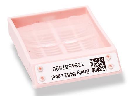
8 Cassette
9 Fixation This is the process by which the constituents of cells and tissue are fixed in a physical and chemical state so that they will withstand subsequent treatment with various reagents with minimum loss of architecture This is achieved by exposing the tissue to chemical compounds: fixatives Fixatives prevent autolysis and bacterial decomposition and preserves tissue in their natural state and fix all components
10 Tissue fixatives Buffered formalin (light microscope preparation) Buffered gluteraldehyde (electron microscope preparation) Osmium tetraoxide (electron microscope preparation, preserve and stain) Zenker s formal saline Bowen s fluid

11 No fixative will penetrate a piece of tissue thicker than 1 cm.

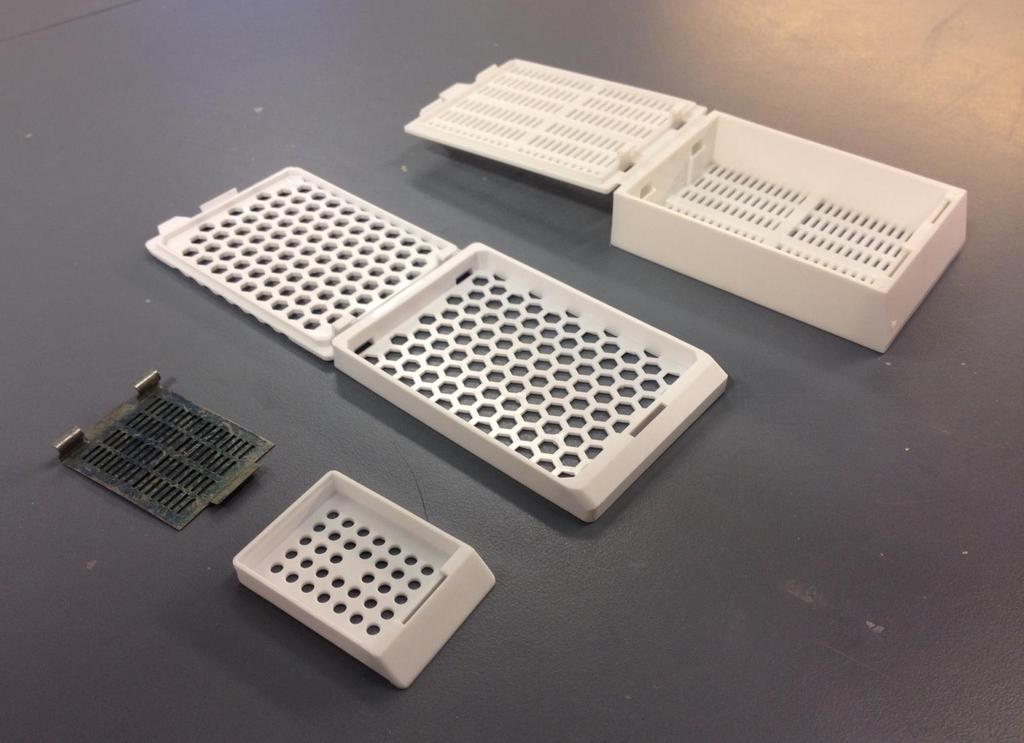
12 Specimen is placed in porous cassettes

13 Cassettes are collected in fixatives 10% formalin 1mm/hour fixation
14 Processing
15 Tissue Processing In order to cut thin sections of the tissues, it should have suitable hardness and consistency when presented to the knife edge. These properties can be imparted by infiltrating and surrounding the tissue with paraffin wax, various types of resins or by freezing. This process is called tissue processing.
16 Tissue Processing It can be subdivided into: a) Dehydration b) Clearing c) Impregnation (infiltration)
17 Types of tissue processing There are two types : 1. Manual Tissue Processing 2. Mechanical Tissue Processing
18 Manual Tissue Processing In this process the tissue is changed from one container of reagent to another by hand Note: The processing, whether manually or mechanically, involves the same steps and reagents in same sequence
19 Mechanical Tissue Processing In this the tissue is moved from one jar to another by mechanical device Timings are controlled by a timer which can be adjusted in respect to hours and minutes Temperature is maintained around 60 ºC Automatic tissue processor: Overnight 12 Baths 16 hours

20 Tissues processor

21 Tissue basket
22 Dehydration (removal of water) It is the process in which the water content in the tissue to be processed is completely removed by passing the tissue through increasing concentrations of dehydrating agents Tissues are dehydrated by using increasing strength of alcohol; e.g. 70%, 90% and 100% Water is replaced by diffusion
23 During dehydration water in tissue has been replaced by alcohol The next step alcohol should be replaced by paraffin wax As paraffin wax is not alcohol soluble, we replace alcohol with a substance in which wax is soluble. This step is called clearing.
24 Clearing Replacing the dehydrating fluid with a fluid that is totally miscible with both the dehydrating fluid (alcohol) and the embedding medium (wax) Some clearing agents: - Xylene - Toluene - Chloroform - Benzene
25 Impregnation (infiltration): The tissue is kept in a wax bath containing molten paraffin wax
26 The duration of the procedure can be noted down as: Fixation % Formalin saline (I) for 1.5 hours % Formalin saline (II) for 1.5 hours Dehydration % alcohol for 1 hour % alcohol (I) for 1 hour % alcohol (II) for 1 hour 4. Absolute alcohol (100%) (I) for 1 hour 5. Absolute alcohol (100%) (II) for 1 hour 6. Absolute alcohol (100%) (III) for 1 hour
27 Clearing: 1. Xylene (I) for 1.5 hours 2. Xylene (II) for 1.5 hours Infiltration: 1. Paraffin wax (I) for 1.5 hours 2. Paraffin wax (II) for 1.5 hours

28 Embedding Embedding: is the process by which impregnated tissues are surrounded by a medium such as agar, gelatin, or wax which when solidified will provide sufficient external support during sectioning
29 Embedding: It is done by transferring the tissue to a mould filled with molten wax & is allowed to cool & solidify After solidification, a wax block is obtained which is then sectioned to obtain ribbons


30 Embedding tools Moulds Paraffin wax
31 Embedding Centre
32 Embedding Centre Molten wax (60 C) (reservoir) Used lids Heated chambers Wax flow adjuster Mould Hot surface Cassette Cold plates (-5 C) Wax dispenser
33 General Embedding Procedure

34 Fill the mould with paraffin wax

35 Using warm forceps select the tissue, take care that it does not cool in the air

36 Orienting the tissue in the mould

37 Orientation Of Tissue In The Block Correct orientation of tissue in a mould is the most important step in embedding 1. cross section 2. longitudinal section Incorrect placement of tissues may result in diagnostically important tissue elements being missed or damaged during microtomy

38 Cool the block on the cold plate

39 Blocks on the cold plate

40 Remove the block from the mould


41 Blocks of embedded tissue are usually trimmed to remove the excess wax on the surface
42 Sectioning (Section Cutting) It is the procedure in which the blocks which have been prepared are cut or sectioned and thin strips of uniform thickness are prepared The instrument by which this is done is called as a Microtome
43 TYPES OF MICROTOMES: Rotary microtome Freezing (cryostat) microtome Ultramicrotome
 1-30µm Steel knife Paraffin")
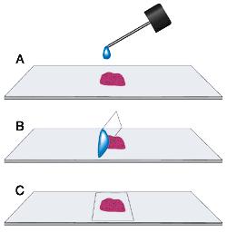
44 Rotary Microtome Micron adjustment (section thickness) 1-30µm Steel knife Paraffin Block
")
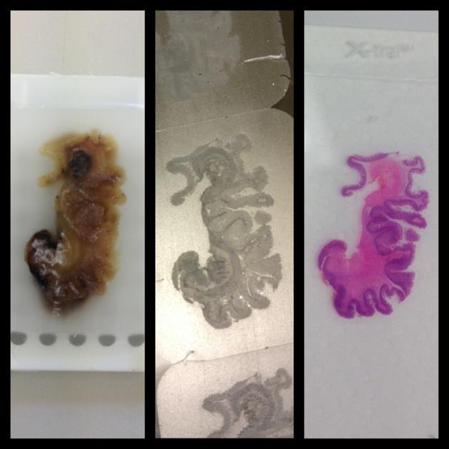
45 Freezing (Cryostat) microtome

46 Ultra Microtome Used for very thin section The typical thickness of tissue cut is between nm for TEM Knife: Diamond or Glass


47 Ultra Microtome Diamond Knife


48 Glass Knife Ultra Microtome

49 Sectioning: Ribbon of sections

50 Tissue floatation bath It is a thermostatically controlled water bath It is maintained at a temperature 5 6 degrees below the melting point of paraffin wax

51 Flattened paraffin sections

52 Taking the floating sections onto slide Adhesives used for fixing the sections on the slides Albumin solution ( Mayor s egg albumin)

53 Taking the section onto slide Flat, no air bubbles, no stretch or breaks
54 Staining Staining is a process by which we give colour to a section. Staining of the section is done to bring out the particular details in the tissue under study There are hundreds of stains available The most commonly used stain in routine practice is Hematoxylin & Eosin stain
55 Staining Classification of Stains: Acid stains (ex. Eosin) Basic stains (ex. Hematoxylin)
56 Acid Dyes In an acidic dye: The basic component is colored and the acid component is colorless Acid dyes stain basic components e.g. eosin stains cytoplasm The color imparted is shade of red
57 Basic Dyes In a basic dye: The acid component is colored and the basic component is colorless Basic dyes stain acidic components e.g. Hematoxylin stains nucleus The color imparted is shade of blue

58 Result : The nucleus stains Blue The cytoplasm stains pink
59 Procedure of staining: There are two types of staining: Manual Staining Automatic staining Slides stained either manually or by automatic stainer, pass through same sequences of reagents.

60 Manual Staining

61 Automatic stainer
62 Mounting Stained section on microscope slide is mounted using mounting medium dissolved in xylene Examples of Mountants : DPX ( Distrene Dibutyl phthalate Xylene ) A coverslip is placed on top, to protect the sample
63
64

65 Light Microscopic Examination
.")
66 Concerning Electron Microscopy Electron beam instead of light Gluteraldehyde fixative Embedding is in hard epoxy plastic Ultramicrotome Diamond or Glass knives Thin section ( µm). Specimen is mounted on a metal grid
PREPARATION OF HISTOLOGICAL SPECIMENS
 PREPARATION OF HISTOLOGICAL SPECIMENS Histo-techniques Preparation of tissue for microscopic examination Series of processes Ultimate aim to make tissue visible as it is Pathology Vs Anatomy Steps vary
Materials and Methods Materials Required for Fixing, Embedding and Sectioning. OCT embedding matrix (Thermo Scientific, LAMB/OCT)
") Page 1 Introduction Tissue freezing and sectioning is a rapid method of generating tissue samples (cryosections) for histological analysis, and obviates the need for wax embedding. The method is popular
Page 1 Introduction Tissue freezing and sectioning is a rapid method of generating tissue samples (cryosections) for histological analysis, and obviates the need for wax embedding. The method is popular
Baraa Ayed AL-Odat. Israa Ayed. Heba kalbouneh
 1 Baraa Ayed AL-Odat Israa Ayed Heba kalbouneh Introduction: "lecture #1" The name " histology " is derived from the Greek words: "histo" means a tissue and "logos" means the study of. So, Histology mean
KCC Path-Core Page 1 of 5
 Instructions for Sample preparation for Paraffin embedding PLEASE NOTE: There is no one-size-fits-all method of tissue preparation for all experimental designs. Before harvesting tissue, you need to assess
Histological preparation of embryonic and adult zebrafish eyes
 Histological preparation of embryonic and adult zebrafish eyes Richard J. Nuckels 1 and Jeffrey M. Gross 1,2,3 1 Section of Molecular Cell and Developmental Biology 2 Institute of Cell and Molecular Biology
Protocol INTRODUCTION RELATED INFORMATION MATERIALS. Histological Preparation of Embryonic and Adult Zebrafish Eyes
 Please cite as: CSH Protocols; 2007; doi:10.1101/pdb.prot4846 Protocol Histological Preparation of Embryonic and Adult Zebrafish Eyes Richard J. Nuckels 1 and Jeffrey M. Gross 1,2,3,4 1 Section of Molecular
PreAnalytiX Supplementary Protocol
 PreAnalytiX Supplementary Protocol Purification of full-length proteins from sections of PAXgene Tissue fixed, paraffin-embedded (PFPE) tissue This protocol describes using the Qproteome FFPE Tissue kit
Web Based Promotion! 10% Off any initial product order. Mention promo code 1204!
 Web Based Promotion! 10% Off any initial product order. Mention promo code 1204! Volume 2, Number 2 1998 FROM PATIENT TO EMBEDDING CENTER IN TWO HOURS OR LESS The single biggest factor in health care today
Boost turnaround time for faster diagnosis
 Boost turnaround time for faster diagnosis Tissue-Tek Xpress x120 Rapid Tissue Processor True rapid continuous tissue processing The Tissue-Tek Xpress x120 is a standardized, turnkey solution combining
Immunofluorescence Staining Protocol for 3 Well Chamber, removable
 Immunofluorescence Staining Protocol for 3 Well Chamber, removable This Application Note presents a simple protocol for the cultivation, fixation, and staining of cells using the 3 Well Chamber, removable.
BIOLOGICAL SAMPLE PREPARATION FOR TEM OBSERVATION. TEM Seminar Nov 16, 2017 Astari Dwiranti, Ph.D
 BIOLOGICAL SAMPLE PREPARATION FOR TEM OBSERVATION TEM Seminar Nov 16, 2017 Astari Dwiranti, Ph.D Why do we need EM for biological samples? (O'Connor and Adams, 2010) Why do we need EM for biological samples?
ab BrdU Immunohistochemistry Kit
 ab125306 - BrdU Immunohistochemistry Kit Instructions for Use For the detection and localization of bromodeoxyuridine incorporated into newly synthesized DNA of actively proliferating cells. This product
Comparing the Quality of Fixation for Gel-based Formalin (Formagel) versus Traditional Liquid-Based Formalin for Immunohistochemistry
 versus Traditional Liquid-Based Formalin for Immunohistochemistry") Comparing the Quality of Fixation for Gel-based Formalin (Formagel) versus Traditional Liquid-Based Formalin for Immunohistochemistry Brian H. Le, M.D., Reading Hospital Reviewed by Michael R. LaFrinere,
Comparing the Quality of Fixation for Gel-based Formalin (Formagel) versus Traditional Liquid-Based Formalin for Immunohistochemistry Brian H. Le, M.D., Reading Hospital Reviewed by Michael R. LaFrinere,
BrdU IHC Kit. For the detection and localization of bromodeoxyuridine incorporated into newly synthesized DNA of actively proliferating cells
 K-ASSAY BrdU IHC Kit For the detection and localization of bromodeoxyuridine incorporated into newly synthesized DNA of actively proliferating cells Cat. No. KT-077 For Research Use Only. Not for Use in
CHAPTER 1 INTRODUCTON. Histopathology- Definition it is a branch of pathology which deals with the
 CHAPTER 1 INTRODUCTON Histopathology- Definition it is a branch of pathology which deals with the study of disease in a tissue section. The tissue undergoes a series of steps before it reaches the examiners
 TABLE OF CONTENTS June 2016 Blades and Sectioning page 4 Cassettes and Accessories page 5 Mounting Media a) Mounting Media b) Frozen Section Media page page 7 8 Paraffin Wax page 9 Safety & Hygiene page
The Children s Hospital of Philadelphia Department of Pathology and Laboratory Medicine
 TheChildren shospitalofphiladelphia DepartmentofPathologyandLaboratoryMedicine Muscle Biopsy - General Instructions The Division of Neuropathology, Department of Pathology and Laboratory Medicine, Children
PreAnalytiX Supplementary Protocol
 PreAnalytiX Supplementary Protocol Purification of total RNA from microdissected PAXgene Tissue fixed, paraffin-embedded (PFPE) and PAXgene Tissue fixed, cryo-embedded (PFCE) tissues This protocol is for
Pinpoint Slide DNA Isolation System Catalog No. D3001
 INSTRUCTIONS Pinpoint Slide DNA Isolation System Catalog No. D3001 Highlights Easily isolates genomic DNA in any targeted microscopic tissue area on a slide. The simple procedure combines Pinpoint tissue
PROTOCOL TO PREPARE PLANTAR FOOTSKIN FOR MORPHOMETRY. I. Removal and Fixation of Plantar Skin (see video)
") PROTOCOL TO PREPARE PLANTAR FOOTSKIN FOR MORPHOMETRY I. Removal and Fixation of Plantar Skin (see video) 1. Sacrifice the animal a. Anaesthetize the animal by placing in a closed chamber with isoflurane.
PROTOCOL TO PREPARE PLANTAR FOOTSKIN FOR MORPHOMETRY I. Removal and Fixation of Plantar Skin (see video) 1. Sacrifice the animal a. Anaesthetize the animal by placing in a closed chamber with isoflurane.
Histopathological techniques The adoption of routine fixation and paraffin wax embedding.
 CIHRT Exhibit P-3359 Page 1 Routine tissue preparation in modern diagnostic histopathology. Bryan R. Hewlett ART, MLT. Histopathological techniques The adoption of routine fixation and paraffin wax embedding.
Electron Microscopy (EM) Grid
 Grid") Anirban Som 25-01-14 Instrumental technique presentation Electron Microscopy (EM) Grid What I will talk about Some basic topics about EM grid Home-made grid preparation Grid cleaning Carbon coating and
Anirban Som 25-01-14 Instrumental technique presentation Electron Microscopy (EM) Grid What I will talk about Some basic topics about EM grid Home-made grid preparation Grid cleaning Carbon coating and
ReliaPrep FFPE gdna Miniprep System
 TECHNICAL MANUAL ReliaPrep FFPE gdna Miniprep System Instructions for Use of Products A2351 and A2352 Revised 12/15 TM352 ReliaPrep FFPE gdna Miniprep System All technical literature is available at: www.promega.com/protocols/
Assessing the Quality of Tissue Processing and the Performance of PelorisTM using the Leica Microsystems Scoring System
 Assessing the Quality of Tissue Processing and the Performance of PelorisTM using the Leica Microsystems Scoring System Geoffrey Rolls, Neville Farmer, Fiona Tarbet Leica Microsystems, Biosystems Division,
ab In situ Apoptosis Detection Kit
 ab206386 In situ Apoptosis Detection Kit Instructions for Use For detection of apoptotic cells. This product is for research use only and is not intended for diagnostic use. Version 8 Last Updated 6 February
Histopathology techniques
 Histopathology techniques Histopathology definition: It is a branch of pathology which deals with the study of disease in a tissue section. The tissue undergoes a series of steps before it reaches the
Histology Fixation Processing Embedding Sectioning Staining. Reliability. Purity. Certainty.
 Histology Fixation Processing Embedding Sectioning Staining Reliability. Purity. Certainty. The Fisher Chemical histology-grade solvents featured in this guide are formulated specifically for histology
ab TripleStain IHC Kit: M&M&R on human tissue (DAB, Red/AP & DAB/Ni)
") ab183287 TripleStain IHC Kit: M&M&R on human tissue (DAB, Red/AP & DAB/Ni) Instructions for Use For the detection of Rabbit and Mouse Primary antibodies on Human tissue or cell samples. This product is
ab183287 TripleStain IHC Kit: M&M&R on human tissue (DAB, Red/AP & DAB/Ni) Instructions for Use For the detection of Rabbit and Mouse Primary antibodies on Human tissue or cell samples. This product is
Guidance Document on Amphibian Thyroid Histology Part 1: Technical guidance for morphologic sampling and histological preparation
 Guidance Document on Amphibian Thyroid Histology Part 1: Technical guidance for morphologic sampling and histological preparation Prepared by: Christiana Grim, OSCP/EPA, USA, May 16, 2007 Contributors:
Manufactured by. Zyagen Barnes Canyon Road San Diego, CA 92121, USA
 Alkaline Phosphatase Immunohistochemistry Detection kits For detection of mouse, rabbit, goat, rat, sheep, chicken, guinea pig, and human primary antibodies Size: 500 Tests Catalog #: AK-011, Mouse Kit
TheraLin. Universal Tissue Fixative Enabling Molecular Pathology
 TheraLin Universal Tissue Fixative Enabling Molecular Pathology TheraLin Universal Tissue Fixative Enabling Molecular Pathology Contents Page # TheraLin Universal Tissue Fixative 3 Introduction 5 Easy
ZytoDot. 2C SPEC HER2/CEN 17 Probe
 ZytoDot 2C SPEC HER2/CEN 17 Probe C-3032-400 C-3032-100 40 (0.4 ml) 10 (0.1 ml) For the detection of the human HER2 gene and alphasatellites of chromosome 17 by chromogenic in situ hybridization (CISH)....
TACS XL TM. In Situ Apoptosis Detection Kit. Catalog Number: TA200. DAB Kit. 30 tests FOR RESEARCH USE ONLY. NOT FOR USE IN DIAGNOSTIC PROCEDURES.
 TACS XL TM In Situ Apoptosis Detection Kit Catalog Number: TA200 DAB Kit 30 tests This package insert must be read in its entirety before using this product. FOR RESEARCH USE ONLY. NOT FOR USE IN DIAGNOSTIC
For research use only. Not for use in diagnostic procedures.
 Dako LSAB 2 System-HRP for use on Rat Specimens Code K0609 Intended use For research use only. Not for use in diagnostic procedures. These instructions apply to the LSAB 2 System-HRP for use on RAT SPECIMENS,
STANDARD OPERATING PROCEDURE
 Effective Date: 12/1/2011 Page 1 of 12 Approval Name/Title Signature Date Authors: Jessica Landis Trial Implementation Manager Immune Tolerance Network Mary C. Philogene Associate Director, QA And Cell
LAMININ. For Immunohistochemical Demonstration of Laminin in Paraffin-embedded and Frozen Human Tissue Sections Stock No. IMMH-7
 LAMININ For Immunohistochemical Demonstration of Laminin in Paraffin-embedded and Frozen Human Tissue Sections Stock No. IMMH-7 TABLE OF CONTENTS BACKGROUND AND PRINCIPLE... 4 REAGENTS AND EQUIPMENT PROVIDED...
celldatasci.com/rnastorm RNAstorm RNA Isolation Kit for FFPE Tissue Samples Sample Kit (20 extractions)
") celldatasci.com/rnastorm info@celldatasci.com RNAstorm RNA Isolation Kit for FFPE Tissue Samples Sample Kit (20 extractions) Support: Email: support@celldatasci.com Phone: 650.285.2376 (option 2) Toll-free:
celldatasci.com/rnastorm info@celldatasci.com RNAstorm RNA Isolation Kit for FFPE Tissue Samples Sample Kit (20 extractions) Support: Email: support@celldatasci.com Phone: 650.285.2376 (option 2) Toll-free:
Order by phone Order on-line Order by fax Website
 Embedding Rings/Capsules Simport, Tissue Capsules These capsules are suitable for holding tissue samples during processing. The lids have a frosted writeon area for sample identification and an open mesh
Quick Ray Manual Tissue Microarrayer. Manual Tissue Microarrayer Quick-Ray
 Manual Tissue Microarrayer Quick-Ray 1 NOTE This user guide describes the instruction of Quick Ray. Review this user guide to avoid injury and prevent damage to this product or any products connected to
Anti-White Spot Syndrome Virus (WSSV) monoclonal antibody. Product no: P13
 monoclonal antibody. Product no: P13") Anti-White Spot Syndrome Virus (WSSV) monoclonal antibody Product no: P13 Product Description The monoclonal antibody (Mab) against the Vp28 protein of White Spot Syndrome Virus (WSSV) is specific for
Anti-White Spot Syndrome Virus (WSSV) monoclonal antibody Product no: P13 Product Description The monoclonal antibody (Mab) against the Vp28 protein of White Spot Syndrome Virus (WSSV) is specific for
ZytoFast HPV type 6/11/16/18/31/33/35 Screening Probe (Bio
 ZytoFast HPV type 6/11/16/18/31/33/35 Screening Probe (Bio iotin in-labeled labeled) T-1044-400 40 (0.4 ml) For the detection of Human Papilloma Virus (HPV) type 6/11/16/18/31/33/35 DNA by chromogenic
Procedure for Immunohistochemical Examination
 Procedure for Immunohistochemical Examination 1. Preparation of Paraffin Block (Separate Attachment 2-2) Disposable bench protection sheet, cutting board, sectioning blade, tweezers, stainless
ZytoLight SPEC FGFR3 Dual Color Break Apart Probe
 ZytoLight SPEC FGFR3 Dual Color Break Apart Probe Z-2170-200 20 (0.2 ml) For the detection of translocations involving the FGFR3 gene at 4p16.3 by fluorescence in situ hybridization (FISH).... In vitro
Fluorescent in-situ Hybridization
 Fluorescent in-situ Hybridization Presented for: Presented by: Date: 2 Definition In situ hybridization is the method of localizing/ detecting specific nucleotide sequences in morphologically preserved
KOS The microwave multifunctional tissue processor you were waiting for. MILESTONE
 KOS The microwave multifunctional tissue processor you were waiting for. Tissue processing Decalcification Special stains Fixation Gross hardening Antigen retrieval In a fraction of the time MILESTONE
(Spin Column) For purification of DNA and RNA from formalin-fixed, paraffin-embedded tissue sections. Instruction for Use
 For purification of DNA and RNA from formalin-fixed, paraffin-embedded tissue sections. Instruction for Use") AmoyDx FFPE DNA/RNA Kit (Spin Column) For purification of DNA and RNA from formalin-fixed, paraffin-embedded tissue sections Instruction for Use Instruction Version: B1.3 Revision Date: September 2015
AmoyDx FFPE DNA/RNA Kit (Spin Column) For purification of DNA and RNA from formalin-fixed, paraffin-embedded tissue sections Instruction for Use Instruction Version: B1.3 Revision Date: September 2015
RNAScope Multiplex Reagent Kit For Tissues
 RNAScope Multiplex Reagent Kit For Tissues Catalog no. R26963 Table 1 Contents and storage Component Amount* Storage** Pretreatment Module Pretreat 2 reagent, 10 4 70 ml Room temperature Pretreat 3 reagent
Segments of the obstructed intestinal loops were fixed in 4% paraformaldehyde
 Supplementary text Supplementary materials and methods Histopathological examination Segments of the obstructed intestinal loops were fixed in 4% paraformaldehyde (PFA) and embedded in paraffin wax with
Electron Microscope Unit The University of Hong Kong SAMPLE PREPARATION TECHNIQUES ON ELECTRON MICROSCOPY
 Electron Microscope Unit The University of Hong Kong SAMPLE PREPARATION TECHNIQUES ON ELECTRON MICROSCOPY (A) Processing of fresh specimen for TEM study 1 Page a) Manual processing procedure for soft tissue
Sample Preparation 3D Cell Explorer. Version 1.1 July 2015 Sabine Bautz
 Sample Preparation 3D Cell Explorer Version 1.1 July 2015 Sabine Bautz Contents 1. Important points to keep in mind for the sample preparation 3 2. Sample preparation specifications and data sheet 4 3.
A Study of Potassium Permanganate 'Fixation' for Electron Microscopy
 241 A Study of Potassium Permanganate 'Fixation' for Electron Microscopy By S. BRADBURY AND G. A. MEEK (From the Department of Human Anatomy, South Parks Road, Oxford) With five plates (figs, i, 2, 4,
Automation in the Histology Lab
 Automation in the Histology Lab Investing in Cost Savings, Faster Turnaround Times, Reduced Contamination, and Standardization Meghan J. Cuddihy, Ph.D. Abigail G. Garrity, M.S. In this white paper Automation
Optimized Protocol for Preparing and Staining LCM Samples from Frozen Tissue and Extraction of High-Quality RNA
 www.arctur.com Optimized Protocol for Preparing and Staining LCM Samples from Frozen Tissue and Extraction of High-Quality RNA Protocol #2 Introduction Laser Capture Microdissection (LCM) enables gene
GeneGel HyRes Starter Kit
 user manual DNA fragment analysis GeneGel HyRes Starter Kit for high-resolution electrophoretic DNA typing um 80-6452-59/Rev. B0/04-02 Page finder Introduction.............................. ii Preparing
HiPer Gel Extraction Teaching Kit (Column Based)
") HiPer Gel Extraction Teaching Kit (Column Based) Product Code: HTBM010 Number of experiments that can be performed: 10 Duration of Experiment Agarose Gel Electrophoresis: 1 hour Protocol: 1 hour Agarose
HiPer Gel Extraction Teaching Kit (Column Based) Product Code: HTBM010 Number of experiments that can be performed: 10 Duration of Experiment Agarose Gel Electrophoresis: 1 hour Protocol: 1 hour Agarose
NanoSteel 3rd Generation AHSS: Auto Evaluation and Technology Expansion
 NanoSteel 3rd Generation AHSS: Auto Evaluation and Technology Expansion Dr. D.J. Branagan Chief Technical Officer & Founder The NanoSteel Company Outline NanoSteel 3 rd Generation AHSS Structural formation
EZ-10 SPIN COLUMN GENOMIC DNA MINIPREPS KIT HANDBOOK
 EZ-0 SPIN COLUMN GENOMIC DNA MINIPREPS KIT HANDBOOK (Bacteria, Plant, Animal, Blood) Version 8 Rev 05/0/03 EZ-0 Genomic DNA Kit Handbook Table of Contents Introduction Limitations of Use Features Applications
THE ASPECTS ABOUT RAPID PROTOTYPING SYSTEM
 THE ASPECTS ABOUT RAPID PROTOTYPING SYSTEM Adrian P. POP 1, Petru UNGUR 1, Gheorghe BEJINARU MIHOC 2 1 University of Oradea, e-mail: adippop@yahoo.com; petru_ungur@yahoo.com; 2 Transilvania University
Investigation of cellular uptake mechanisms by correlative TEM and SIM
 Collaborative Research Center (SFB 1278) Investigation of cellular uptake mechanisms by correlative TEM and SIM Rainer Heintzmann, Fengjiao Ma, Institute of Physical Chemistry Stephanie Höppener, Martin
Overview of Immunohistochemistry. (with a focus on wax-embedded sections)
") Overview of Immunohistochemistry (with a focus on wax-embedded sections) Overview of Immunohistochemistry (with a focus on wax-embedded sections) Overview of Immunohistochemistry IHC is like cooking. There
Overview of Immunohistochemistry (with a focus on wax-embedded sections) Overview of Immunohistochemistry (with a focus on wax-embedded sections) Overview of Immunohistochemistry IHC is like cooking. There
4. The Second-dimensional SDS-PAGE (vertical) Protocol
 Protocol") 4. The Second-dimensional SDS-PAGE (vertical) Protocol I. PURPOSE This procedure outlines the steps that must be carried out in the seconddimension SDS-PAGE using vertical system. II. ENVIRONMENT All work
4. The Second-dimensional SDS-PAGE (vertical) Protocol I. PURPOSE This procedure outlines the steps that must be carried out in the seconddimension SDS-PAGE using vertical system. II. ENVIRONMENT All work
Combined Digoxigenin-labeled in situ hybridization/ Immunohistochemistry protocol (for fixed frozen cryostat sections)
") Combined Digoxigenin-labeled in situ hybridization/ Immunohistochemistry protocol (for fixed frozen cryostat sections) A. Digoxigenin-UTP labeling of crna antisense probe Refer to laboratory protocol and
Combined Digoxigenin-labeled in situ hybridization/ Immunohistochemistry protocol (for fixed frozen cryostat sections) A. Digoxigenin-UTP labeling of crna antisense probe Refer to laboratory protocol and
Protocol. NEXTERION Slide H. Protein application
 Seite: 1/8 1 Introduction... 2 2 Storage and handling... 3 3 General precautions... 3 4 Reagents required... 4 5 Equipment required... 4 6 Protein concentration for spotting... 5 7 Array printing... 5
Activity 2.1. Activity 2.2. Looking at animal cells. Looking at plant cells
 Activity 2.1 Looking at animal cells Skills C1, C2 a source of animal cells, for example some macerated liver or scrapings from the lining of the trachea from a set of sheep or other lungs (obtainable
Anglon Tools Tools for ANGL ROTARY HANDPC Soft rubber pads available in 4 different diameters (ø 0 mm, ø mm, ø 30 mm, ø mm) are available. Pressure se
 are available. Pressure se") inimo One Anglon Tools inimo Anglon System -- Versatility & Performance inimo Anglon angle grinders, with angles of 90 and degrees, are suitable for polishing flat, curved and end surfaces. Great for areas
inimo One Anglon Tools inimo Anglon System -- Versatility & Performance inimo Anglon angle grinders, with angles of 90 and degrees, are suitable for polishing flat, curved and end surfaces. Great for areas
Improved Monitoring of P. aeruginosa on Agar Plates
 Electronic Supplementary Material (ESI) for Analytical Methods. This journal is The Royal Society of Chemistry 2015 Improved Monitoring of P. aeruginosa on Agar Plates SUPPLEMENTAL INFORMATION T. A. Webster,
Rapid Aerobic Count. Interpretation Guide. 3M Food Safety 3M Petrifilm Rapid Aerobic Count Plate
 3M Food Safety 3M Petrifilm Rapid Aerobic Count Plate Rapid Aerobic Count Interpretation Guide The 3M Petrifilm Rapid Aerobic Count Plate is a sample-ready culture medium system which contains nutrients,
ZytoLight SPEC ABL2 Dual Color Break Apart Probe
 ZytoLight SPEC ABL2 Dual Color Break Apart Probe Z-2200-200 20 (0.2 ml) For the detection of translocations involving the ABL2 gene at 1q25.2 by fluorescence in situ hybridization (FISH).... In vitro diagnostic
QuickExtract FFPE RNA Extraction Kit
 QuickExtract FFPE RNA Extraction Kit Cat. Nos. QFR82805 and QFR82050 Connect with Epicentre on our blog (epicentral.blogspot.com), Facebook (facebook.com/epicentrebio), and Twitter (@EpicentreBio). www.epicentre.com
truxtrac FFPE DNA microtube Kit - Bead Purification (24)
") truxtrac FFPE DNA microtube Kit - Bead Purification (24) Adaptive Focused Acoustics (AFA) -based DNA extraction and purification from Formalin-Fixed, Paraffin-Embedded (FFPE) Tissue using Magnetic Beads
truxtrac FFPE DNA microtube Kit - Bead Purification (24) Adaptive Focused Acoustics (AFA) -based DNA extraction and purification from Formalin-Fixed, Paraffin-Embedded (FFPE) Tissue using Magnetic Beads
Phosphohistone-H3 (PHH3)
") Rev. 0.0 v3 Phosphohistone-H3 (PHH3) Rabbit Polyclonal Antibody PRODUCT AVAILABILITY Cat. No. Description 369A-14 0.1 ml, concentrate 369A-15 0.5 ml, concentrate 369A-16 1.0 ml, concentrate 369A-17 1.0
Rev. 0.0 v3 Phosphohistone-H3 (PHH3) Rabbit Polyclonal Antibody PRODUCT AVAILABILITY Cat. No. Description 369A-14 0.1 ml, concentrate 369A-15 0.5 ml, concentrate 369A-16 1.0 ml, concentrate 369A-17 1.0
MICROBIOLOGICAL TOOLS FOR QUALITY ASSURANCE IN HATCHERY: Laboratory Methods
 Issue No.29 / March 2010 MICROBIOLOGICAL TOOLS FOR QUALITY ASSURANCE IN HATCHERY: Laboratory Methods By Dr Vincent TURBLIN, Deputy Regional Market Manager Poultry - CEVA Animal Health Asia Pacific Most
Specimen Preparation Technique for a Microstructure Analysis Using the Focused Ion Beam Process
 Specimen Preparation Technique for a Microstructure Analysis Using the Focused Ion Beam Process by Kozue Yabusaki * and Hirokazu Sasaki * In recent years the FIB technique has been widely used for specimen
DNA RESTRICTION ANALYSIS
 DNA RESTRICTION ANALYSIS In this experiment, DNA from the bacteriophage Lambda (48,502 base pairs in length) is cut with a variety of restriction enzymes and the resulting fragments are separated using
Cell & Tissue Staining Kit
 Cell & Tissue Staining Kit For the detection of goat, mouse, rabbit, rat, or sheep primary IgG Antibodies Size: 50 Tests HRP-DAB System Goat Kit (Catalog Number CTS008) Mouse Kit (Catalog Number CTS002)
Stellaris RNA FISH Protocol for Simultaneous IF + FISH in Adherent Cells
 Stellaris RNA FISH Protocol for Simultaneous IF + FISH in Adherent Cells General Protocol & Storage Product Description A set of Stellaris RNA FISH Probes is comprised of up to 48 singly labeled oligonucleotides
Reagents for Hospitals. Medical and Research Laboratories
 Reagents for Hospitals Medical and Research Laboratories Reagents for Hospitals Summary About us 4 Medical Laboratories 6 Microscopy / p. 7 Indroduction / p. 7 Sample Processing / p. 8 Getting the sample
MODEL 1051 TEM Mill ION MILLING. Ion milling is used on physical science. specimens to reduce thickness to electron
 MODEL 1051 TEM Mill A state-of-the-art ion milling and polishing system offering reliable, high performance specimen preparation. It is compact, precise, and consistently produces high-quality transmission
Preparation thin blood films and Giemsa staining
 TLM_PRO_026 Version No.001 Page 1 of 5 Department: Clinical sciences Unit: Tropical Laboratory Medicine Preparation thin blood films and Giemsa staining Project/study: Not applicable 1. Scope and application
Lymph node biopsy specimens
 CE Update Anatomic Pathology Achieving Technical Excellence in Lymph Node Specimens: An Update Carolyn Beard, HTL(ASCP); Kay Nabers, HTL(ASCP); Mary C. Bowling, MT(ASCP); and Costan W. Berard, MD Lymph
2. Crystallization. A. Background
 2. Crystallization A. Background Crystallization is one of several available techniques available to purify organic compounds. Unlike other techniques, however, crystallization is specific to the purification
EZ-10 SPIN COLUMN GENOMIC DNA MINIPREPS KIT HANDBOOK
 EZ-0 SPIN COLUMN GENOMIC DNA MINIPREPS KIT HANDBOOK (Bacteria, Plant, Animal, Blood) Version 5.0 Rev 03/25/205 Table of Contents Introduction 2 Limitations of Use 2 Features 2 Applications 2 Storage 2
ZytoLight Dual Color Dual Fusion Probe
 ZytoLight SPEC ETV6/RUNX1 Dual Color Dual Fusion Probe Z-2157-200 20 (0.2 ml) For the detection of the translocation t(12;21)(p13;q22) by fluorescence in situ hybridization (FISH).... In vitro diagnostic
Whole Mount IHC Protocol
 Whole Mount IHC Protocol Authors: Ruth Sullivan, Ryan Trevena and Kyle Wegner Creation Date: 03/17/2016 All steps should be conducted with gentle agitation on an orbital shaker, unless otherwise instructed.
Ethanol Assay Kit (Colorimetric/Fluorometric) For the rapid, sensitive and accurate measurement of ethanol levels in various samples.
 For the rapid, sensitive and accurate measurement of ethanol levels in various samples.") ab65343 Ethanol Assay Kit (Colorimetric/Fluorometric) Instructions for Use For the rapid, sensitive and accurate measurement of ethanol levels in various samples. This product is for research use only
ab65343 Ethanol Assay Kit (Colorimetric/Fluorometric) Instructions for Use For the rapid, sensitive and accurate measurement of ethanol levels in various samples. This product is for research use only
RAC. Interpretation Guide. Rapid Aerobic Count Plate
 Interpretation Guide The 3M Petrifilm is a sample-ready-culture medium system which contains nutrients, a cold-water-soluble gelling agent and a dual-sensing indicator technology that facilitates aerobic
Agarose Gel Electrophoresis Lab
 Agarose Gel Electrophoresis ACTIVITY AT A GLANCE Goal: This lab will determine the presence or absence of PCR products and uantify the size (length of the DNA molecule) of the products. Learning Objectives:
RNAscope 2.5 LS Duplex Reagent Kit User Manual for BDZ 11. For use with Leica Biosystems BOND RX System
 RNAscope 2.5 LS Duplex Reagent Kit User Manual for BDZ 11 For use with Leica Biosystems BOND RX System For Research Use Only. Not for diagnostic use. 322440-USM Effective Date 03312017 For Research Use
NAHITA MICROTOME MICROTOME ro r tar otary micr y mic o r t o ome tome semi-aut semi-au omatic tomatic
 NAHITA MICROTOME rotary microtome semi-automatic Nahita semi-automatic microtome has been designed with the most advanced technology for routine or research applications in the field of biology, medicine
truxtrac FFPE DNA microtube Kit Column Purification (25)
") PROTOCOL truxtrac FFPE DNA microtube Kit Column Purification (25) Adaptive Focused Acoustics (AFA) -based DNA extraction and purification from Formalin-Fixed, Paraffin-Embedded Tissue using columns Product
PROTOCOL truxtrac FFPE DNA microtube Kit Column Purification (25) Adaptive Focused Acoustics (AFA) -based DNA extraction and purification from Formalin-Fixed, Paraffin-Embedded Tissue using columns Product
Tooling gelcoat MGS F 300
 3-15 Tooling gelcoat MGS Hardener MGS page Characteristics 15 Application 16 Specifications 20 Processing details 20 Mixing ratios 20 DMA 21 Content Application tooling Characteristics Operational temperature
Lab: Blood Smear and RBC Count
 2014-09- 01 Blood Page 1 of 5 Lab: Blood Smear and RBC Count Aim: Learn to count cells, observe and identify different blood cells in a smear, quantify their proportions and count RBCs per µl (mm^3) using
Department of Microbiology, Lab 016 instructions
 Protocol for standard FISH and DOPE-FISH for prokaryotes (slightly modified from Amann, 1995, note, for other modifications or other microorganisms like eukaryotes, consult special literature or check
Procedure for Stool Routine & Microscopy
 Procedure for Stool Routine & Microscopy : To perform Gross & Physical examination of Stool Variables : Specimen collection, Labelling of specimen, Name, Identifying number, date, time Gross Examination
SER: Seminal Fluid ABAcard p30 Immunoassay Test
 Immunoassay Test Principle Equipment Reagents Controls Quality control The ABAcard p30 Immunoassay Test is a one-step test for the detection of p30. The test devices use a conjugated dye labeled antibody
Protocol. Nexterion Slide A+ MPX 16. DNA-application
 Seite: 1/12 1 Introduction...2 2 Storage and handling...3 3 General precautions...3 4 Reagents required...3 5 Equipment required...4 6 Array printing...4 7 Printing guidelines...5 8 DNA immobilization...6
4.1 DETERMINATION OF SPECIFIC GRAVITY
 TESTS ON CEMENT 94 4.1 DETERMINATION OF SPECIFIC GRAVITY STANDARD IS: 4031-1988. DEFINITION Specific Gravity is defined as the ratio of the mass of the cement to the mass of an equal volume of kerosene.