X-RAY DIFFRACTION. X- Ray Sources Diffraction: Bragg s Law Crystal Structure Determination
|
|
|
- Henry Washington
- 5 years ago
- Views:
Transcription
1 X-RAY DIFFRACTION X- Ray Sources Diffraction: Bragg s Law Crystal Structure Determination Part of MATERIALS SCIENCE & ENGINEERING A Learner s Guide AN INTRODUCTORY E-BOOK Anandh Subramaniam & Kantesh Balani Materials Science and Engineering (MSE) Indian Institute of Technology, Kanpur anandh@iitk.ac.in, URL: home.iitk.ac.in/~anandh Elements of X-Ray Diffraction B.D. Cullity & S.R. Stock Prentice Hall, Upper Saddle River (001) Recommended websites:
2 What will you learn in this sub-chapter? How to produce monochromatic X-rays? How does a crystal scatter these X-rays to give a diffraction pattern? Bragg s equation What determines the position of the XRD peaks? Answer) the lattice. What determines the intensity of the XRD peaks? Answer) the motif. How to analyze a powder pattern to get information about the lattice type? (Cubic crystal types). What other uses can XRD be put to apart from crystal structure determination? Grain size determination Strain in the material
3 Some Basics For electromagnetic radiation to be diffracted* the spacing in the grating (~a series of obstacles or a series of scatterers) should be of the same order as the wavelength. In crystals the typical interatomic spacing ~ -3 Å** so the suitable radiation for the diffraction study of crystals is X-rays. Hence, X-rays are used for the investigation of crystal structures. Neutrons and Electrons are also used for diffraction studies from materials. Neutron diffraction is especially useful for studying the magnetic ordering in materials. ** If the wavelength is of the order of the lattice spacing, then diffraction effects will be prominent. ** Lattice parameter of Cu (a Cu ) = 3.61 Å d hkl is equal to a Cu or less than that (e.g. d 111 = a Cu /3 =.08 Å) Click here to know more about this
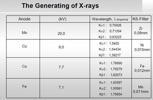
4 Generation of X-rays X-rays can be generated by decelerating electrons. Hence, X-rays are generated by bombarding a target (say Cu) with an electron beam. The resultant spectrum of X-rays generated (i.e. X-rays versus Intensity plot) is shown in the next slide. The pattern shows intense peaks on a broad background. The intense peaks can be thought of as monochromatic radiation and be used for X-ray diffraction studies. Beam of electrons Target X-rays An accelerating (or decelerating) charge radiates electromagnetic radiation
5 Mo Target impacted by electrons accelerated by a 35 kv potential shows the emission spectrum as in the figure below (schematic) K Intense peak, nearly monochromatic X-ray sources with different for doing XRD studies Intensity White radiation K Characteristic radiation due to energy transitions in the atom Target Metal Of K radiation (Å) Mo 0.71 Cu 1.54 Co 1.79 Fe 1.94 Cr Wavelength () The high intensity nearly monochromatic K x-rays can be used as a radiation source for X-ray diffraction (XRD) studies a monochromator can be used to further decrease the spread of wavelengths in the X-ray
6
 Fluorescent X-rays Electrons Scattered X-rays Compton recoil Photoelectrons Coherent From bound charges Incoherent (Compton modified) From loosely bound")
7 When X-rays hit a specimen, the interaction can result in various signals/emissions/effects. The coherently scattered X-rays are the ones important from a XRD perspective. Incident X-rays SPECIMEN Absorption (Heat) Fluorescent X-rays Electrons Scattered X-rays Compton recoil Photoelectrons Coherent From bound charges Incoherent (Compton modified) From loosely bound charges Transmitted beam Click here to know more X-rays can also be refracted (refractive index slightly less than 1) and reflected (at very small angles)
8 Diffraction Click here to Understand Diffraction Now we shall consider the important topic as to how X-rays interact with a crystalline array (of atoms, ions etc.) to give rise to the phenomenon known as X- ray diffraction (XRD). Let us consider a special case of diffraction a case where we get sharp [1] diffraction peaks. Diffraction (with sharp peaks) (with XRD being a specific case) requires three important conditions to be satisfied: Coherent, monochromatic, parallel waves (with wavelength ). Crystalline array of scatterers* with spacing of the order of (~). Fraunhofer diffraction geometry Aspects related to the wave Coherent, monochromatic, parallel wave Diffraction pattern with sharp peaks Aspects related to the material Crystalline*, ** Aspects related to the diffraction set-up (diffraction geometry) Fraunhofer geometry [1] The intensity- plot looks like a function. * A quasicrystalline array will also lead to diffraction with sharp peaks (which we shall not consider in this text). ** Amorphous material will give diffuse peak.
9 Some comments and notes The waves could be: electromagnetic waves (light, X-rays ), matter waves** (electrons, neutrons ) or mechanical waves (sound, waves on water surface ). Not all objects act like scatterers for all kinds of radiation. If wavelength is not of the order of the spacing of the scatterers, then the number of peaks obtained may be highly restricted (i.e. we may even not even get a single diffraction peak!). In short diffraction is coherent reinforced scattering (or reinforced scattering of coherent waves). In a sense diffraction is nothing but a special case of constructive (& destructive) interference. To give an analogy the results of Young s double slit experiment is interpreted as interference, while the result of multiple slits (large number) is categorized under diffraction. Fraunhofer diffraction geometry implies that parallel waves are impinging on the scatteres (the object), and the screen (to capture the diffraction pattern) is placed far away from the object. Click here to know more about Fraunhofer and Fresnel diffraction geometries ** With a de Broglie wavelength
10 XRD the first step A beam of X-rays directed at a crystal interacts with the electrons of the atoms in the crystal. The electrons oscillate under the influence of the incoming X-Rays and become secondary sources of EM radiation. The secondary radiation is in all directions. The waves emitted by the electrons have the same frequency as the incoming X-rays coherent. The emission can undergo constructive or destructive interference. Incoming X-rays Secondary emission Schematics Oscillating charge re-radiates In phase with the incoming x-rays Sets Electron cloud into oscillation Sets nucleus into oscillation Small effect neglected
11 Some points to recon with We can get a better physical picture of diffraction by using Laue s formalism (leading to the Laue s equations). However, a parallel approach to diffraction is via the method of Bragg, wherein diffraction can be visualized as reflections from a set of planes. As the approach of Bragg is easier to grasp we shall use that in this elementary text. We shall do some intriguing mental experiments to utilize the Bragg s equation (Bragg s model) with caution. Let us consider a coherent wave of X-rays impinging on a crystal with atomic planes at an angle to the rays. Incident and scattered waves are in phase if the: i) in-plane scattering is in phase and ii) scattering from across the planes is in phase. Incident and scattered waves are in phase if In plane scattering is in phase Scattering from across planes is in phase
12 Let us consider in-plane scattering A B Atomic Planes X Y There is more to this Click here to know more and get introduced to Laue equations describing diffraction Extra path traveled by incoming waves AY Extra path traveled by scattered waves XB These can be in phase if incident = scattered A B But this is still reinforced scattering and NOT reflection X Y


 =")

.")
13 BRAGG s EQUATION Let us consider scattering across planes Click here to visualize constructive and destructive interference A portion of the crystal is shown for clarity- actually, for destructive interference to occur many planes are required (and the interaction volume of x-rays is large as compared to that shown in the schematic). The scattering planes have a spacing d. Ray- travels an extra path as compared to Ray-1 (= ABC). The path difference between Ray-1 and Ray- = ABC = (d Sin + d Sin) = (d.sin). For constructive interference, this path difference should be an integral multiple of : n = d Sin the Bragg s equation. (More about this sooner). The path difference between Ray-1 and Ray-3 is = (d.sin) = n = n. This implies that if Ray-1 and Ray- constructively interfere Ray-1 and Ray-3 will also constructively interfere. (And so forth).


14 The previous page explained how constructive interference occurs. How about the rays just of Bragg angle? Obviously the path difference would be just off as in the figure below. How come these rays go missing? Click here to understand how destructive interference of just of-bragg rays occur Interference of Ray-1 with Ray- Note that they almost constructively interfere!
15 Reflection versus Diffraction Though diffraction (according to Bragg s picture) has been visualized as a reflection from a set of planes with interplanar spacing d diffraction should not be confused with reflection (specular reflection). Reflection Occurs from surface Takes place at any angle Diffraction Occurs throughout the bulk Takes place only at Bragg angles ~100 % of the intensity may be reflected Small fraction of intensity is diffracted Note: X-rays can ALSO be reflected at very small angles of incidence
16 Understanding the Bragg s equation n = d Sin The equation is written better with some descriptive subscripts: n d Sin Cu K hkl hkl n is an integer and is the order of the reflection (i.e. how many wavelengths of the X-ray go on to make the path difference between planes). Bragg s equation is a negative statement If Bragg s eq. is NOT satisfied NO reflection can occur If Bragg s eq. is satisfied reflection MAY occur (How?- we shall see this a little later). The interplanar spacing appears in the Bragg s equation, but not the interatomic spacing a along the plane (which had forced incident = scattered ); but we are not free to move the atoms along the plane randomly click here to know more. For large interplanar spacing the angle of reflection tends towards zero as d increases, Sin decreases (and so does ). The smallest interplanar spacing from which Bragg diffraction can be obtained is / maximum value of is 90, Sin is 1 from Bragg equation d = /.
17 Order of the reflection (n) For Cu K radiation ( = 1.54 Å) and d 110 =. Å n Sin = n/d º First order reflection from (110) º Second order reflection from (110) planes 110 Also considered as first order reflection from (0) planes 0 Relation between d nh nk nl and d hkl d d d Cubic crystal hkl nhnk nl nhnk nl a h k l a ( nh) ( nk) ( nl) a dhkl n h k l n e.g. d d a 8 a d d
 n d sin hkl d hkl sin n d nhnk nl d hkl 1 n d sin nh nk nl Hence,")

 to")
 planes).")
18 In XRD n th order reflection from (h k l) is considered as 1 st order reflection from (nh nk nl) n d sin hkl d hkl sin n d nhnk nl d hkl 1 n d sin nh nk nl Hence, (100) planes are a subset of (00) planes d d d d Important point to note: In a simple cubic crystal, 100, 00, 300 are all allowed reflections. But, there are no atoms in the planes lying within the unit cell! Though, first order reflection from 00 planes is equivalent (mathematically) to the second order reflection from 100 planes; for visualization purposes of scattering, this is better thought of as the later process (i.e. second order reflection from (100) planes).
 is converted to angular information (the angle of diffraction, Bragg ).")
19 Funda Check How is it that we are able to get information about lattice parameters of the order of Angstroms (atoms which are so closely spaced) using XRD? Diffraction is a process in which linear information (the d-spacing of the planes) is converted to angular information (the angle of diffraction, Bragg ). If the detector is placed far away from the sample (i.e. R in the figure below is large) the distances along the arc of a circle (the detection circle) get amplified and hence we can make easy measurements.


20 Forward and Back Diffraction Here a guide for quick visualization of forward and backward scattering (diffraction) is presented
21 Funda Check What is (theta) in the Bragg s equation? is the angle between the incident x-rays and the set of parallel atomic planes (which have a spacing d hkl ). Which is 10 in the above figure. It is NOT the angle between the x-rays and the sample surface (note: specimens could be spherical or could have a rough surface).
22 The missing reflections We had mentioned that Bragg s equation is a negative statement: i.e. just because Bragg s equation is satisfied a reflection may not be observed. Let us consider the case of Cu K radiation ( = 1.54 Å) being diffracted from (100) planes of Mo (BCC, a = 3.15 Å = d 100 ). d100 Sin100 Sin d100 (3.15) But this reflection is absent in BCC Mo The missing reflection is due to the presence of additional atoms in the unit cell (which are positions at lattice points) which we shall consider next The wave scattered from the middle plane is out of phase with the ones scattered from top and bottom planes. I.e. if the green rays are in phase (path difference of ) then the red ray will be exactly out of phase with the green rays (path difference of /).
23 Continuing with the case of BCC Mo However, the second order reflection from (100) planes (which is equivalent to the first order reflection from the (00) planes is observed 1.54 nd order 1 Sin d nd ~ 9.67 order This is because if the green rays have a path difference of then the red ray will have path difference of which will still lead to constructive interference!
24 Important points Presence of additional atoms/ions/molecules in the UC at lattice points or as a part of the motif can alter the intensities of some of the reflections Some of the reflections may even go missing Position of the reflections / peaks tells us about the lattice type. The Intensities tells us about the motif.
25 Intensity of the Scattered waves Bragg s equation tells us about the position of the intensity peaks (in terms of ) but tells us nothing about the intensities. The intensities of the peaks depend on many factors as considered here. Scattering by a crystal can be understood in three steps A Electron Polarization factor B Atom To understand the scattering from a crystal leading to the intensity of reflections (and why some reflections go missing), three levels of scattering have to be considered: 1) scattering from electrons ) scattering from an atom 3) scattering from a unit cell Click here to know the details Atomic scattering factor (f) Structure Factor (F): The resultant wave scattered by all atoms of the unit cell The Structure Factor is independent of the shape and size of the unit cell; but is dependent on the position of the atoms/ions etc. within the cell C Unit cell (uc) Structure factor (F) Click here to know more about Structure factor calculations & Intensity in powder patterns
26 The concept of a Reciprocal lattice and the Ewald Sphere construction: Reciprocal lattice and Ewald sphere constructions are important tools towards understanding diffraction. (especially diffraction in a Transmission Electron Microscope (TEM)) A lattice in which planes in the real lattice become points in the reciprocal lattice is a very useful one in understanding diffraction. click here to go to a detailed description of these topics. Click here to know more about Reciprocal Lattice & Ewald Sphere construction
27 Selection / Extinction Rules As we have noted before even if Bragg s equation is satisfied, reflections may go missing this is due to the presence of additional atoms in the unit cell. The reflections present and the missing reflections due to additional atoms in the unit cell are listed in the table below. Click here to see the derivations Structure factor calculations Bravais Lattice Reflections which may be present Reflections necessarily absent Simple all None Body centred (h + k + l) even (h + k + l) odd Face centred h, k and l unmixed h, k and l mixed End centred (C centred) h and k unmixed h and k mixed Bravais Lattice SC BCC FCC DC Allowed Reflections All (h + k + l) even h, k and l unmixed Either, h, k and l are all odd or all are even & (h + k + l) divisible by 4
28 Allowed reflections in SC*, FCC*, BCC* & DC crystals Cannot be expressed as (h +k +l ) * lattice decorated with monoatomic/monoionic motif h + k + l SC FCC BCC DC , , , ,
29 The ratio of (h + k + l ) derived from extinction rules (previous page) As we shall see soon the ratios of (h + k + l ) is proportional to Sin which can be used in the determination of the lattice type SC BCC FCC DC Note that we have to consider the ratio of only two lines to distinguish FCC and DC. I.e. if the ratios are 3:4 then the lattice is FCC. But, to distinguish between SC and BCC we have to go to 7 lines!
30 Crystal structure determination As diffraction occurs only at specific Bragg angles, the chance that a reflection is observed when a crystal is irradiated with monochromatic X-rays at a particular angle is small (added to this the diffracted intensity is a small fraction of the beam used for irradiation). The probability to get a diffracted beam (with sufficient intensity) is increased by either varying the wavelength () or having many orientations (rotating the crystal or having multiple crystallites in many orientations). The three methods used to achieve high probability of diffraction are shown below. Monochromatic X-rays Many s (orientations) Powder specimen POWDER METHOD λ fixed θ variable Panchromatic X-rays Single LAUE TECHNIQUE λ variable θ fixed Monochromatic X-rays Varied by rotation ROTATING CRYSTAL METHOD λ fixed θ rotated Only the powder method (which is commonly used in materials science) will be considered in this text.
31 THE POWDER METHOD sin ) ( l k h sin 4 ) ( a l k h ) ( 4sin l k h a l k h a d Cubic d Sin sin 4 l k h a Cubic crystal In the powder method the specimen has crystallites (or grains) in many orientations (usually random). Monochromatic* X-rays are irradiated on the specimen and the intensity of the diffracted beams is measured as a function of the diffracted angle. In this elementary text we shall consider cubic crystals. (1) () () in (1) * In reality this is true only to an extent
 The coherent x-ray beam is diffracted by these crystallites at various angles")

")
32 POWDER METHOD In the powder sample there are crystallites in different random orientations (a polycrystalline sample too has grains in different orientations) The coherent x-ray beam is diffracted by these crystallites at various angles to the incident direction All the diffracted beams (called reflections ) from a single plane, but from different crystallites lie on a cone. Depending on the angle there are forward and back reflection cones. A diffractometer can record the angle of these reflections along with the intensities of the reflection The X-ray source and diffractometer move in arcs of a circle- maintaining the Bragg reflection geometry as in the figure (right) Different cones for different reflections


are considered].")


33 How to visualize the occurrence of peaks at various angles It is somewhat difficult to actually visualize a random assembly of crystallites giving peaks at various angels in a XRD scan. The figures below are expected to give a visual feel for the same. [Hypothetical crystal with a = 4Å is assumed with =1.54Å. Only planes of the type xx0 (like (100,110)are considered]. Random assemblage of crystallites in a material As the scan takes place at increasing angles, planes with suitable d, which diffract are picked out from favourably oriented crystallites h hkl d Sin()
34 Determination of Crystal Structure from versus Intensity Data in Powder Method In the power diffraction method a versus intensity (I) plot is obtained from the diffractometer (and associated instrumentation). The intensity is the area under the peak in such a plot (NOT the height of the peak). The information of importance obtained from such a pattern is the relative intensities and the absolute value of the intensities is of little importance (for now). I is really diffracted energy (as Intensity is Energy/area/time). A table is prepared as in the next slide to tabulate the data and make calculations to find the crystal structure (restricting ourselves to cubic crystals for the present). Powder diffraction pattern from Al Radiation: Cu K, = 1.54 Å Increasing d Increasing
35 Determination of Crystal Structure from versus Intensity Data The following table is made from the versus Intensity data (obtained from a XRD experiment on a powder sample (empty starting table of columns is shown below- completed table shown later). n Intensity Sin Sin ratio
")
36 Powder diffraction pattern from Al Radiation: Cu K, = 1.54 Å Note: This is a schematic pattern In real patterns peaks or not idealized peaks broadened Increasing splitting of peaks with g ( 1 & peaks get resolved in the high angle peaks) Peaks are all not of same intensity No brackets are used around the indexed numbers (the peaks correspond to planes in the real space)
37 Powder diffraction pattern from Al Radiation: Cu K, = 1.54 Å Note: Peaks or not idealized peaks broadened Increasing splitting of peaks with g Peaks are all not of same intensity In low angle peaks K 1 & K peaks merged K 1 & K peaks resolved in high angle peaks (in and 400 peaks this can be seen)
, grain size etc. broadening.")
38 Funda Check How are real diffraction patterns different from the ideal computed ones? We have seen real and ideal diffraction patterns. In ideal patterns the peaks are functions. Real diffraction patterns are different from ideal ones in the following ways: Peaks are broadened Could be due to instrumental, residual non-uniform strain (microstrain), grain size etc. broadening. Peaks could be shifted from their ideal positions Could be due to uniform strain macrostrain. Relative intensities of the peaks could be altered Could be due to texture in the sample. Funda Check What is the maximum value of possible (experimentally)? Ans: 90 At = 90 the reflected ray is opposite in direction to the incident ray. Beyond this angle, it is as if the source and detector positions are switched. max is 180.
39 Funda Check What will determine how many peaks I will get? 1) smaller the wavelength of the X-rays, more will be the number of peaks possible. From Bragg s equation: [=dsin], (Sin) max will correspond to d min. (Sin) max =1. Hence, d min =/. Hence, if is small then planes with smaller d spacing (i.e. those which occur at higher values) will also show up in a XRD patter (powder pattern). Given that experimentally cannot be greater than 90. ) Lattice type in SC we will get more peaks as compared to (say) FCC/DC. Other things being equal. 3) Lower the symmetry of the crystal, more the number of peaks (e.g., in tetragonal crystal the 100 peak will lie at a different as compared to the 001 peak). dsin Sin max d min d min
40 Determination of Crystal Structure (lattice type) from versus Intensity Data Let us assume that we have the versus intensity plot from a diffractometer To know the lattice type we need only the position of the peaks (as tabulated below) Solved example 1 # Sin Sin ratio Index d From the ratios in column 6 we conclude that FCC a Using d Sin 1.54 d111 Sin o a 4.04A Al Note that Sin cannot be > 1 We can get the lattice parameter which correspond to that for Al Note ( h k l ) sin Note: Error in d spacing decreases with so we should use high angle lines for lattice parameter calculation Click here to know more XRD_lattice_parameter_calculation.ppt
41 Solved example Another example Given the positions of the Bragg peaks we find the lattice type Sin Sin Ratios of Sin Dividing Sin by 0.134/3 = Whole number ratios FCC

42 Comparison of diffraction patterns of SC, BCC & B structures Click here More Solved Examples on XRD Click here
43 Funda Check What happens when we increase or decrease? We had pointed out that ~ a is preferred for diffraction. Let us see what happens if we drastically increase or decrease. (This is only a thought experiment!!) Aluminium = 1.54 Å = 3 Å = 0.1 Å hkl d Sin() Sin() Sin() With Cu K = 1.54 Å If we ~double we get too few peaks If we make small all the peaks get crowded to small angles And the detector may not be able to resolve these peaks if they come too close!
44 Applications of XRD Bravais lattice determination Lattice parameter determination We have already seen these applications Determination of solvus line in phase diagrams Long range order Crystallite size and Strain Click here to know more Determine if the material is amorphous or crystalline Next slide
45 Intensity Intensity Intensity Crystal Schematic of difference between the diffraction patterns of various phases Sharp peaks Diffraction angle () Monoatomic gas No peak Diffraction angle () Liquid / Amorphous solid 0 Diffraction angle () Diffuse Peak
46 Actual diffraction pattern from an amorphous solid Diffuse peak from Cu-Zr-Ni-Al-Si Metallic glass Note Sharp peaks are missing Broad diffuse peak survives the peak corresponds to the average spacing between atoms which the diffraction experiment picks out (XRD patterns) courtesy: Dr. Kallol Mondal, MSE, IITK
 d Cubic crystal hkl a h k l As h,k, l increases, d decreases we could have planes with infinitesimal")

47 Funda Check What is the minimum spacing between planes possible in a crystal? How many diffraction peaks can we get from a powder pattern? Let us consider a cubic crystal (without loss in generality) d Cubic crystal hkl a h k l As h,k, l increases, d decreases we could have planes with infinitesimal spacing d 13 a 10 With increasing indices the interplanar spacing decreases a a d d 10 a 1 a The number of peaks we obtain in a powder diffraction pattern depends on the wavelength of x-ray we are using. Planes with d < / are not captured in the diffraction pattern. These peaks with small d occur at high angles in diffraction pattern. d 1 a 5 d 11 a
48
Identification of Crystal Structure and Lattice Parameter. for Metal Powders Using X-ray Diffraction. Eman Mousa Alhajji
 Identification of Crystal Structure and Lattice Parameter for Metal Powders Using X-ray Diffraction Eman Mousa Alhajji North Carolina State University Department of Materials Science and Engineering MSE
Atomic Densities. Linear Density. Planar Density. Linear Density. Outline: Planar Density
 Atomic Densities Outline: Atomic Densities - Linear Density - Planar Density Single- vs poly- crystalline materials X-ray Diffraction Example Polymorphism and Allotropy Linear Density Number of atoms per
Diffraction Basics. The qualitative basics:
 The qualitative basics: Diffraction Basics Coherent scattering around atomic scattering centers occurs when x-rays interact with material In materials with a crystalline structure, x-rays scattered in
Atomic Densities. Linear Density Number of atoms per length whose centers lie on the direction vector for a specific crystallographic direction.
 Atomic Densities Linear Density Number of atoms per length whose centers lie on the direction vector for a specific crystallographic direction. Planar Density Number of atoms per unit area that are centered
9/29/2014 8:52 PM. Chapter 3. The structure of crystalline solids. Dr. Mohammad Abuhaiba, PE
 1 Chapter 3 The structure of crystalline solids 2 Home Work Assignments HW 1 2, 7, 12, 17, 22, 29, 34, 39, 44, 48, 53, 58, 63 Due Sunday 12/10/2014 Quiz # 1 will be held on Monday 13/10/2014 at 11:00 am
9/28/2013 9:26 PM. Chapter 3. The structure of crystalline solids. Dr. Mohammad Abuhaiba, PE
 Chapter 3 The structure of crystalline solids 1 2 Why study the structure of crystalline solids? Properties of some materials are directly related to their crystal structure. Significant property differences
Fundamentals of X-ray diffraction and scattering
 Fundamentals of X-ray diffraction and scattering Don Savage dsavage@wisc.edu 1231 Engineering Research Building (608) 263-0831 X-ray diffraction and X-ray scattering Involves the elastic scattering of
9/16/ :30 PM. Chapter 3. The structure of crystalline solids. Mohammad Suliman Abuhaiba, Ph.D., PE
 Chapter 3 The structure of crystalline solids 1 Mohammad Suliman Abuhaiba, Ph.D., PE 2 Home Work Assignments HW 1 2, 7, 12, 17, 22, 29, 34, 39, 44, 48, 53, 58, 63 Due Sunday 17/9/2015 3 Why study the structure
Experiment 2b X-Ray Diffraction* Optical Diffraction Experiments
 * Experiment 2b X-Ray Diffraction* Adapted from Teaching General Chemistry: A Materials Science Companion by A. B. Ellis et al.: ACS, Washington, DC (1993). Introduction Inorganic chemists, physicists,
Chapter 3 Basic Crystallography and Electron Diffraction from Crystals. Lecture 9. Chapter 3 CHEM Fall, L. Ma
 Chapter 3 Basic Crystallography and Electron Diffraction from Crystals Lecture 9 Outline The geometry of electron diffraction Crystallography Kinetic Theory of Electron diffraction Diffraction from crystals
LECTURE 7. Dr. Teresa D. Golden University of North Texas Department of Chemistry
 LECTURE 7 Dr. Teresa D. Golden University of North Texas Department of Chemistry Diffraction Methods Powder Method For powders, the crystal is reduced to a very fine powder or microscopic grains. The sample,
Carbon nanostructures. (http://www.mf.mpg.de/de/abteilungen/schuetz/index.php?lang=en&content=researchtopics&type=specific&name=h2storage)
") Carbon nanostructures (http://www.mf.mpg.de/de/abteilungen/schuetz/index.php?lang=en&content=researchtopics&type=specific&name=h2storage) 1 Crystal Structures Crystalline Material: atoms arrange into a
Carbon nanostructures (http://www.mf.mpg.de/de/abteilungen/schuetz/index.php?lang=en&content=researchtopics&type=specific&name=h2storage) 1 Crystal Structures Crystalline Material: atoms arrange into a
This lecture is part of the Basic XRD Course.
 This lecture is part of the Basic XRD Course. Basic XRD Course 1 A perfect polycrystalline sample should contain a large number of crystallites. Ideally, we should always be able to find a set of crystallites
It is instructive however for you to do a simple structure by hand. Rocksalt Structure. Quite common in nature. KCl, NaCl, MgO
 Today the structure determinations etc are all computer -assisted It is instructive however for you to do a simple structure by hand Rocksalt Structure Quite common in nature KCl, NaCl, MgO 9-1 Typical
X-ray diffraction
 2.2.3.- X-ray diffraction 2.2.3.1.- Origins and fundamentals of the technique The first experimental evidence concerning x-ray diffraction was given by Max von Laue who in 1912 demonstrated that x-rays
Lesson 1 X-rays & Diffraction
 Lesson 1 X-rays & Diffraction Nicola Döbelin RMS Foundation, Bettlach, Switzerland February 11 14, 2013, Riga, Latvia Electromagnetic Spectrum X rays: Wavelength λ: 0.01 10 nm Energy: 100 ev 100 kev Interatomic
Materials Lab 1(MT344) X-ray Diffractometer Operation and Data Analysis. Instructor: Dr. Xueyan Wu ( 吴雪艳 )
 X-ray Diffractometer Operation and Data Analysis. Instructor: Dr. Xueyan Wu ( 吴雪艳 )") Materials Lab 1(MT344) X-ray Diffractometer Operation and Data Analysis Instructor: Dr. Xueyan Wu ( 吴雪艳 ) Goals To give students a practical introduction into the use of X-ray diffractometer and data collection.
Materials Lab 1(MT344) X-ray Diffractometer Operation and Data Analysis Instructor: Dr. Xueyan Wu ( 吴雪艳 ) Goals To give students a practical introduction into the use of X-ray diffractometer and data collection.
The object of this experiment is to test the de Broglie relationship for matter waves,
 Experiment #58 Electron Diffraction References Most first year texts discuss optical diffraction from gratings, Bragg s law for x-rays and electrons and the de Broglie relation. There are many appropriate
INGE Engineering Materials. Chapter 3 (cont.)
") Some techniques used: Chapter 3 (cont.) This section will address the question how do we determine the crystal structure of a solid sample? Electron microscopy (by direct and indirect observations) Scanning
Some techniques used: Chapter 3 (cont.) This section will address the question how do we determine the crystal structure of a solid sample? Electron microscopy (by direct and indirect observations) Scanning
Structure of silica glasses (Chapter 12)
") Questions and Problems 97 Glass Ceramics (Structure) heat-treated so as to become crystalline in nature. The following concept map notes this relationship: Structure of noncrystalline solids (Chapter 3)
Questions and Problems 97 Glass Ceramics (Structure) heat-treated so as to become crystalline in nature. The following concept map notes this relationship: Structure of noncrystalline solids (Chapter 3)
X-Ray Diffraction. Nicola Pinna
 X-Ray Diffraction Nicola Pinna Department of Chemistry, CICECO, University of Aveiro, 3810-193 Aveiro, Portugal. School of Chemical and Biological Engineering, College of Engineering, Seoul National University
Strain. Two types of stresses: Usually:
 Stress and Texture Strain Two types of stresses: microstresses vary from one grain to another on a microscopic scale. macrostresses stress is uniform over large distances. Usually: macrostrain is uniform
X-ray Diffraction (XRD)
") هب انم خدا X-ray Diffraction (XRD) 1.0 What is X-ray Diffraction 2.0 Basics of Crystallography 3.0 Production of X-rays 4.0 Applications of XRD 5.0 Instrumental Sources of Error 6.0 Conclusions Bragg s
هب انم خدا X-ray Diffraction (XRD) 1.0 What is X-ray Diffraction 2.0 Basics of Crystallography 3.0 Production of X-rays 4.0 Applications of XRD 5.0 Instrumental Sources of Error 6.0 Conclusions Bragg s
Single crystal X-ray diffraction. Zsolt Kovács
 Single crystal X-ray diffraction Zsolt Kovács based on the Hungarian version of the Laue lab description which was written by Levente Balogh, Jenő Gubicza and Lehel Zsoldos INTRODUCTION X-ray diffraction
Thin Film Scattering: Epitaxial Layers
 Thin Film Scattering: Epitaxial Layers 6th Annual SSRL Workshop on Synchrotron X-ray Scattering Techniques in Materials and Environmental Sciences: Theory and Application May 29-31, 2012 Thin films. Epitaxial
UNIVERSITY OF OSLO. Faculty of Mathematics and Natural Sciences
 Page 1 UNIVERSITY OF OSLO Faculty of Mathematics and Natural Sciences Exam in MENA3100 Characterization of materials Day of exam: 12th. June 2015 Exam hours: 14:30 This examination paper consists of 5
Advanced Methods for Materials Research. Materials Structure Investigations Materials Properties Investigations
 Advanced Methods for Materials Research Materials Structure Investigations Materials Properties Investigations Advanced Methods for Materials Research 1. The structure and property of sample and methods
X-RAY DIFFRACTIO N B. E. WARREN
 X-RAY DIFFRACTIO N B. E. WARREN Chapter 1 X-Ray Scattering by Atom s 1.1 Classical scattering by a free electron 1 1.2 Polarization by scattering 4 1.3 Scattering from several centers, complex representation
UNIT V -CRYSTAL STRUCTURE
 UNIT V -CRYSTAL STRUCTURE Solids are of two types: Amorphous and crystalline. In amorphous solids, there is no order in the arrangement of their constituent atoms (molecules). Hence no definite structure
Thin Film Scattering: Epitaxial Layers
 Thin Film Scattering: Epitaxial Layers Arturas Vailionis First Annual SSRL Workshop on Synchrotron X-ray Scattering Techniques in Materials and Environmental Sciences: Theory and Application Tuesday, May
LECTURE 8. Dr. Teresa D. Golden University of North Texas Department of Chemistry
 LECTURE 8 Dr. Teresa D. Golden University of North Texas Department of Chemistry Practical applications for lattice parameter measurements: -determine composition (stoichiometry) of the sample -determine
3.091 Introduction to Solid State Chemistry. Lecture Notes No. 5 X-RAYS AND X-RAY DIFFRACTION
 3.091 Introduction to Solid State Chemistry Lecture Notes No. 5 X-RAYS AND X-RAY DIFFRACTION * * * * * * * * * * * * * * * * * * * * * * * * * * * * * * * * * * * * * * * * * * * * * * * * * * Sources
Example: Compute the wavelength of a 1 [kg] block moving at 1000 [m/s].
![Example: Compute the wavelength of a 1 [kg] block moving at 1000 [m/s].](/thumbs/95/123245062.jpg "Example: Compute the wavelength of a 1 [kg] block moving at 1000 [m/s].") Example: Calculate the energy required to excite the hydrogen electron from level n = 1 to level n = 2. Also calculate the wavelength of light that must be absorbed by a hydrogen atom in its ground state
Example: Calculate the energy required to excite the hydrogen electron from level n = 1 to level n = 2. Also calculate the wavelength of light that must be absorbed by a hydrogen atom in its ground state
11.3 The analysis of electron diffraction patterns
 11.3 The analysis of electron diffraction patterns 277 diameter) Ewald reflecting sphere, the extension of the reciprocal lattice nodes and the slight buckling of the thin foil specimens all of which serve
Metallic crystal structures The atomic bonding is metallic and thus non-directional in nature
 Chapter 3 The structure of crystalline solids Hw: 4, 6, 10, 14, 18, 21, 26, 31, 35, 39, 42, 43, 46, 48, 49, 51, 56, 61 Due Wensday 14/10/2009 Quiz1 on Wensday 14/10/2009 Why study the structure of crystalline
TEM and Electron Diffraction Keith Leonard, PhD (1999) U. Cincinnati
 U. Cincinnati") TEM and Electron Diffraction Keith Leonard, PhD (1999) U. Cincinnati Electron Microscopes: Electron microscopes, such as the scanning electron microscope (SEM) and transmission electron microscope (TEM)
TEM and Electron Diffraction Keith Leonard, PhD (1999) U. Cincinnati Electron Microscopes: Electron microscopes, such as the scanning electron microscope (SEM) and transmission electron microscope (TEM)
Diffraction: Powder Method
 Diffraction: Powder Method Diffraction Methods Diffraction can occur whenever Bragg s law λ = d sin θ is satisfied. With monochromatic x-rays and arbitrary setting of a single crystal in a beam generally
Lecture C4b Microscopic to Macroscopic, Part 4: X-Ray Diffraction and Crystal Packing
 Lecture C4b Microscopic to Macroscopic, Part 4: X-Ray Diffraction and Crystal Packing X-ray Diffraction Max von Laue won the 1914 Nobel Prize for his discovery of the diffraction of x-rays by crystals.
An Introduction to X-Ray Powder Diffraction. credits to: Scott A Speakman, Patrick McArdle Edited by Di Cicco 2014
 An Introduction to X-Ray Powder Diffraction credits to: Scott A Speakman, Patrick McArdle Edited by Di Cicco 2014 LATTICE ARRAYS AND BRAVAIS LATTICES Crystalline materials differ from amorphous materials
Travaux Pratiques de Matériaux de Construction
 Travaux Pratiques de Matériaux de Construction Section Matériaux 6 ème semestre 2009 Etude de Matériaux Cimentaire Par Diffraction des Rayons X Responsable: Silke Ruffing E-Mail: silke.ruffing@epfl.ch
Fundamentals of Crystalline State and Crystal Lattice p. 1 Crystalline State p. 2 Crystal Lattice and Unit Cell p. 4 Shape of the Unit Cell p.
 Fundamentals of Crystalline State and Crystal Lattice p. 1 Crystalline State p. 2 Crystal Lattice and Unit Cell p. 4 Shape of the Unit Cell p. 7 Crystallographic Planes, Directions, and Indices p. 8 Crystallographic
Characterization of Materials Using X-Ray Diffraction Powder Diffraction
 Praktikum III, Fall Term 09 Experiment P1/P2; 23.10.2009 Characterization of Materials Using X-Ray Diffraction Powder Diffraction Authors: Michael Schwarzenberger (michschw@student.ethz.ch) Philippe Knüsel
Workshop RIETVELD REFINEMENT OF DIFFRACTION PATTERNS Program Monday June 1st, Introduction to Rietveld refinement S.
 Workshop RIETVELD REFINEMENT OF DIFFRACTION PATTERNS Program Monday June 1st, 2009 9.00 13.00 Introduction to Rietveld refinement S.Enzo Università di Sassari X-ray diffraction for bulk samples and thin
X-RAY DIFFRACTION in POWDERS
 X-RAY DIFFRACTION in POWDERS PURPOSE: To learn x-ray powder-pattern diffraction techniques, to verify Vegard's law for copper-nickel alloys, to determine the nickel content in some American and Canadian
Basic X-ray Powder Diffraction (XRPD)
") Basic X-ray Powder Diffraction (XRPD) Solid-State, Material Science Crystalline (Scattering : diffraction) Non-crystalline (Scattering) Analytical Tool Qualitative and Quantitative Analysis Quantitative
Basic X-ray Powder Diffraction (XRPD) Solid-State, Material Science Crystalline (Scattering : diffraction) Non-crystalline (Scattering) Analytical Tool Qualitative and Quantitative Analysis Quantitative
A Brief History of XRD 1895: Röntgen discovers X-Rays received the first Nobel prize in physics in 1901
 X-ray Diffraction A Brief History of XRD 1895: Röntgen discovers X-Rays received the first Nobel prize in physics in 1901 1912: Laue diffracts X-Rays from single crystal 1914 Nobel prize in Physics 1912:
SECTION A. NATURAL SCIENCES TRIPOS Part IA. Friday 4 June to 4.30 MATERIALS AND MINERAL SCIENCES
 NATURAL SCIENCES TRIPOS Part IA Friday 4 June 1999 1.30 to 4.30 MATERIALS AND MINERAL SCIENCES Answer five questions; two from each of sections A and B and one from section C. Begin each answer at the
X-Ray Diffraction Analysis
 162402 Instrumental Methods of Analysis Unit III X-Ray Diffraction Analysis Dr. M. Subramanian Associate Professor Department of Chemical Engineering Sri Sivasubramaniya Nadar College of Engineering Kalavakkam
Physics 6180: Graduate Physics Laboratory. Experiment CM5: X-ray diffraction and crystal structures
 Physics 6180: Graduate Physics Laboratory Experiment CM5: X-ray diffraction and crystal structures References: Preston and Dietz, Expt. 10 pp. 180-197 Eisberg and Resnick, Quantum Physics, Sec. 9 Kittel,
Instrument Configuration for Powder Diffraction
 Instrument Configuration for Powder Diffraction Advanced X-ray Workshop S.N. Bose National Centre for Basic Sciences, 14-15/12/2011 Innovation with Integrity Overview What is the application? What are
(iii) Describe how you would use a powder diffraction pattern of this material to measure
 Describe how you would use a powder diffraction pattern of this material to measure") Supplemental Problems for Chapter 5 100 45.29 Intensity, au 80 60 40 20 38.95 65.98 30 40 50 60 70 2!, 1) The figure above shows a schematic diffraction pattern for a cubic material, recorded with an X-ray
Supplemental Problems for Chapter 5 100 45.29 Intensity, au 80 60 40 20 38.95 65.98 30 40 50 60 70 2!, 1) The figure above shows a schematic diffraction pattern for a cubic material, recorded with an X-ray
A Brief History of XRD 1895: Röntgen discovers X-Rays received the first Nobel prize in physics in 1901
 X-ray Diffraction A Brief History of XRD 1895: Röntgen discovers X-Rays received the first Nobel prize in physics in 1901 1912: Laue diffracts X-Rays from single crystal 1914 Nobel prize in Physics 1912:
X-ray diffraction and structure analysis Introduction
 Teknillisen fysiikan ohjelmatyö X-ray diffraction and structure analysis Introduction Oleg Heczko 120 100 80 118 12-5 125 Ni-Mn-Ga (298K) SQRT(Intensity) 60 40 20 015 200 123 12-7 20-10 20,10 20-8 040
X-RAY POWDER DIFFRACTION XRD
 X-RAY POWDER DIFFRACTION XRD for the analyst Getting acquainted with the principles Martin Ermrich nλ = 2d sin θ Detlef Opper The Analytical X-ray Company X-RAY POWDER DIFFRACTION XRD for the analyst Getting
Key crystallographic concepts: Theory of diffraction. (Crystallography y without tears, Part 1)
") Protein Crystallography (3) Key crystallographic concepts: Theory of diffraction. (Crystallography y without tears, Part 1) Cele Abad-Zapatero University of Illinois at Chicago Center for Pharmaceutical
Protein Crystallography (3) Key crystallographic concepts: Theory of diffraction. (Crystallography y without tears, Part 1) Cele Abad-Zapatero University of Illinois at Chicago Center for Pharmaceutical
AP 5301/8301 Instrumental Methods of Analysis and Laboratory Lecture 5 X ray diffraction
 1 AP 5301/8301 Instrumental Methods of Analysis and Laboratory Lecture 5 X ray diffraction Prof YU Kin Man E-mail: kinmanyu@cityu.edu.hk Tel: 3442-7813 Office: P6422 Lecture 5: Outline Review on crystallography
CRYSTAL LATTICE. Defining lattice: Mathematical construct; ideally infinite arrangement of points in space.
 CRYSTAL LATTICE How to form a crystal? 1. Define the structure of the lattice 2. Define the lattice constant 3. Define the basis Defining lattice: Mathematical construct; ideally infinite arrangement of
GEOLOGY 333 LAB 14. Lab Final Exam See information sheet for details
 GEOLOGY 333 LAB 14 X-RAY DIFFRACTION OF EVERYDAY MATERIALS Lab Final Exam See information sheet for details! Next week during Lab (10 am - noon, May 2, 69 CAB).! 25% of Lab grade, out of 65 points plus
بسم هللا الرحمن الرحیم. Materials Science. Chapter 3 Structures of Metals & Ceramics
 بسم هللا الرحمن الرحیم Materials Science Chapter 3 Structures of Metals & Ceramics 1 ISSUES TO ADDRESS... How do atoms assemble into solid structures? How does the density of a material depend on its structure?
X-RAY DIFFRACTION in POWDERS
 X-RAY DIFFRACTION in POWDERS PURPOSE: To learn x-ray powder-pattern diffraction techniques, to verify Vegard's law for copper-nickel alloys, to determine the nickel content in some American and Canadian
Lesson 1 Good Diffraction Data
 Lesson 1 Good Diffraction Data Nicola Döbelin RMS Foundation, Bettlach, Switzerland Digital Diffractometers Transmission Geometry Debye-Scherrer Geometry Reflective Geometry Bragg-Brentano Geometry Glass
Uses of Powder Diffraction. Diffraction
 Powder X-ray X Diffraction Brendan J. Kennedy School of Chemistry The University of Sydney Uses of Powder Diffraction Qualitative Analysis Identification of single-phase materials Identification of multiple
Fundamentals of Crystalline State p. 1 Introduction p. 1 Crystalline state p. 2 Crystal lattice and crystal structure p. 4 Shape of the unit cell p.
 Preface p. xvii Fundamentals of Crystalline State p. 1 Introduction p. 1 Crystalline state p. 2 Crystal lattice and crystal structure p. 4 Shape of the unit cell p. 6 Content of the unit cell p. 7 Asymmetric
X-Ray Diffraction by Macromolecules
 N. Kasai M. Kakudo X-Ray Diffraction by Macromolecules With 351 Figures and 56 Tables Kodansha ~Springer ... Contents Preface v Part I Fundamental 1. Essential Properties of X-Rays................. 3 1.1
A - Transformation of anatase into rutile
 Exercise-Course-XRD.doc 1/12 04/06/2012 A - Transformation of anatase into rutile Anatase and rutile are two distinct phases of titanium dioxide TiO 2. The stable phase is rutile. 1. Structural study Anatase:
Electron Microscopy. Dynamical scattering
 Electron Microscopy 4. TEM Basics: interactions, basic modes, sample preparation, Diffraction: elastic scattering theory, reciprocal space, diffraction pattern, Laue zones Diffraction phenomena Image formation:
Travaux Pratiques de Matériaux de Construction. Etude de Matériaux Cimentaires par Diffraction des Rayons X sur Poudre
 Travaux Pratiques de Matériaux de Construction Section Matériaux 6 ème semestre 2015 Etude de Matériaux Cimentaires par Diffraction des Rayons X sur Poudre Study Cementitious Materials by X-ray diffraction
Powder X-ray Diffraction. Brendan J. Kennedy School of Chemistry The University of Sydney
 Powder X-ray Diffraction Brendan J. Kennedy School of Chemistry The University of Sydney State of the Art on Earth1912 Bragg s X-ray tube Laue X-ray Diffractometer State of the Art on Mars 2012 Prototype
Physical structure of matter. Monochromatization of molybdenum X-rays X-ray Physics. What you need:
 X-ray Physics Physical structure of matter Monochromatization of molybdenum X-rays What you can learn about Bremsstrahlung Characteristic radiation Energy levels Absorption Absorption edges Interference
Introduction to Powder Diffraction/Practical Data Collection
 Durham University Chemistry Department Introduction to Powder Diffraction/Practical Data Collection Dr Ivana Evans Durham, January 2007 Durham Outline Information in a powder pattern What is diffraction
Earth & Planetary Science Applications of X-Ray Diffraction: Advances Available for Research with our New Systems
 Earth & Planetary Science Applications of X-Ray Diffraction: Advances Available for Research with our New Systems James R. Connolly Dept. of Earth & Planetary Sciences University of New Mexico 401/501
High Resolution X-ray Diffraction
 High Resolution X-ray Diffraction Nina Heinig with data from Dr. Zhihao Donovan Chen, Panalytical and slides from Colorado State University Outline Watlab s new tool: Panalytical MRD system Techniques:
X-rays were discovered by the German physicist
 Calculating Crystal Structure and Lattice Parameters Using X-ray Diffraction Robert Welch Abstract Certain materials, such as Molybdenum and NaCl, have repeating crystal structures with lattice parameters
Lecture C4a Microscopic to Macroscopic, Part 4: X-Ray Diffraction and Crystal Packing
 Lecture C4a Microscopic to Macroscopic, Part 4: X-Ray Diffraction and Crystal Packing X-ray Diffraction Max von Laue won the 1914 Nobel Prize for his discovery of the diffraction of x-rays by crystals.
Why does the growth rate slow down as a precipitate thickens during diffusion-controlled growth?
 Part II: Worked Examples H. K. D. H. Bhadeshia Question 14 Why does the growth rate slow down as a precipitate thickens during diffusion-controlled growth? The surface of a metal can be nitrided to form
DIFFRACTION METHODS IN MATERIAL SCIENCE. PD Dr. Nikolay Zotov Tel Room 3N16.
 DIFFRACTION METHODS IN MATERIAL SCIENCE PD Dr. Nikolay Zotov Tel. 0711 689 3325 Email: zotov@imw.uni-stuttgart.de Room 3N16 Lecture 5 OUTLINE OF THE COURSE 0. Introduction 1. Classification of Materials
Chapter 3 Structure of Crystalline Solids
 Chapter 3 Structure of Crystalline Solids Crystal Structures Points, Directions, and Planes Linear and Planar Densities X-ray Diffraction How do atoms assemble into solid structures? (for now, focus on
Spreadsheet Applications for Materials Science
 Spreadsheet Applications for Materials Science Introduction to X-ray Powder Diffraction Introduction X-ray powder diffraction is a powerful analytical technique that is widely used in many fields of science
X-Ray Analytical Methods
 X-Ray Analytical Methods X-rays were discovered by W.C. Röentgen in 1895, and led to three major uses: X-ray radiography is used for creating images of light-opaque materials relies on the relationship
Powder X-ray Diffraction
 Powder X-ray Diffraction The construction of a simple powder diffractometer was first described by Hull in 1917 1 which was shortly after the discovery of X-rays by Wilhelm Conrad Röntgen in1895 2. Diffractometer
X-ray diffraction. Talián Csaba Gábor University of Pécs, Medical School Department of Biophysics
 X-ray diffraction Talián Csaba Gábor University of Pécs, Medical School Department of Biophysics 2012.10.11. Outline of the lecture X-ray radiation Interference, diffraction Crystal structure X-ray diffraction
Now, let s examine how atoms are affected as liquids transform into solids.
 Now, let s examine how atoms are affected as liquids transform into solids. 1 Before we deal with PROPERTIES of materials, it s beneficial to remember where we have come from, and where we are going. Later,
Materials Science and Engineering
 Introduction to Materials Science and Engineering Chap. 3. The Structures of Crystalline Solids How do atoms assemble into solid structures? How does the density of a material depend on its structure?
SOLID STATE
 SOLID STATE Short Answer Questions: 1. Derive Bragg s equation? Ans. Bragg s equation: W.H. Bragg has proposed an equation to explain the relation between inter planar distance (d) and wave length ( λ
Lesson 3 Sample Preparation
 Lesson 3 Sample Preparation Nicola Döbelin RMS Foundation, Bettlach, Switzerland January 14 16, 2015, Bern, Switzerland Repetition: Bragg-Brentano Diffractometer Typical Configuration (with Kβ filter)
X-RAY DIFFRACTION IN SEMICONDUCTOR INDUSTRY AND RESEARCH
 X-RAY DIFFRACTION IN SEMICONDUCTOR INDUSTRY AND RESEARCH M. Leszczyński High Pressure Research Center UNIPRESS, Sokolowska 29/37, 01 142 Warsaw, Poland, e-mail: mike@unipress.waw.pl ABSTRACT The paper
Characterisation of materials using x-ray diffraction and X-ray powder diffraction. Cristina Mercandetti Nicole Schai
 P1 and P2 Characterisation of materials using x-ray diffraction and X-ray powder diffraction Cristina Mercandetti Nicole Schai Supervised by Taylan Oers and Pawel Kuczera Report ETH Zurich 2012 TABLE OF
General Objective. To develop the knowledge of crystal structure and their properties.
 CRYSTAL PHYSICS 1 General Objective To develop the knowledge of crystal structure and their properties. 2 Specific Objectives 1. Differentiate crystalline and amorphous solids. 2. To explain nine fundamental
IF YOUR ONLY SINGLE CRYSTAL IS NOT REALLY SINGLE
 THE RIGAKU JOURNAL VOL. 12 / NO.1 / 1995 IF YOUR ONLY SINGLE CRYSTAL IS NOT REALLY SINGLE L. W. FINGER Geophysical Laboratory and Center for High-Pressure Research, 5251 Broad Branch Road, N.W. Washington,
Basic Solid State Chemistry, 2 nd ed. West, A. R.
 Basic Solid State Chemistry, 2 nd ed. West, A. R. Chapter 1 Crystal Structures Many of the properties and applications of crystalline inorganic materials revolve around a small number of structure types
Signals from a thin sample
 Signals from a thin sample Auger electrons Backscattered electrons BSE Incident beam secondary electrons SE Characteristic X-rays visible light 1-100 nm absorbed electrons Specimen electron-hole pairs
Packing of atoms in solids
 MME131: Lecture 6 Packing of atoms in solids A. K. M. B. Rashid Professor, Department of MME BUET, Dhaka Today s topics Atomic arrangements in solids Points, directions and planes in unit cell References:
STUDY & ANALYSIS OF ALUMINIUM FOIL AND ANATASE TITANIUM OXIDE (TiO2) USING TRANSMISSION ELECTRON MICROSCOPY
 USING TRANSMISSION ELECTRON MICROSCOPY") STUDY & ANALYSIS OF ALUMINIUM FOIL AND ANATASE TITANIUM OXIDE (TiO2) USING TRANSMISSION ELECTRON MICROSCOPY Ayush Garg Department of Chemical and Materials Engineering, University of Auckland, Auckland,
STUDY & ANALYSIS OF ALUMINIUM FOIL AND ANATASE TITANIUM OXIDE (TiO2) USING TRANSMISSION ELECTRON MICROSCOPY Ayush Garg Department of Chemical and Materials Engineering, University of Auckland, Auckland,
Crystallographic Textures Measurement
 Crystallographic Textures Measurement D. V. Subramanya Sarma Department of Metallurgical and Materials Engineering Indian Institute of Technology Madras E-mail: vsarma@iitm.ac.in Macrotexture through pole
Microstructural Characterization of Materials
 Microstructural Characterization of Materials 2nd Edition DAVID BRANDON AND WAYNE D. KAPLAN Technion, Israel Institute of Technology, Israel John Wiley & Sons, Ltd Contents Preface to the Second Edition
TEM imaging and diffraction examples
 TEM imaging and diffraction examples Duncan Alexander EPFL-CIME 1 Diffraction examples Kikuchi diffraction Epitaxial relationships Polycrystalline samples Amorphous materials Contents Convergent beam electron
Topic 2-1: Lattice and Basis Kittel Pages: 2-9
 Topic 2-1: Lattice and Basis Kittel Pages: 2-9 Summary: We begin our introduction of crystal structure by defining a few terms. The first is translational symmetry which explains the periodicity of a crystal.
FIRST MIDTERM EXAM Chemistry March 2011 Professor Buhro
 FIRST MIDTERM EXAM Chemistry 465 1 March 2011 Professor Buhro Signature Print Name Clearly ID Number: Information. This is a closed-book exam; no books, notes, other students, other student exams, or any
Background Statement for SEMI Draft Document 5945 New Standard: Test Method for Determining Orientation of A Sapphire Single Crystal
 Background Statement for SEMI Draft Document 5945 New Standard: Test Method for Determining Orientation of A Sapphire Single Crystal Notice: This background statement is not part of the balloted item.