Spatially resolved crystal domain identification: Implementing Laue-mapping technique on the M4 TORNADO spectrometer
|
|
|
- Gervase Harvey
- 5 years ago
- Views:
Transcription

1 Spatially resolved crystal domain identification: Implementing Laue-mapping technique on the M4 TORNADO spectrometer Bruker Nano Analytics, Berlin, Germany Webinar, July 14 th, 2016 Innovation with Integrity

2 Presenters Dr. Roald Tagle Sr. Application Scientist micro-xrf with Bruker Nano Analytics Analytical chemist with background in material science, geo- and cosmo-chemistry, art and conservation with 10 years experience in micro- XRF and electron probe micro analysis


3 Energy-dispersive XRF The M4 Tornado Rh X-ray source with poly capillary lens, 25 µm spot size Double video 10x, 100x 30 mm 2 SDD Servo XYZ-table 260 mm Vacuum chamber 7/29/2016 3

4 Micro-XRF Data Cube PTS Position Tagged Spectroscopy the data cube 7/29/2016 4
5 Energy-dispersive XRF signal-to-noise What we want: X-rays hit atoms and eject electrons from inner shells Subsequently electrons from outer shells fill the vacancies and in the process emit characteristic fluorescence X-rays The energy-dispersive detector detects the characteristic fluorescence What also happens: Primary X-rays hit outer electrons, transfer some energy and change direction Compton scattering The primary x-rays stimulate electron vibration Secondary X-rays of the same energy are isotropically emitted (Rayleigh-scattering) Rayleigh scattering in crystals (regularly ordered atoms) yields interference into different angles Bragg diffraction peaks 7/29/2016 5
6 Bragg s law Diffractometry Bragg s law: nnλλ = 2dd sinθθ Monochromatic X-ray source one wavelength λλ collimators Parallel incident beam θ θ Well-known incident and diffracted angle θθ/2θθ 2θ Information on crystal structure (lattice spacing dd) 7/29/2016 6
7 Bragg s law M4 Tornado a Diffractoscope Bragg s law: nnλλ = 2dd sinθθ polychromatic X-ray source All wavelengths λλ between 0.05 nm and 1.5 nm polycapillary optic large area SDD Divergent (focused) incident beam Large accepted diffraction angle αα divergent beam αα There will be a crystalline structure for which one of the many incident wavelengths can be diffracted in the direction of the detector Bragg peaks will be visible but the crystallographic information cannot be extracted (at the moment) 7/29/2016 7

8 Energy-dispersive XRF Diffractions peaks in a µ-xrf spectrum Diffraction peaks are related to the structural properties of the sample. In this case the aluminum crystal is responsible for disturbing signals. Without filter With strong filter Aluminum samples are strongly affected due to the large information depth in the light matrix as well as the crystallinity of the material. The diffraction peaks hamper the quantification of the trace elements in the samples due to overlap with fluorescence peaks. Rh tube lens 50 kv 600 µa Al100/Ti50/Cu s vacuum 7/29/2016 8
9 Laue-mapping technique on the M4 TORNADO spectrometer Spatially resolved crystal domain identification Guguschev, C., Tagle, R., Juda, U., & Kwasniewski, A. (2015). Microstructural investigations of SrTiO 3 single crystals and polysilicon using a powerful new X-ray diffraction surface mapping technique. Journal of Applied Crystallography,48(6), /29/2016 9


10 Diffractoscopy Single crystal (Si) Without filter 50 kv 600 µa 80 µm, 4 ms Vacuum Time 1 h 7/29/


11 Diffractoscopy Twin crystal (NdGaO 3 ) Without filter 50 kv 600 µa 26 µm, 10 ms Vacuum Time 2 h 7/29/

12 Diffractoscopy multi-crystalline silicon Without filter 50 kv 600 µa 80 µm, 8 ms Vacuum Time 1:45 h 7/29/

13 Examples 7/29/
 Measurement without filter to detect diffraction peaks allowing to determine size and distribution of crystalline domains in the sample. Slower cooling leads to larger crystallites.")
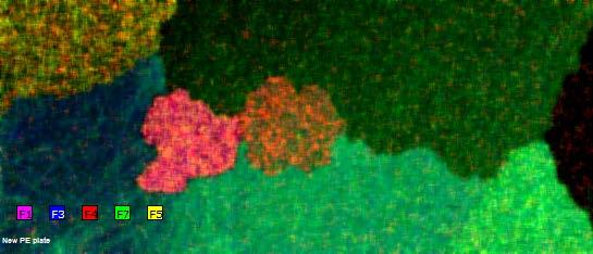
14 Welding joint cooling speed gradient 1) Measurement with filter: optimized for determining the composition of the sample as the diffraction peaks are strongly reduced. 2) Measurement without filter to detect diffraction peaks allowing to determine size and distribution of crystalline domains in the sample. Slower cooling leads to larger crystallites. Temperature gradient visible. Al100/Ti50/Cu25 50 kv 600 µa 15 µm, 3 ms Vacuum Time 14 h Without filter 50 kv 100 µa 20 µm, 3 ms Vacuum Time 8 h 7/29/

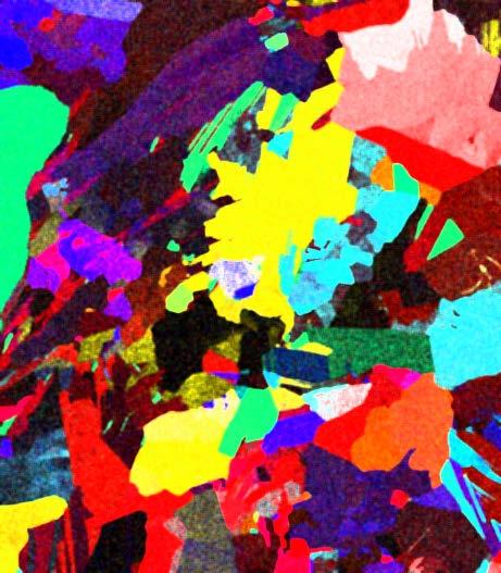
15 Aluminum blocks implications on trace element analysis and material properties Without filter 50 kv 600 µa 100 µm, 50 ms Vacuum Time 10:30 h 7/29/2016 Without filter 50 kv 600 µa 100 µm, 15 ms Vacuum Time 1 h 15

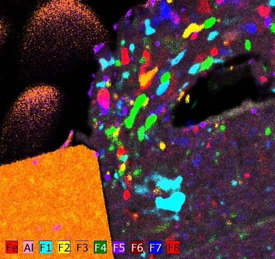
16 Crystal growth Identification of crystals domain development during controlled crystal growth process. The measurement allows to identify all mayor crystal domains in the Without filter 50 kv 600 µa 33 µm, 20 ms Vacuum Time 47 h Ganschow et al. (2016), Conditions for the growth of Fe1xO crystals using the micro-pulling-down technique, Journal of Crystal Growth, 450, /29/


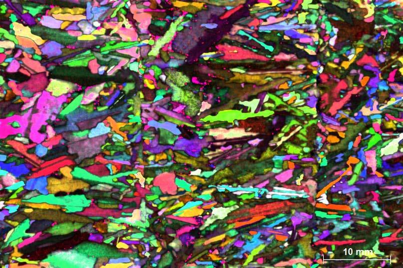
17 Multicrystalline Silicon growth process and domain size Fluorescence and diffraction information in one data set Crystal domains disjunction from metal enrichments and impurity horizons Without filter 50 kv 600 µa 50 µm, 100 ms Vacuum Time 36 h 7/29/
18 L I V E 7/29/
19 Webinar Q&A Questions, Thoughts or Comments? If you have questions or want to contact us during the Webinar, please type your questions, thoughts, or comments in the Q&A box and press Submit. We ask for your understanding if we do not have time to discuss all comments and questions within the session. Any unanswered questions or comments will be answered and discussed by e- mail or in another Webex session. 19
20 Copyright 2014 Bruker Corporation. All rights reserved. Innovation with Integrity