Mobile Computed Tomography
|
|
|
- Michael Shelton
- 5 years ago
- Views:
Transcription
1 Mobile Computed Tomography U. Ewert, B. Redmer, C. Rädel BAM Berlin 1


2 Radiography in in Nuclear Power Stations Task: Non destructive measurement of flaws in cross sections of austenitic welds X-ray tube Control and Power supply Manipulation rail 2


3 Development of of a Mechanised X-Ray Inspection System X-ray tube Camera 3
4 New Tasks for Radiography Task: --Reliable detection of of planar defects There exist exist always one one direction with with optimum contrast for for planar defects! 4

5 New Tasks for Radiography Task: --Reliable detection of of planar defects Linear scan of X-ray tube for sizing of cracks or lack of fusion X-ray tube There There exist exist always always one one direction with with optimum contrast for for planar planar defects! 5

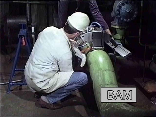
6 Mechanized Tomographic X-Ray System in in a Power Station In-field test: Setup of the scanning unit and adjustment of the digital array detector 6


7 Mechanized Tomographic X-Ray System In-field test: Control unit for measurements in a power station Environment for the measurements in a power station 7


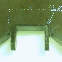
8 Metallographic transversal section, etched destructive Results Untreated metallic surface after mechanical cut non-destructive Non-destructive tomographic section Austenitic Austenitic weld weld sample sample Metallography 8.1mm Tomography Tomography 7.7mm 7.7mm 8
9 Innovations included: New flat X-ray tube New digital array detector on basis of CdTe C-MOS line Camera New tomographic reconstruction algorithms Special manipulator 9
10 Innovations included: New flat X-ray tube 10
")


11 Bipolar Extra Flat X-ray Tube MCT240 Window for fan beam geometry (±45 ) Technical Data Energy 240 kev Current/Power 2,5 ma / 600W Focal spot 0,5 x 0,5 mm Weight ca. 7 kg Manufactured by Röntgen-Technik Warrikow (rtw) 11
12 Innovations included: New digital array detector on basis of CdTe 12
: Oy Ajat Ltd Tietotie 3 02150 ESPOO Finnland www.ajat.")
13 Direct Converting Detector Array Dynamic Imaging Camera (DIC 100): Oy Ajat Ltd Tietotie ESPOO Finnland 13


14 Resolution Enhancement by by Direct X-Ray Conversion TFT-Structure on am-si CMOS ASIC 14
15 Innovations included: New tomographic reconstruction algorithms 15
16 3D-Reconstruction Limited view problem Data acquisition of radiometric images Acquisition of geometry Limited view problem Reconstruction: volume restoration Reconstruction: surface restoration Data fusion Image presentation 16


17 Results: Austenitic Pipe 3D-Planar Tomography Transversal section scan: Camera trip trough surface Pipe 156 x 20 mm, Austenit Tube voltage 190 kv 17


")
18 Planar Computed Tomography vs. High Contrast Sensitivity RT Planar Tomogram Film (D4) Flachdetektor (Hamamatsu) 18
19 European Network of Inspection and Quality (ENIQ) Method has been selected for a German pilot qualification study Tests are successfully performed at test specimen of German nuclear power stations Certificates were issued Quelle: Neundorf,
20 German ENIQ - Pilot study Technical justification and collection of documentation Requirements for instrumentation and personal Description of qualification procedure Written procedure 20
21 Value of difference of indication and measured dimension of the discontinuity Differenz der Tiefenausdehnung "Planartomografie - as function of opening of flaw (with of discontinuity) Metallografie" Value of difference of metallography and tomography depth [mm] Betrag der Differenz (mm) 2 1,8 1,6 1,4 1,2 1 0,8 0,6 0,4 0,2 0 Metallographie vs. Planartomographie Measurement uncertainty Width Fehlerbreite of flaw (µm) [µm] Line camera Digital detector array 21
22 Inspection of Composites of Aircraft Components 22
23 CT of of large components Mobile Tomosynthesis 23
24 Planar Tomography with Simple Means Rail Composite plate Detector X-ray X-ray tube tube is is shifted shifted parallel parallel to to the the object object to to inspect inspect Measurement of of few few hundred projections. Reconstruction of of 3D-image. 24

25 Mobile CT of of Fibre Composites 25


26 Crack indication Resin nest Planar imperfection Incomplete filled X-ray exposure 26
27 Conclusions: Planar tomography (also coplanar laminography) generates non destructive cross sectional images containing all planar and volumetric indications in the observation angle. The depth and length of planar discontinuities (crack, lack of fusion) with an opening width of > 25 µm can be reconstructed with an uncertainty of ± 1 mm. The spatial resolution is sufficient to detect longitudinal discontinuities with an quarter of the pixel size..welds of austenitic and ferritic materials were successfully examined in a European pilot study on the basis of the European Network for Inspection and Qualification (ENIQ). Certificates were issued. The system is furthermore suitable for the inspection of large fibre composite parts in the aircraft industry. Detector and X-ray tube at a control rail can be mounted independently from each other. The maximum penetrated thickness of steel amounts to 50 mm. 27
28 End 28