Immune system IgGs. Carla Cortinas, Eva Espigulé, Guillem Lopez-Grado, Margalida Roig, Valentina Salas. Group 2
|
|
|
- Ethelbert Francis
- 5 years ago
- Views:
Transcription
1 Immune system IgGs Carla Cortinas, Eva Espigulé, Guillem Lopez-Grado, Margalida Roig, Valentina Salas Group 2
2 Index 1. Introduction Immunoglobulins IgG formation IgG subclasses Structural classification Structural analysis Β-strands CDRs CDRs classification CDR1 L1 CDR2 L2 CDR3 L3 CDR4 H1 CDR5 H2 CDR6 H3 3. IgG antigen interaction HIV Vaccinia Conclusions Bibliography Multiple choice questions
3 Introduction
4 Introduction - Immunoglobulins IgG2 1IGT

5 Introduction - Immunoglobulins Ig are formed by 4 chains: Heavy chain Light chain Heavy chain Light chain IgG2 1IGT


6 Introduction - Immunoglobulins Ig are formed by different regions: Papain IgG2 1IGT
, Chromosome 2 (κ LC) and Chromosome 22 (λ")
 and in the DNA section")
7 Introduction - IgG formation - Ig chains are encoded in: Chromosome 14 (HChains), Chromosome 2 (κ LC) and Chromosome 22 (λ LC). - Ig variability is generated by somatic recombination. - CDRs are encoded in the V Domain (HC) and in the DNA section between V and J (LC).
8 Introduction - IgG IgG are formed by different regions: VC CH1 VL CL CH2 CH3 IgG2 1IGT
9 Introduction - IgG subclasses IgG amino acidic identity: 90% Function Response Structure IgG1 IgG2 Differences? IgG3 IgG4
10 Introduction - IgG subclasses: function IgG1 IgG2 Proteins +++ +/ * Polysaccharides /- +/- Allergens + - (-) - IgG3 IgG4


11 Introduction - IgG subclasses: response IgG1 IgG3 They can activate the complement response trough C1b In response to T-heplers through MHC-II in B-cells IgG1 the most abundant one IgG3 the 1st in viral infections
12 Introduction - IgG subclasses: response IgG2 Reacts to polysaccharides without T-helpers Most of bacterial capsular polysaccharides Allergies after a peptide exposure repetition Modulated by IL-10 Important against helminths and philaries IgG4
13 Introduction - IgG subclasses: structure Flexibility affects antigen-binding capacity and immune complex formation. The hinge has different flexibility in the different IgG subclasses. IgG3 > IgG1 > IgG4 > IgG2 Factors that determine the hinge flexibility: Length of the molecule Poly-proline helix Amino Acids disulfide bonds
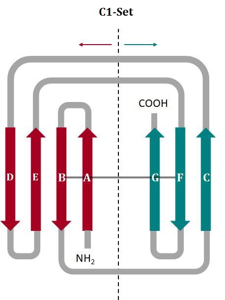
14 Introduction - Structural classification SCOP Classification CATH Classification - Class: All beta protein - Class: Mainly Beta - Fold: Immunoglobulin-like beta-sandwich - Architecture: Sandwich - Topology: Immunoglobulin like - Homologous Superfamily: Immunoglobulins - Superfamily: Immunoglobulin - Family: - V set domains C1 set domains C2 set domains I set domains

15 Introduction - Structural classification: Class IgG have a typical structure: Beta-sheet Helix Loop IgG2 1IGT
16 Introduction - Structural classification SCOP Classification CATH Classification - Class: All beta protein - Class: Mainly Beta - Fold: Immunoglobulin-like beta-sandwich - Architecture: Sandwich - Topology: Immunoglobulin like - Homologous Superfamily: Immunoglobulins - Superfamily: Immunoglobulin - Family: - V set domains C1 set domains C2 set domains I set domains


17 Introduction - Structural classification: fold and architecture Diagram of the immunoglobulin sandwich structure in the light chain: Constant domain Variable domain IgG2 1IGT


18 Introduction - Structural classification: fold and architecture Diagram of the immunoglobulin fold structure in the light chain: disulfide bond Constant domain IgG2 1IGT

19 Introduction - Structural classification: fold and architecture Diagram of the immunoglobulin fold structure in the light chain: disulfide bond Variable domain IgG2 1IGT

20 Introduction - Structural classification: fold and architecture Diagram of the immunoglobulin fold structure light domain: hydrophobic core Hydrophilic Hydrophobic IgG2 1IGT
21 Introduction - Structural classification: fold and architecture Diagram of the immunoglobulin fold structure light chain: IgG2 1IGT
22 Introduction - Structural classification SCOP Classification CATH Classification - Class: All beta protein - Class: Mainly Beta - Fold: Immunoglobulin-like beta-sandwich - Architecture: Sandwich - Topology: Immunoglobulin like - Homologous Superfamily: Immunoglobulins - Superfamily: Immunoglobulin - Family: - V set domains C1 set domains C2 set domains I set domains

23 Introduction - Structural classification: family

24 Introduction - Structural classification: family
25 IgG structural analysis


26 IgG structural analysis: β - strands - Fc Heavy chain CH2 CH3

27 IgG structural analysis: β - strands - Fab Light chain VL CL
28 IgG structural analysis: β - strands - Fab Heavy chain VC CH1

29 IgG structural analysis: CDRs - Fab light chain CDR L2 CDR L1 CDR L1 CDR L2 CDR L3 CDR L3 IgG1 3HC0

30 IgG structural analysis: CDRs - Fab heavy chain CDR H1 IgG1 3HC0 CDR H2 CDR H2 CDR H3 CDR H3 CDR H1
31 IgG structural analysis: CDR and canonical structures Canonical structures are determined by: Residues at key sites Chothia et al. classification The length of the loop Light chain Heavy chain CDR Canonical Structures CDR Canonical Structures L1 κ1-6 λ1-4 H1 3 L2 1 H2 4 H3 NO L3 κ1-6 λ1-2
32 CDR L1
33 IgG structural analysis: CDR L1 IgG 5KVL

34 IgG structural analysis: CDR L1 IgG 5KVL Canonical structure: K L1 1

35 IgG structural analysis: CDR L1 IgG 5U3K

36 IgG structural analysis: CDR L1 IgG 5U3K Hydrogen Bonds ILE 29 O.... N TYR 32 ASN 28 O..... N ARG 30 Canonical structure: K L1 2a TYR 32

37 IgG structural analysis: CDR L1 IgG 5U3K Hydrogen Bonds ASP 31 O..... N ARG 100B ASP 31 O..... N ARG 100B ASP 31 O..... O TYR 100J H3 - interaction TYR 100

38 IgG structural analysis: CDR L1 IgG 2CMR

39 IgG structural analysis: CDR L1 IgG 2CMR Hydrogen Bonds ILE 29 O..... N TRP 32 Canonical structure: K L1 2a

40 IgG structural analysis: CDR L1 IgG1 5WHK

41 IgG structural analysis: CDR L1 IgG1 5WHK Hydrogen Bonds γ-turn VAL 29 O..... N TYR 32 THR 25 N..... O ASN 33 Canonical structure: ---

42 IgG structural analysis: CDR L1 IgG2 4M1G

43 IgG structural analysis: CDR L1 IgG2 4M1G Hydrogen Bonds SER 30 O..... N TYR 33 Canonical structure: K L1 2a
44 IgG structural analysis: CDR L1 - superimposition 3HC0 RMSD: 4.23
45 CDR L2

46 IgG structural analysis: CDR L2 IgG 2CMR

47 IgG structural analysis: CDR L2 IgG 2CMR 49 53


48 IgG structural analysis: CDR L2 IgG 5KLV Length of the loop Hydrogen Bonds TYR 49 N..... O 53 TYR 49 O..... N 53 IgG1 3HC0


49 IgG structural analysis: CDR L2- superimposition 3HC0 5KVL RSMD: 0.13
50 CDR L3

51 IgG structural analysis: CDR L3 IgG 5KVL

52 IgG structural analysis: CDR L3 Canonical structure: K L3 1 Hydrogen bonds: GLN 91 O N SER 93 GLN 91 O N SER 94 IgG2 4M1G

53 IgG structural analysis: CDR L3 IgG 2CMR Canonical structure: K L3 1 Hydrogen bonds: GLN 90 O N SER 92 GLN 90 O N ASN 93 GLN 90 N...O PRO 95

54 IgG structural analysis: CDR L3 IgG 5KVL Canonical structure: K L3 1 Hydrogen bonds: GLN 90 O N ALA 92 GLN 90 N...O PRO 95 SER 93 O...N ARG 96


55 IgG structural analysis: CDR L3 Canonical structure: K L3 1 IgG2 4M1G IgG 2CMR IgG 5KVL
56 IgG structural analysis: CDR L3 - superimposition 3HC0 5KVL RMSD: 0.306
57 CDR H1

58 IgG structural analysis: CDR H1 IgG2 4M1G

59 IgG structural analysis: CDR H1 IgG2 4M1G Hydrogen bonds SER 28 O N ASP 31 SER 28 O... O THR 30 GLY 33 O... N HIS 98 Interaction with H3

60 IgG structural analysis: CDR H1 IgG1 3HC0

61 IgG structural analysis: CDR H1 IgG1 3HC0 Hydrogen bonds No internal interactions TYR 33 O. N SER 99 Interaction with H3

62 IgG structural analysis: CDR H1 Hydrogen bonds THR 28 O ASN 31 N PHE 33 O... N ARG 99 Interaction with H3 IgG3 4HDI

63 IgG structural analysis: CDR H1 - superimposition 3HC0 RMSD: 0.356
64 CDR H2

65 IgG structural analysis: CDR H2 IgG1 5WHK

66 IgG structural analysis: CDR H2 Canonical structure: H2 3A Hydrogen bonds GLY 52 O N GLY 55 SER 53 O N GLY 56 GLY 52 N... O GLY 57 IgG1 5WHK

67 IgG structural analysis: CDR H2 IgG1 3HC0

68 IgG structural analysis: CDR H2 Canonical structure: H2 2A IgG1 3HC0 Hydrogen bonds TYR 52 O N 55 ASN ASN 55 O N HIS 57

69 IgG structural analysis: CDR H2 IgG2 4M1G

70 IgG structural analysis: CDR H2 Canonical structure: H2 1 Hydrogen bonds TRP 52 N O 56 THR TRP 52 O N 55 GLY IgG2 4M1G


71 IgG structural analysis: CDR H2 - superimposition 3HC0 RMSD: 2.416
72 CDR H3

73 IgG structural analysis: CDR H3 Heavy chain
74 IgG structural analysis: CDR H3

75 IgG structural analysis: CDR H3 IgG1 5WHK

76 IgG structural analysis: CDR H3 Bulged IgG1 5WHK

77 IgG structural analysis: CDR H3 IgG1 5WHK Hydrogen bonds TRP 106 N O SER 104 Conserved

78 IgG structural analysis: CDR H3 IgG1 4S2S

79 IgG structural analysis: CDR H3 IgG1 4S2S No bulged

80 IgG structural analysis: CDR H3 Hydrogen bonds ARG 102 N O ASP 104 TYR 100 O.O ASP 104 Interaction with L3 SER 50 O O ASP 104 IgG1 4S2S

81 IgG structural analysis: CDR H3 IgG2 4M1G

82 IgG structural analysis: CDR H3 IgG2 4M1G No bulged

83 IgG structural analysis: CDR H3 Interactions with light chain: - HIS 35 N.... O TYR 102 Near L1 - TYR 50 O....O ASN 103 Near L2 IgG2 4M1G


84 IgG structural analysis: CDR H3 - superimposition 3HC0 RMSD: 1,42
85 IgG-Antigen interaction
86 HIV

87 IgG-Antigen interaction: HIV - H1 interaction IgG 5U3K

88 IgG-Antigen interaction: HIV - H1 interaction IgG 5U3K Interactions with H1: - ASN 31 O... N ASN ASN 31 N O TRP 670

89 IgG-Antigen interaction: HIV - H2 interaction IgG 5U3K


90 IgG-Antigen interaction: HIV - H2 interaction IgG 5U3K Hydrogen bonds: LYS 52C N O TRP 666 LYS 52C N O LEU 669 Salt Bridge: ARG 52A N O ASP 674 ARG 52A N O ASP 674

91 IgG-Antigen interaction: HIV - H3 interaction IgG 5U3K

92 IgG-Antigen interaction: HIV - H3 interaction IgG 5U3K Interactions with H3: - ARG 683 N...O TRP ARG 683 N...O TRP 100
93 Vaccinia virus

94 IgG-Antigen interaction: Vaccinia virus - L3 interaction IgG2 4M1G

95 IgG-Antigen interaction: Vaccinia virus - L3 interaction Interactions with L3: - GLN 173 N...ILE 95 O - GLN 173 O...ILE 95 N - VAL 175 N...SER 93 O IgG2 4M1G

96 IgG-Antigen interaction: Vaccinia virus - H2 interaction IgG2 4M1G

97 IgG-Antigen interaction: Vaccinia virus - H2 interaction IgG2 4M1G Interactions with H2: Gly 54 N O Asp 115 Thr 56 N O Asp 115 Thr 56 O...N Asp 115 Thr 56 O N Gln 117
98 Conclusions
99 Conclusions Comparing the amino acid sequences from many different immunoglobulins we can observe that beta strands are conserved while loops are variable. Five of the six CDRs usually have a small number of main chain conformations, called canonical structures. Following with CDRs, although they have a big amount of possible canonical structure classes, only a few of them appear with a high prevalence in the nature. Although CDRs are hypervariable regions, analyzing the amino acids sequence of the IgG we can predict what their structure/position in space will be.
100 Conclusions L2, L3 and H1 are the most conserved CDRs whereas L1 and H2 have more variability. Even though H3 is the most hypervariable CDR and no canonical structures have been identified in this region, it appears to have a limited repertoire of conformations. CDRs are important for antigen recognition. However, we have seen that in our case only H2 interacts with both Vaccinia and HIV.
101 Bibliography
102 Bibliography Al-Lazikani B, Lesk A, Chothia C. Standard conformations for the canonical structures of immunoglobulins. Edited by I. A. Wilson. Journal of Molecular Biology. 1997;273(4): Morea V, Tramontano A, Rustici M, Chothia C, Lesk A. Conformations of the third hypervariable region in the VH domain of immunoglobulins 1 1Edited by I. A. Wilson. Journal of Molecular Biology. 1998;275(2): North B, Lehmann A, Dunbrack R. A New Clustering of Antibody CDR Loop Conformations. Journal of Molecular Biology. 2011;406(2): Vargas-Madrazo, E., Lara-Ochoa, F. and Carlos Almagro, J. (1995). Canonical Structure Repertoire of the Antigen-binding Site of Immunoglobulins Suggests Strong Geometrical Restrictions Associated to the Mechanism of Immune Recognition. Journal of Molecular Biology, 254(3), pp Bra nde n C, Tooze J. Introduction to protein structure. 2nd ed. New York, NY: Garland Pub.; Vidarsson G, Dekkers G, Rispens T. IgG Subclasses and Allotypes: From Structure to Effector Functions. Frontiers in Immunology. 2014;5. Owen, J., Punt, J., Stranford, S. and Jones, P. (2013). Kuby immunology. 7th ed. New York: W.H. Freeman. Cuesta Á, Sainz-Pastor N, Bonet J, Oliva B, Álvarez-Vallina L. Multivalent antibodies: when design surpasses evolution. Trends Biotechnol. 2010;28(7): Schroeder H, Cavacini L. Structure and function of immunoglobulins. J Allergy Clin Immunol. 2010;125(2):S41-S52.
103 Multiple choice questions
104 Multiple choice questions 1. a) b) c) d) e) Canonical structures Are located in the CDRs Are determined by the loop length and by the presence of certain residues at key positions The two previous ones are correct Are located in both beta strands and loops All the above are correct 2. a) b) c) d) e) Which of the CDRs is the most hypervariable? L1 L3 H2 H3 H1 3. Following SCOP classification, immunoglobulins: a) Belong to all alpha proteins b) Belong to mainly beta class c) The two previous ones are correct d) Have four families: C2 set domain, C1 set domain, I set domain and V set domains e) All the above are correct
105 Multiple choice questions 4. The beta strands from immunoglobulin domains: a) Are not conserved in structure b) Are as conserved as loops in structure c) Are less conserved than loops in structure d) Are more conserved than loops in structure e) Are the less conserved in sequence 5. Which of the L3 CDR canonical structures is the most common? a) 1 b) 2 c) The two previous ones are correct d) All the L3 CDR canonical structures are presented equally e) None of the above are correct 6. CDR canonical structures can be identified by: a) The number of residues of the loop (length) b) Residues at key sites c) The two previous ones are correct d) The number of Arginine residues e) All the above are correct
106 Multiple choice questions 7. The correct statement about the immunoglobulins is a) Polar residues are located in the surface b) Polar residues are located only around the core c) The two previous ones are correct d) Hydrophobic residues are located in the surface e) None of the above are correct 8. The correct statement about the L2 is a) L2 has 4 canonical structures b) L2 has 2 canonical structures c) The two previous ones are correct d) L2 has 1 canonical structure e) None of the above are correct 9. CDRs are encoded... a) In the DNA regions between the V and J sites in the genome b) In the spacing DNA between the Variable and Constant coding regions of the light chain c) The V regions of the light chain d) Between the D and J regions coding for the heavy chain e) In the Hypervariability coding region of the Immunoglobulin light chain
107 Multiple choice questions 10. The correct statement about CDRs is a) They are important for the antigen recognition. b) H2 has more variability than L2 and can interact with different kinds of antigens. c) The two previous are correct. d) Only H1 is involved in antigen recognition. e) None of the above are correct.
108 Thank you for your attention!