STRUCTURE, DYNAMICS AND INTERACTIONS OF PROTEINS BY NMR SPECTROSCOPY
|
|
|
- Morris McGee
- 5 years ago
- Views:
Transcription


1 STRUCTURE, DYNAMICS AND INTERACTIONS OF PROTEINS BY NMR SPECTROSCOPY Constantin T. Craescu INSERM & Institut Curie - Recherche Orsay, France
2 A SHORT INTRODUCTION TO PROTEIN STRUCTURE
3 Chemical composition of bacterial and mammalian cells Component Bacteria Mammals (%) H 2 O Inorganic ions 1 1 Metabolites 3 3 Proteins DNA+RNA Phospholipids 2 5 Sugars 2 2


4 Proteins have multiple vital functions Alcohol dehydrogenase oxidizes alcohols to aldehydes or ketones Insulin controls the amount of sugar in the blood Haemoglobin carries oxygen

5 Proteins are biopolymers peptide bond aa residue Φ Ψ ω ω
6 Amino acid: the basic unit of proteins NH 3+ Amino group R C H α COO - Carboxylic acid group Different side chains, R, determine the properties of 20 amino acids. An α-amino acid
 Threonine (T) Tyrosine (Y) Cysteine (C) Asparatic acid (D) Glutamic acid (E) Lysine (K) Arginine (R) Histidine (H) White: Hydrophobic, Green: Hydrophilic, Red: Acidic, Blue:")
7 There are 20 different R Glycine (G) Alanine (A) Valine (V) Isoleucine (I) Leucine (L) Proline (P) Methionine (M) Phenylalanine (F) Tryptophan (W) Asparagine (N) Glutamine (Q) Serine (S) Threonine (T) Tyrosine (Y) Cysteine (C) Asparatic acid (D) Glutamic acid (E) Lysine (K) Arginine (R) Histidine (H) White: Hydrophobic, Green: Hydrophilic, Red: Acidic, Blue: Basic


8 Proteins are formed by the linear condensation of a number of amino R 1 NH 3 C COO ー H H 2 O R 1 acids R 2 NH 3 C COO ー H R 2 H 2 O R 3 A carboxylic acid condenses with an amino group with the release of a water NH 3 A C CO H F Peptide bond G N NH C CO S T H D Peptide bond K NH C CO G S H A The amino acid sequence is called primary structure
9 Gene is protein s s blueprint, genome is life s s blueprint DNA Protein Gene Genome Gene Gene Gene Gene Gene Gene Gene Gene Gene Gene Gene Gene Gene Gene Protein Protein Protein Protein Protein Protein Protein Protein Protein Protein Protein Protein Protein Protein

10 Each protein has a unique structure Amino acid sequence NLKTEWPELVGKSVEE AKKVILQDKPEAQIIVL PVGTIVTMEYRIDRVR LFVDKLDNIAEVPRVG Folding!
, NH(i+1), C α (i+1)")

11 The peptide unit : C α (i), C=O(i), NH(i+1), C α (i+1) is planar The global folding is defined by Φ and Ψ angles
 without Gly")
12 Ramachandran theoretical prediction Experimental values (PDB) without Gly residues
13 Levels of structural organization

14 Secondary structure: α helix Φ= -60, Ψ=-50

 variants")
15 Secondary structure: helix α ( ) variants π
16 Helical profiles hydrophobe amphiphile polaire
17 Antiparallel β sheet Φ= -140 Ψ=135
18 Parallel β sheet Φ= -120 Ψ=115
19 The loops connect the secondary structure elements The β-hairpin
20 Tertiary structure Aspartate transcarbamylase Flavodoxin Plastocyanin Antiparallel 4 strands Parallel 5 strands Antiparallel 8 strands
21 Many proteins are organized in domains Epidermal growth factor Serine protease Domaine Kringle Ca 2+ -binding domain


22 Graphical representation Sticks Ribbon Balls and sticks
23 Bibliography Carl Branden & John Tooze: Introduction to protein structure, Garland Publ., 1998 Arthur M. Lesk: Protein Architecture, IRL Press, 1991 Janet M. Thornton: Protein Science 10, 3-11 (2001) Journals: Structure Protein Structure Nature Structural Biology Proteins: Structure, Function and Genetics Journal of Molecular Biology
24 NMR OF PROTEINS - Structure - Dynamics - Interactions - Folding - Functional mechanism
25
26 Intrinsic Sensitivity of Nuclei Nucleus γ % Natural Relative Abundance Sensitivity 1 H 2.7 x C 6.7 x N -2.7 x P 1.1 x Prepare samples enriched in these nuclei
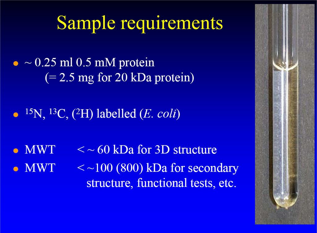
27 Biomolecules Have Many Signals 1 H NMR Spectrum of Ubiquitin ~75 residues, ~500 1 H resonances Terminology: signals are overlapped
28 Quality of the sample The ideal sample, 12.5 kda Oligomerization, 16 kda Structural flexibility, 10 kda
29 Protein structure determination by NMR Spectral assignment Collection of distance and angle restraints Calculation of the structures by restraint molecular dynamics
30 Premises for NMR structure determination - resonance frequency depends on the chemical nature of the group containing the nucleus - the chemical shift depends on the conformation - nuclear Overhauser effect contains interatom distance information the coupling constant is a function of a dihedral angle
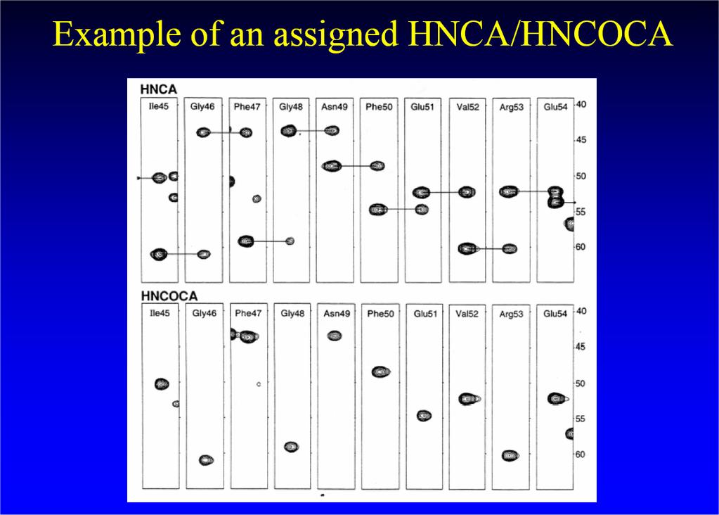
31 Resonance assignment of protein spectra

32 1 H spectrum in H 2 O at 500 MHz Spectral assignment
33 Amino acids have specific chemical shifts and COSY patterns TOCSY

34
35 Sequential assignment strategy using proton NMR
36 Resonance assignment using proton NMR COSY spectrum NOESY spectrum superposed in the fingerprint region
37 Multi-Dimensional NMR H α H β H N If 2D cross peaks overlap go to 3D or 4D..

38 General 2D experiment with magnetization transfer

39 A 2D heteronuclear experiment
40
41 Advantages of Heteronuclear nd NMR Uses a second nucleus to resolve overlap of the first: chemical shift of each nucleus is characteristic/sensitive to different factors More information to identify resonances Less sensitive to MW because this strategy uses large 1 and 2-bond scalar couplings
42 Multi-Dimensional Heteronuclear 15 N- 1 H HSQC NMR F1 Chemical Shift ( 15 N) F2 Chemical Shift ( 1 H)

43 Dispersion of 2D peaks in a 3D spectrum 2D NOESY


44 Proteins larger than ~15 kda Problems: peak superposition, linewidth Solution: double labeling: 13 C 15 N, run tri-nuclear experiments
45
46 Collection of distance and angle restraints. distannces (NOEs). chemical shifts. angles
47 noe NOESY gives distance information


48 The chemical shifts depends on the structure
49
50 Coupling constants / dihedral angle HN Φ ω Hα Karplus equation

51 The calculated structure depends on the number and quality of the exp. restraints


52 The structure calculation: finding the minimum of the potential function V Physical potential Pseudopotential (exp. restraints)
53
54 The Ca 2+ loaded regulatory domain C-CaVP CaVP (W81-S161) rmsd (moyenne) = 0.7 Å ; (Φ, Ψ) très favorable = 83 % Théret et al. Biochemistry 39, (2000)

55 Flowchart of the NMR structure determination procedure
56 Overall comparison of the two methods