Imaging Researcher s Workshop Annotation and Imaging Markup. Introduction
|
|
|
- Arline Wiggins
- 5 years ago
- Views:
Transcription
 Chief Imaging VA Maryland")
1 Imaging Researcher s Workshop Annotation and Imaging Markup Eliot Siegel, M.D. Professor and Vice Chair University of Maryland Department of Diagnostic Radiology and Nuclear Medicine Director MIRTL (Maryland Imaging Research and Technologies Lab) Chief Imaging VA Maryland Healthcare System Introduction One of the major original goals of cabigwas to determine out how to create a system that would enable extraction of data for research or clinical decision support that would: Allow access to a variety of types and sources of data including genomic, proteomic, clinical, lab, demographic, and diagnostic imaging Take advantage of analytic potential of grid computing to combine and cross-reference these for analysis for research and clinical care 1
2 Introduction to the cabig in Vivo Imaging Workspace cabig in vivo Imaging workspace established April 2005 a little more than a year after the establishment of the other cabig workspaces NCI funded effort by far the biggest and most productive effort in imaging informatics today Subject matter experts from around country with representation from major Universities, informatics experts, industry, NCI Review of Relevant Workspace Projects XIP, AIM, Middleware, NBIA 2
 XIP Host Adapter DICOM, HL7, and othercagrid Services via services per IHE ProfilesImaging Middleware WG 23 WG 23 WG 23 Host-Specific Plug-in")
 Grid computing has received surprisingly little attention.")
3 XIP Application Builder Medical Imaging Workstation XIP Class Library Auto Conversion Tool XIP Application XIP Modules Host Independent ITK VTK XIP LIB XIP Host... (Can be replaced with any DICOM WG23- compatible Host) XIP Host Adapter DICOM, HL7, and othercagrid Services via services per IHE ProfilesImaging Middleware WG 23 WG 23 WG 23 Host-Specific Plug-in Libraries Rapid application development environment for diagnostic imaging tasks that researchers and others use to create targeted workflows customized for specific projects WG 23 Distribute Web-based Application Standalone Application Imaging Middleware (including GridCAD and Virtual PACS) Grid computing has received surprisingly little attention. One application has been to allow multiple computers to work in parallel on a single task such as CAD detection of lung nodules or to give multiple opinions using multiple algorithms Middleware software is used to create interoperability between DICOM devices and the cagrid which uses a service oriented architecture 3

4 NBIA: National Cancer Imaging Archive Initially designed as repository for LIDC and RIDER CT lung nodule studies Expanded to include multiple additional types of image collections with role based security to share with public or a selected group or to support ongoing clinical trials or other reader studies Open source and free Meant to be federated to create virtual database across multiple instances of NCIA software E1 NBIA Demo: Home Page 4
5 Slide 8 E1 We should try to avoid confusion in this slide between our own NCIA which is "now offering we-based image visualization and mark-up" and the NCIA suite of software which does not Eliot, 3/2/2008


6 Slides Courtesy Adam Flanders Daniel Rubin 5


7 6


8 7

9 Create An AIM Annotation 8
.")
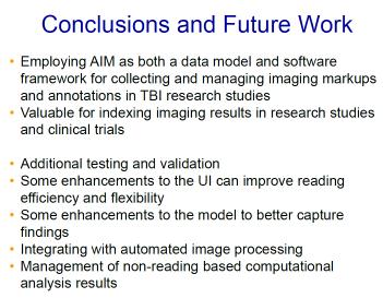
10 The user selected the Thoracic aortic aneurysm measurement protocol (arrowhead), below which a list of required measurements isdisplayed (short straight arrow). As the user manipulates the 3D volume and performs measurements according to the protocol, annotations with text labels are automatically applied to the image (long straight arrow). Additional descriptive RadLex terms can be applied to each annotation using the Observation text box (curved arrow). When all measurements have been completed, annotation data are exported in AIM-XML format by selecting the appropriate entry (*) from the dropdown menu under Generate Report. The data are then automatically sent to the reporting software and may also be sent to a research database. Exporting quantitative imaging data in AIM-XML format AIM report-generating function An AIM-XML file is created that includes quantitative and descriptive data in a standardized format. 9


11 Screen shot of reporting software (M*Modal) shows how quantitative data (blue) are automatically imported into the reporting template from the AIM-XML file After the quantitative data are imported into the template, the radiologist dictates the qualitative findings and finalizes the report He or she is able to complete the report without having to type or dictate the quantitative findings, thereby reducing the chances for error and streamlining work flow. Lung nodule follow-up work flow Lung nodules that are part of a comparison study are automatically segmented, with maximal diameters and volumes calculated with a single button click These data are exported to an AIM-XML file that is then sent to the reporting software The reporting software incorporates the data into a reporting template and performs simple calculations, such as doubling time and percent change in lesion size (blue), and can automatically report information using a variety of standard criteria (WHO [World Health Organization] or RECIST [Response Evaluation Criteria in Solid Tumors] criteria). 10

12 11


13 12


14 13


15 14


16 15


17 16


18 17


19 18

20 19
21 Ongoing Work Translation of proprietary LIDC and RIDER annotations to AIM format in NBIA Creation of high level AIM model for images Applied to: Radiology Nuclear Medicine Pathology Cell biology Baggage screening Satellite reconnaissance imagery Etc. Creation of API For those using software such as Slicer, NIH Image, Clinical trial purpose build applications, commercial software Initially for RECIST measurements Will be generalized 20