This is the author's accepted version of the manuscript.
|
|
|
- Jessie Ray
- 5 years ago
- Views:
Transcription
1 This is the author's accepted version of the manuscript. The definitive version is published in Nature Communications Online Edition: 2015/4/16 (Japan time), doi: /ncomms7780. The final version published is available online at

2 Nedd4-induced mono-ubiquitination of IRS-2 enhances IGF-I signaling and mitogenic activity (by Fukushima et al.) Supplementary Figures and Legends Supplementary Fig. 1 Supplementary Fig. 1 Identification of Nedd4 as an IRS-2-associated protein in camp-treated FRTL-5 cells. (a) FRTL-5 cells were treated with 1 mm dibutyryl camp for 24 h, and the lysates were subjected to immunoprecipitation with anti-irs-2 antibody. As a negative control, the antigen peptide was mixed with anti-irs-2 antibody prior to immunoprecipitation. Immunoprecipitates were subjected to SDS-PAGE and silver stain. Representative data is shown, and arrowheads indicate bands of IRS-2-associated proteins. (b) p120 was subjected to "in gel" digestion with trypsin, and the peptides were analyzed with MALDI-TOF MS. Peptides attributed to E3 ubiquitin ligase Nedd4 are shown (#matched peaks/#subjected peaks, 8/24; sequence cover 12%). (c) Lysates of camp-treated FRTL-5 cells were subjected to immunoprecipitation and immunoblotting using the indicated antibodies. Non-specific IgG was used as a negative control. (d) HEK293 cells overexpressing indicated proteins were serum starved. Cell lysates were subjected to immunoprecipitation and immunoblotting. -1-


3 Supplementary Fig. 2 Supplementary Fig. 2 The association of Nedd4 with IRS-2 (related to Fig. 1) (a) The association of the Nedd4 N-terminal region and the IRS-2 PH/PTB domain in cell-free binding assay. HEK293T cells overexpressing IRS-2 deletion mutants were serum-starved, and the lysates were subjected to pull-down analysis using GST-Nedd (b) Putative intramolecular binding sites in the Nedd4 C2 domain and HECT domain. Blue, basic residues; red, acidic residues. (c) Determination of the intramolecular binding sites in Nedd4 C2 domain and HECT domain. HEK293T cells overexpressing indicated proteins were serum-starved. Cell lysates were subjected to immunoprecipitation and immunoblotting. Nedd (C854S), a mutant of the ubiquitin ligase active site, was used because intact Nedd was unstable. -2-


4 Supplementary Fig. 3-3-
5 Supplementary Fig. 3 Ubiquitination of IRS-2 by Nedd4 (related to Fig. 2) (a) Effects of Nedd4 overexpression on IGF-IR, IRS-1 and IRS-2 ubiquitination. HEK293T expressing indicated proteins were serum-starved, followed by IGF-I stimulation (100 ng/ml, 1 min). Cell lysates were denatured, followed by immunoprecipitation and immunoblotting using the indicated antibody. (b) IRS-2 peptides with ubiquitin remnant detected by LC-MS/MS analysis. IRS-2 immunoprecipitates derived from HEK293T cells expressing Nedd4 and IRS-2 were treated with indicated proteases, and the resulted peptides with ubiquitin remnant motif (K-ε-GG) were enriched using anti-ubiquitin remnant motif antibody-conjugated beads. The sample was subjected to LC-MS/MS analysis. Peptides attributed to IRS-2 are shown. Lower case letters k in peptide sequences indicate ubiquitinated Lys residues. (c) Representative MS/MS spectrum of the peptide DFLSHHLK 1331 [GG]. (d, e) Quantitative MS analysis results. IRS-2 immunoprecipitates derived from HEK293T cells expressing IRS-2 (-Nedd4) or IRS-2 and Nedd4 (+Nedd4) were treated with trypsin and Asp-N. Peptides with ubiquitin remnant motif were enriched, and subjected to LC-MS/MS analysis. Chromatograms of several product ions (different colors) derived from DFLSHHLK 1331 [GG] are shown in (d). Fragment ions used for PRM are shown in (e). The area under the curves (AUCs) were summed and shown in Fig. 2i. -4-
6 Supplementary Fig. 4-5-
7 Supplementary Fig. 4 Regulation of IGF-I signaling by Nedd4 (related to Fig. 3) (a, b) Effects of Nedd4 overexpression on IGF-I-dependent IGF-IR and IRS-1 tyrosine phosphorylation. HEK293 cells overexpressing Nedd4 or Nedd4 C854S together with IGF-IR-FLAG (a) or FLAG-IRS-1 (b) were serum-starved, followed by IGF-I stimulation (100 ng/ml, 1 min). Lysates were subjected to immunoprecipitation and immunoblotting as indicated. In (b), densitometric analyses were performed, and tyrosine phosphorylation levels of IGF-IRβ and IRS-1 were normalized to their protein levels in immunoprecipitates. p85 PI3K bound to IRS-1 was normalized to IRS-1 levels in immunoprecipitates. The graph shows means ± SD of three independent experiments., significant difference from control (P<0.05, one-way ANOVA followed by Tukey-Kramer test). (c) Effects of Nedd4 overexpression on cell surface IGF-IR levels. HEK293 cells overexpressing Nedd4 or Nedd4 C854S together with IGF-IR-FLAG were serum-starved, followed by IGF-I stimulation (100 ng/ml, 1 min). Cell surface proteins were biotinylated, followed by immunoprecipitation and blotting with indicated antibodies or streptavidin-hrp. Densitometric analyses were performed, and biotinylated IGF-IRβ were normalized to IGF-IRβ levels in immunoprecipitates. The graph shows means ± SD of four independent experiments. (d) Effects of Nedd4 overexpression on Grb10 levels. HEK293 cells overexpressing IRS-2 alone, or IRS-2 and Nedd4 were serum-starved. Grb10 levels were measured by immunoblotting. The graph shows show means ± SD of three independent experiments. (e) Effects of the deletion of Nedd4 N-terminal region on IRS-2 tyrosine phosphorylation. HEK293 cells overexpressing IRS-2 and myc-nedd4 (wild type or aa ) were serum-starved, followed by IGF-I stimulation (100 ng/ml, 1 min). Cell lysates were subjected to immunoprecipitation and immunoblotting using the indicated antibodies. -6-
 (a) Effects of the IRS-2-ubiquitin chimeric protein and its mutant, (b) Nedd4 overexpression, and (c) the deletion")

8 Supplementary Fig. 5 Supplementary Fig. 5 Association of IRS-2 with ubiquitin binding proteins (related to Fig. 4) (a) Effects of the IRS-2-ubiquitin chimeric protein and its mutant, (b) Nedd4 overexpression, and (c) the deletion of Epsin1 UIM motifs on the association of IRS-2 with the indicated ubiquitin binding proteins. HEK293T cells overexpressing indicated proteins were serum-starved. Lysates were subjected to immunoprecipitation and immunoblotting as indicated. -7-
9 Supplementary Fig. 6 Supplementary Fig. 6 Regulation of IGF-I signaling by Nedd4 in thyrocytes (related to Fig. 5) (a, b) The association of Nedd4 with IRS-2 or IGF-IR. FRTL-5 cells were treated with 1 mm dibutyryl camp for 24 h (a, b), and then treated with 100 ng/ml IGF-I for 1 min (a). Cell lysates were subjected to immunoprecipitation and immunoblotting as indicated. In (b), the antigen peptide was mixed with anti-irs-2 antibody prior to immunoprecipitation as a negative control. (c) Effects of Nedd4 knockdown on Grb10 levels. Cells transfected with Nedd4 sirnas were treated with dibutyryl camp. Grb10 levels were measured by immunoblotting. (d) Effects of Nedd4-2 knockdown on IGF-I-induced IRS-2 tyrosine phosphorylation. Cells transfected with Nedd4-2 sirnas were treated with dibutyryl camp followed by IGF-I stimulation as described above. Cell lysates were subjected to immunoprecipitation and immunoblotting. -8-
10 Supplementary Fig. 7 Supplementary Fig. 7 Lack of Nedd4-2 expression in PC-3 cells (related to Fig. 6) FRTL-5 cells and PC-3 cells were transfected with Nedd4-2 sirnas or control RNAs. Cell lysates were subjected to immunoblotting using the indicated antibodies. -9-
 (a) Sequence information of the 5 -UTR Irs2 -Venus capped RNA used for the validation of zebrafish Irs2 MO.")
 Efficient knockdown of Irs-2 by Irs2 MO.")
11 Supplementary Fig. 8 Supplementary Fig. 8 Confirmation of Irs2 knockdown and Nedd4 expression in zebrafish embryo (related to Fig. 7) (a) Sequence information of the 5 -UTR Irs2 -Venus capped RNA used for the validation of zebrafish Irs2 MO. The RNA contains a sequence of zebrafish Irs2 mrna 5 -UTR (black letter) and translation start site (25 base, red letter), fused with Venus fluorescent protein-cording sequence (green letter). Irs2 MO target sequence is shown as blue shaded. (b) Efficient knockdown of Irs-2 by Irs2 MO. 5 -UTR Irs2 -Venus mrna or Venus mrna was co-injected with either Irs2 MO or control MO into 1-2 cell stage embryos. Bright field images and Venus fluorescence at 22 hpf are shown. bars, 1.0 mm. (c) Successful Nedd4 expression. Nedd4-Venus mrna or Venus mrna was co-injected with either Irs2 MO or control MO into 1-2 cell stage embryos. Cell lysate was prepared from 12 hpf embryos, and subjected to immunoblotting. -10-


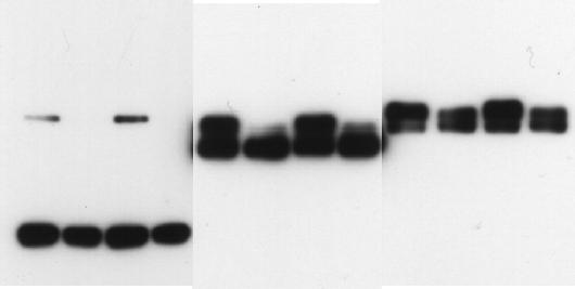
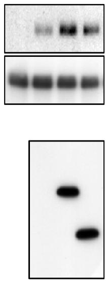
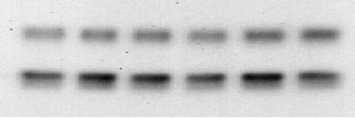
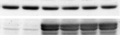
12 Supplementary Fig. 9 Supplementary Fig. 9 Uncropped scans of blots shown in Fig. 1b, 1c, 1d, 2b, 2c, 2d, 2e and 2g. Rectangles delimit cropped areas used in the indicated panels. -11-


13 Supplementary Fig. 10 Supplementary Fig. 10 Uncropped scans of blots shown in Fig. 3a, 3c, 3d and 3e. Rectangles delimit cropped areas used in the indicated panels. -12-

14 Supplementary Fig. 11 Supplementary Fig. 11 Uncropped scans of blots shown in Fig. 4a, 4b, 4c and 4d. Rectangles delimit cropped areas used in the indicated panels. -13-

15 Supplementary Fig. 12 Supplementary Fig. 12 Uncropped scans of blots shown in Fig. 5a, 5b, 5c, 5d and 5e. Rectangles delimit cropped areas used in the indicated panels. -14-

16 Supplementary Fig. 13 Supplementary Fig. 13 Uncropped scans of blots shown in Fig. 6a, 6b, 6c and 6d. Rectangles delimit cropped areas used in the indicated panels. -15-