Lab. 7: Serological Tests ELISA. 320 MIC Microbial Diagnosis 320 MBIO PRACTICAL. Amal Alghamdi 2018
|
|
|
- Corey Powers
- 5 years ago
- Views:
Transcription


1 Lab. 7: 320 MIC Microbial Diagnosis Serological Tests ELISA. 320 MBIO PRACTICAL Amal Alghamdi

2 Infection and Immunity Serology is the study of immune bodies in human blood. These are products of the defense mechanisms against disease-causing organisms in the body. The antigen actually comes first, in that the antigen is the substance which "induce" the body to produce antibodies. As we all know, the antibody is the substance which fights the invading organism. Antibodies take many forms because there are many forms of antigens which can invade the body. 2
3 What is the principle of Serological test? It involved with antibody-antigen response. Serology can involve a number of laboratory techniques, to diagnose various disease conditions. Definition: Serology is a blood test to detect the presence of antibodies against a microorganism. Certain microorganisms stimulate the body to produce antibodies during an active infection. 3
 e.g. Enzyme linked immune sorbent assay (ELISA).")
.")
4 Classification of antigen-antibody interactions: A- Primary serological tests: (Marker techniques) e.g. Enzyme linked immune sorbent assay (ELISA). Immune florescent antibody technique (IFAT). Radio immune assay (RIA). B- Secondary serological tests: e.g. Agglutination tests. Complement fixation tests (CFT). Precipitation tests. Serum neutralization tests (SNT). Toxin-antitoxin test. 5
5 Antibodies Antibodies are immune system-related proteins called immunoglobulin's, abbreviated (Ig) produced by white blood cell called a B cell as a primary immune defense against foreign agents (antigen). Each antibody has a region that binds specifically to a particular antigen which it neutralizes. 6

6 Antibodies Antibodies are grouped based on their mode of action, Some of which are as follows: 1. Agglutinins. Antibodies are divided into five major classes; IgM, IgG, IgA, IgD, and IgE 2. Bacteriolyins. 3. Haemolysins. 4. Precipitins. 6

7 The Five Classes of Antibodies Abb. Presence Function Structure IgG Gamma glubulin Monomer Blood IgM Pentamer in structure 10% in blood IgA Dimer 15% in blood concentrates in body fluids; Found in mucous, RT Also found in saliva, tears, and breast milk. Responsible for the secondary immuno response The only antibody capable of crossing the placenta to give passive immunity to the fetus. Responsible for the Primary immune response Eliminates pathogens in the early stages of B cellmediated immunity before there is sufficient IgG. Guard the entrances of the body. IgA - IgM is the largest antibody; it tends to remain in the blood, where it can lead to efficient killing of bacteria. IgD Monomer 1% in blood Surface ofb-cells Functions mainly as an antigen receptor on B cells that have not been exposed to antigens. It has been shown to activate basophils and mast cells to produce antimicrobial factors. IgE Monomer 0.1% in blood Skin tissue Binds to allergens and triggers histamine release from mast cells and basophils, and is involved in allergy. Also protects against parasitic worms. 9

8 Antibody Production : The Antibody: An immunoglobulin, a specialized immune protein, produced because of the presence of an antigen into the body, and which possesses the remarkable ability to combine with the very speecific antigen. The antibody recognizes and bind to the antigenic determinant region of the antigen. 8
9 Antibody Production : Specific antibodies are produced by injecting an antigen into a mammal, such as a mice. Blood isolated from these animals contains polyclonal antibodies multiple antibodies that bind to the same antigen in the serum, which can now be called antiserum. Antiserum is : A blood serum containing polyclonal antibodies. Antiserum is used to give passive immunity to many diseases. For example, Passive antibody transfusion from a previous human survivor is the only known effective treatment for Ebola virus infection. 14

10 Antigens A substance that when introduced into the body stimulates the production of an antibody. Immunoassay A laboratory technique that makes use of the binding between an antigen and its homologous antibody in order to identify and quantify the specific antigen or antibody in a sample. Analyte The sample being analyzed and in immunoassays the analyte is either Antibody orantigen. 10


11 Serological Test - ELISA Enzyme Linked Elisa Assay Immune Sorbent
12 What is ELISA? ELISA is a biochemical technique used mainly in immunology to detect the presence of an antibody or an antigen in a sample. Its is a diagnostic tool in medicine and plant pathology. In simple terms, in ELISA an unknown amount of antigen is place, to a surface, and then a specific antibody is washed over the surface so that it can bind the antigen. This antibody is linked to an enzyme, and in the final step a substance is added that the enzyme can convert to some detectable signal. A type of Serological Test; it is uses Antibodies and color change to detectdisease. These types of testes are called: Immunoassays. 12
.")
13 Immunoassays are based on detectable interactions between antigen and antibodies such as : 1. Precipitation 2. Agglutination 3. Complement fixation ELISA take a advantage of the strong and specific attachment that occurs between an antibody and antigen (imumunosorbent). It is used to detect the presence of an antibody or an antigen in asample. An ELISA test uses components of the immune system and chemicals to detect immune responses in the body (for example, to infectious microbes). 13

14 Principle Antigens from sample are attached to the surface then Specific antibody is applied over the surface so: 1. Can bind to each other. 2. This antibody is linked to an enzyme. 3. Color changed in the substrate. 15


15 Purpose The purpose of an ELISA is: To determine if a particular protein is present in a sample and if so, how much; (Quantitative + Qualitative) 16


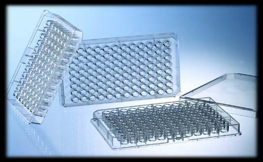
16 There two main variations on this method you can determine how much antibody is in a sample. you can determine how much protein is bound by an antibody. It uses a 96-well plate to measure a protein or substance based on an antigen/antibody reaction. 17
17 Steps Involved in an ELISA Bind the protein or antigen to the plate. Then you block the plate to get rid of any non specific binding sites. Incubate with the primary antibody which is specific for the antigen. Secondary antibody that is linked with an Enzyme is allowed to bind with the primary antibody. Use a Substrate for the enzyme which will cause color to be released. 24

 Indirect")
18 Types of ELISA: Competitive ELISA The technique is divided into three types Sandwich ELISA (Direct ELISA) Indirect ELISA 19
19 1- Competitive ELISA The labeled antigen competes for primary antibody binding sites with the sample antigen (unlabeled). The more antigen in the sample, the less labeled antigen is retained in the well and the weaker the signal. 20

20 1- Competitive ELISA 21

21 2- Sandwich ELISA 1. The plate is coated with a capture antibody. 2. sample is added, and any antigen present binds to capture antibody; 3. Detecting antibody is added, and binds to antigen; 4. Enzyme-linked secondary antibody is added, and binds to detecting antibody; 5. Substrate is added, and is converted by enzyme to detectable form. 22
 detecting antibody is added,")
 substrate is added, and is converted by enzyme to detectable")
22 2- Sandwich ELISA (1) Plate is coated with a capture antibody; (2) sample is added, and any antigen present binds to capture antibody; (3) detecting antibody is added, and binds to antigen; (4) enzyme-linked secondary antibody is added, and binds to detecting antibody; (5) substrate is added, and is converted by enzyme to detectable form. 30
23 3- Indirect ELISA 1. The protein antigen to be tested for is added to each well of ELISA plate, where it is given time to adhere to the plastic through charge interactions. 2. A solution of non-reacting protein is added to block any plastic surface in the well. 3. Then the serum is added, which contains a mixture of the serum antibodies, of unknown concentration, some of which may bind specifically to the test antigen that is coating the well. 4. Afterwards, a secondary antibody is added, which will bind to the antibody bound to the test antigen in the well. This secondary antibody often has an enzyme attached to it a substrate for this enzyme is then added. 5. This substrate changes color upon reaction with the enzyme. 32


24 3- Indirect ELISA 6. The color change shows that secondary antibody has bound to primary antibody, which strongly implies that the donor has had an immune reaction to the test antigen. 7. The higher the concentration of the primary antibody that was present in the serum, the stronger the color change. 8. Spectrometer is used to give quantitative values for color strength. 33
25 3- Indirect ELISA Indirect ELISA is a two-step ELISA which involves two binding process of primary antibody and labeled secondary antibody. The primary antibody is incubated with the antigen followed by the incubation with the secondary antibody. However, this may lead to nonspecific signals because of cross-reaction that the secondary antibody may bring about. 1. Micro-well plates are incubated with antigens, washed up and blocked with BSA. 2. Samples with antibodies are added and washed. 3. Enzyme linked secondary antibody are added and washed. 4. A substrate is added, and enzymes on the antibody elicit a chromogenic or fluorescent 34 signal.
26 Material & Method Before starting the work read kit instruction carefully. The 96 well plate is labeled carefully and the first wells are used to draw the standard curve. The sample is added to plate in duplicate or triplicate and then the mean result is calculated. The quality control sample which is provided with the kit is treated as the test samples. After reading the results the standard curve is drawn were the concentration is blotted on the X-axis and the absorbance on the Y-axis. 35
. Coating buffer. Washing buffer.")

27 For example: Salmonella typhimurium Kit content Culture Salmonella typhimurium ( heated for 30 min at 56 C in a water bath). Coating buffer. Washing buffer. Blocking buffer. Patients serum. Alkaline phosphate -labeled -anti bodies. BCIP/NBT substrate. Flat-bottom microliter plate. Micropipette tips Latex gloves. Facemask 37

28 Procedure Absorption - nm Concentration ng/ml 36 29


29 1- Add 100 ul coating buffer to each well of one row ( wells 1-12) of the micrometer plate. 2- Add 100 ul of Salmonella typhimurium to each well. 3- Seal the wells with a strip of plastic tape, and refrigerate the plate at 5 C for1-7 days. 4- Remove your plate from the refrigerator and carefully remove the tape. 5- Shakethe inverted plate with a quick shake to remove the liquid into disinfectant. 30


 9- incubate the plate at 35 C for")

30 6- Fill the wells with washing buffer and shake to remove, Wash two more times. 7- Add 100 ul blocking buffer, Leave for min. 8- Preform dilution of the patient serum by placing 100 ul in the first well, Mix up and dawn three times. (Continue the dilution until u have reached the 11 th well) 9- incubate the plate at 35 C for 60 min. 10- Shake the inverted plate with a quick shake to remove the contents. Wash three times with washing buffer as described in step 5. 40
.")


31 11- Add 100 ul of alkaline phosphatelabeled anti- antibody to each well (1-12). 12- Seal the wells with tape and incubate the plate at 35 C for 45 min. 13- Plates can be sealed and stored at 5 C until next lab period. 14- Remove the tape carefully shake out the contents, and wash the wells three times with washing buffer. 15- Add 100 ul of the alkaline phosphate substrate (BCIP/NBT) to each well in the row. 41


32 16- Leave at room temp. for min until color develops; well 12 will be colorless. 17- Record the result. 33


33 Results: The highest dil. with a blue color is the endpoint. The titer is the reciprocal of the dil. of the endpoint. 34


34 Any Questions 45