Diagnosis and detection of different genotypes of Paenibacillus larvae, the causal agent of American Foulbrood disease
|
|
|
- Gabriel Booth
- 5 years ago
- Views:
Transcription


1 OIE SYMPOSIUM ON EMERGING INFECTIOUS AGENTS IN HONEY BEES AND OIE-LISTED DISEASES 45th APIMONDIA International Apicultural Congress Istanbul, Turkey, 2017 Diagnosis and detection of different genotypes of Paenibacillus larvae, the causal agent of American Foulbrood disease Laboratorio de Referencia de loque Americana Unidad de Bacteriología CIDEFI Facultad de Ciencias Agrarias y Forestales Universidad Nacional de La Plata, Argentina Dr. Adriana M. Alippi- Research Scientist CIC
2 American Foulbrood -AFB- AFB is the most contagious and destructive infectious disease affecting the larval and pupal stages of honeybees. Classified within the OIE list and considered to be of socioeconomic impact and significant in the international trade of bees and bee products. Causal agent: Paenibacillus larvae- spore-forming Gram (+) Due to its highly contagious nature is one of the few bee diseases capable of killing a colony There is no seasonal outbreak of AFB it occurs at any time of the year when brood is present. Bacterial spores survive for decades remaining viable and resist adverse conditions (desiccation, high temperatures, UV ) AFB occurs throughout the world and impact not only in apicultural economy but also in pollination rates.

3 Healthy brood

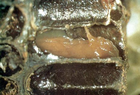

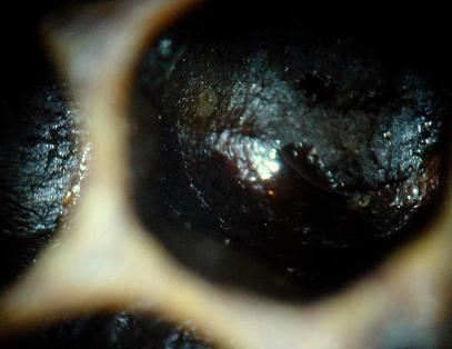

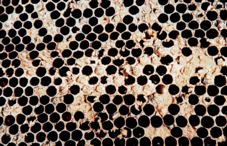
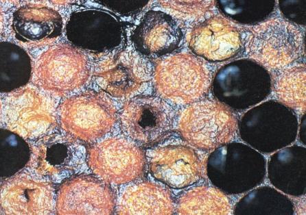
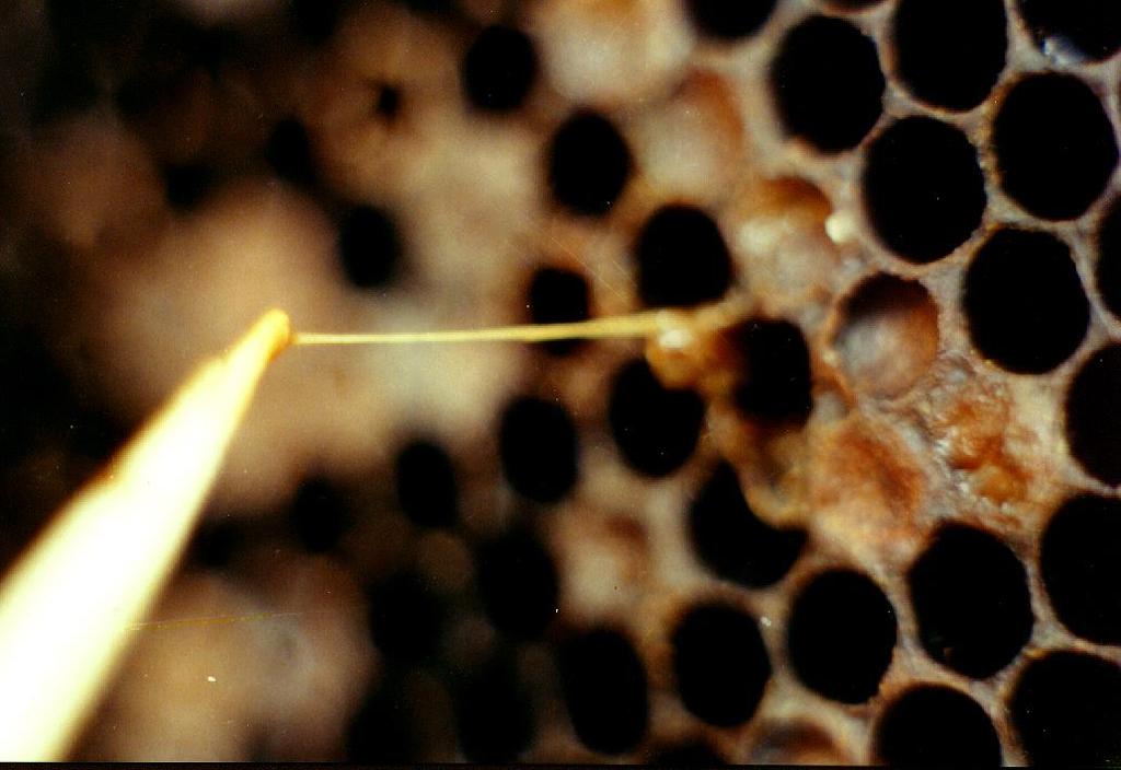
4 Clinical Signs
 Gram stain (larval")


5 DIAGNOSIS MICROSCOPY Simple stain (larval remains) Gram stain (larval remains and pure culture) ISOLATION Semi-selective media+antibiotics (Nalidixic acid and Pipemidic acid) ) IMMUNOLOGICAL TESTS AFB Diagnostic Test kit DETECTION BY PCR Conventional PCR: Specific primers PL1/PL2 Real Time PCR 970 pb
6 Different techniques to identify Paenibacillus larvae Technique Principle Samples Advantages Disadvantages Cultivation Germination and growth of spores in solid medium Brood, honey, bees, debris, pollen, etc. Facilitates tracing infect ion sources Suitable for AFB detection programs Allows testing spore viability Permits quantification Additional identification step Semi-selective media usually required to avoid contamination Microscopy Morphological identification of spores Brood Rapid diagnosis Low cost Smears in the field Only for confirmation of clinical diseased larvae Biochemical profiling Identification based on microbiological laboratory tests Bacterial colonies Traditional approach for most laboratories Commercial kits First step of bacterial isolation Time consuming PCR Amplification of specific bacterial DNA Bacterial colonies, brood, honey, bees, Fast Permits rapid confirmation without cultivation Needs special equipment Can identify unviable spores Immunotechniques Different tools based on specific antigen-antibody interactions Brood Rapid AFB diagnosis Commercialized device confirm clinical AFB in the field Utility of each test is highly dependent of the specificity of antibody used
 Without Ø + cicloheximide (ERIC")
7 Isolation and cultivation from diseased larvae º C (ERIC I) Without Ø + cicloheximide (ERIC II)

 Without Ø +")
8 Isolation of viable spores of P. larvae from honey samples 1:1 45 ºC 37 ºC º C (ERIC I) Without Ø + cycloheximide (ERIC II) Without Ø + Cicloheximide (ERIC II)


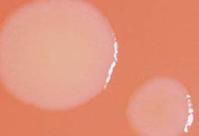
9 Paenibacillus larvae growing on semi-selective medium MYPGP + Nalidixic acid + Pipemidic acid
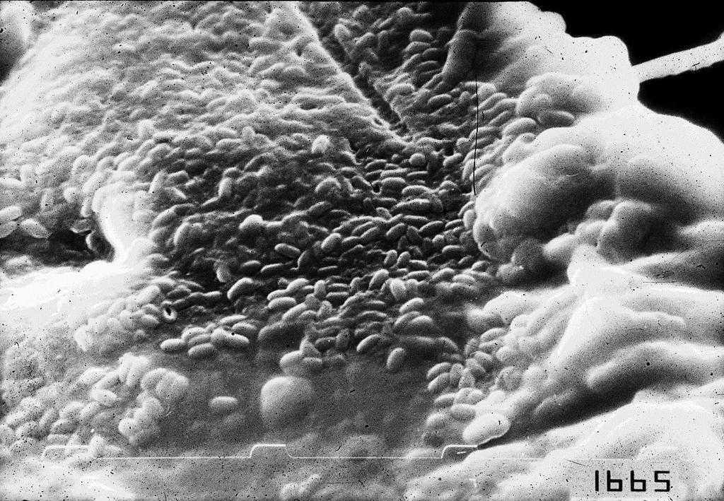
10 Gram stain

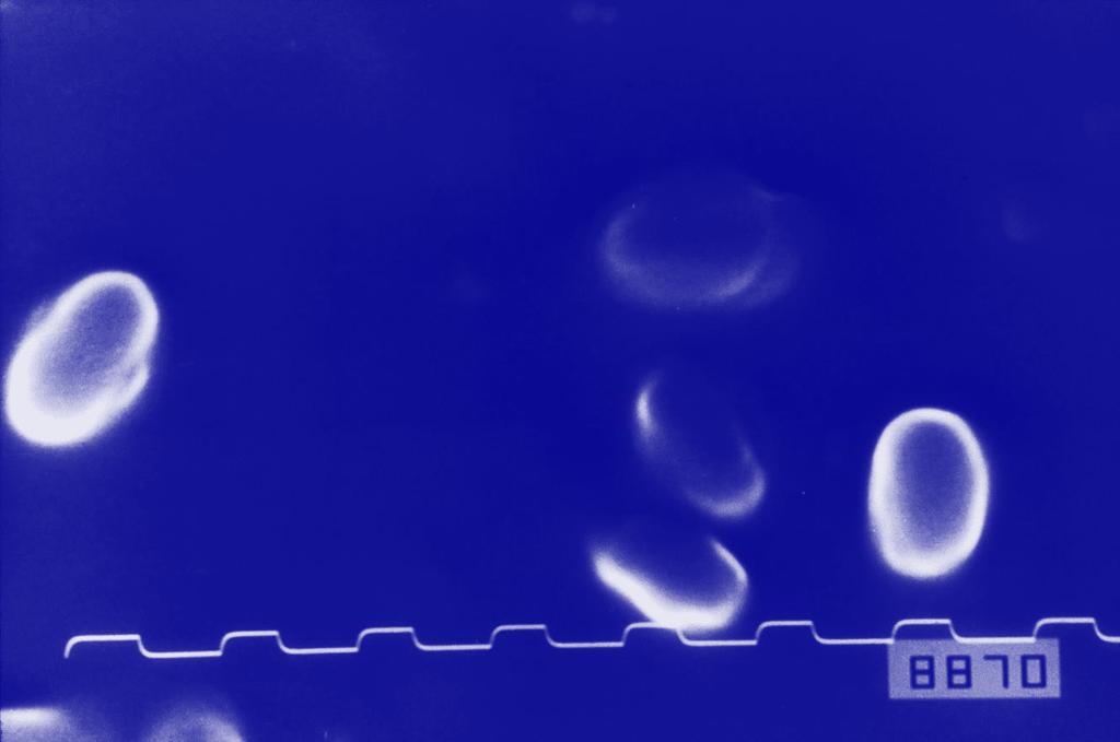
11 Schaeffer-Fulton stain for spores


12 Nitrate reductase Casein hydrolysis Positive Negative Catalase (-) (+) (-) (+) (-) (+) Oxidase

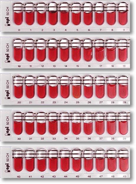
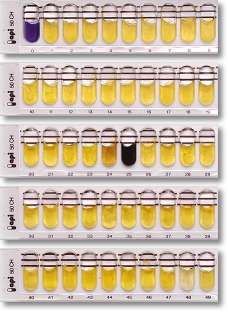
13 API 50 CH + 20E


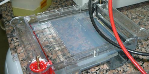

14 Honey samples Isolated DNA PCR Preparation Amplification in thermocycler Agarose gel Electrophoresis
15 PCR 16S rdna by specific primers Pl1/Pl2 973 bp

16 rep-pcr with primers BOX A1R, REP and ERIC

17 <BOX A1R- PCR pb 700 pb Paenibacillus larvae strains

18 ERIC ERIC I ERIC IV 900 pb

19 Photo: Genersch et al., 2006 International Journal of Systematic and Evolutionary Microbiology (2006), 56,


20 Colony morphology on Columbia sheet blood agar plates ERIC I ERIC II

21 Colony morphology on MYPGP agar plates ERIC I ERIC II

22 Colony morphology of P.larvae cultivated on sheep blood agar plates ERIC I ERIC II ERIC III ERIC IV Photo: Genersch et al., 2006 International Journal of Systematic and Evolutionary Microbiology (2006), 56:


23 Photos: Genersch and co-workers ERIC I ERIC II ERIC III ERIC IV

24 Application of multilocus sequence typing (MLST) for the analysis of population diversity of P. larvae From: Morrissey et al., 2014 ERIC I ERIC II From: Morrissey, et al.,2015. Environmental Microbiology 17: ERIC III ERIC IV
 PLOS ONE 12(5): e0176831 ERIC I ERIC III and")
25 Presentations of MLST schemes From: Krongdang et al. (2017) PLOS ONE 12(5): e ERIC I ERIC III and ERIC IV
: e90914.doi:10.")
26 Djukic M. et al., PLoS ONE 9(3): e90914.doi: /journal.pone
27 Pathogenesis of P. larvae Adapted From: Poppinga & Genersch, Curr. Opinion in Insect. Scie. 10: Transmission of spores to larvae through nurse bees Degradation of larval cadaver and sporulation of P. larvae 2 Ingestion of bacterial spores Invasive phase Noninvasive phase 5 3 Breaching of the epithelium and invasion of the haemocoel Larval death 4 Germination of spores Proliferation of P. larvae in the midgut lumen


28 Pathogenesis of P. larvae Adapted From: Poppinga & Genersch, Curr. Opinion in Insect. Scie. 10: Epithelium Peritrophic matrix Midgut lumen ERIC I and II ERIC I ERIC II Secondary metabolites ERIC I Chitindegrading enzymes ERIC II Toxins??????? S-layer proteins Hemocoel of infected larva
29

30

31
32


33 Thanks for the attention!!!! Questions??