Nature Structural & Molecular Biology: doi: /nsmb.3428
|
|
|
- Sandra York
- 5 years ago
- Views:
Transcription
1
2 Supplementary Figure 1 Biochemical characterization of the monou and oligou activity switch of TUT4(7). (a) Mouse TUT4 and human TUT7 were assayed for monou and Lin28-dependent oligou addition activities for group II pre-let-7g (1 nt 3 -end overhang) and group I pre-let-7g substrate with a pre-existing 2 nt overhang. (b) Sequence and secondary structure prediction (mfold) of pre-let-7g used in the activity assays. The GGAG element within the terminal loop is highlighted (red shadow). Pre-let-7g is a group II mirna, containing a 1 nt 3 -end overhang. The green box highlights the stem region, which was the basis for design of dsrna mimics for crystallization. (c) Shown is the stem region of pre-let-7g with a pre-existing 2 nt overhang end structure, as used in the activity assays. (d) Shown is the sequence of the palindromic RNA used in crystallization to obtain the CM-dsRNA structure. (e) Domain layout of mouse TUT4 (mtut4). The Lin28-interating module (LIM) and catalytic module (CM) are indicated. The LIM is composed of the CCHH zinc finger motif (pink) and inactive NTD1 (light purple, white hash marks). The CM contains the zinc knuckle domains ZK1-ZK3 (purple) and the active NTD2 (green). The mtut4 truncation constructs analyzed in this study are labeled (mt1- mt7). We found that construct mt1 (truncation of C-terminal domain up to ZK3) has the same activity as full-length mtut4, but is easier to purify and more stable. We therefore used mt1 as the backbone for other mutant constructs used in this study. (f) MonoU addition time course (0-15 minutes) assay conducted with the indicated mtut4 truncation construct and pre-let-7g. Reactions were resolved to single nucleotide resolution on 10% sequencing gels. 1 nt vs. 2nt products are labeled. (g) Lin28-dependent oligou addition time course (0-15 minutes) assays conducted with the indicated mtut4 truncation construct and pre-let-7g (pre-incubated with mouse Lin28). Reactions were resolved to single nucleotide resolution on 10% sequencing gels. OligoU and monou products are labeled as 1) oligou, 2) oligou short, and 3) monou. Gels shown are representative of three technical replicates. Uncropped source gels and replicate gels are shown in Supplementary Data Set 1.
3 Supplementary Figure 2 Analysis of Lin28 domains controlling the TUT4 oligou switch. (a) Domain layout of mtut4. The construct used for the experiments presented in this figure is indicated (mt1). (b) Domain layout of full length mouse Lin28 (CSD, brown and CCHCx2, blue) and mutant constructs used for experiments presented in panels c-g (ml1- ml5). Point mutants of the Lin28 CCHC zinc-binding motifs (ml3, C139A, C142A) and (ml4, C161A, C164A) are indicated by a red X. (c) OligoU addition assay of mt1 and pre-let-7g pre-incubated with ml1. (d) OligoU addition assay of mt1 and pre-let-7g pre-incubated with ml2. (e) OligoU addition assay of mt1 and pre-let-7g pre-incubated with ml3. (f) OligoU addition assay of mt1 and pre-let-7g preincubated with ml4. (g) OligoU addition assay of mt1 and pre-let-7g pre-incubated with ml5. Shown in each panel is a schematic of mtut4 (green and light purple oval), pre-let-7g (black cartoon), and the Lin28 construct (CSD, brown and CCHCx2, blue) used in the corresponding assay. The product generated in each reaction is represented by either oligou tail (orange ribbon cartoon) or monou addition (orange U). Reactions were resolved to single nucleotide resolution on 10% sequencing gels. OligoU and monou products are labeled for each gel. Gels shown are representative of three technical replicates. Uncropped source gels and replicate gels are shown in Supplementary Data Set 1.

4
5 Supplementary Figure 3 Structure of human TUT7 CM. (a) The structure of CM-apo (collected at the Zn 2+ absorption edge, λ=1.28 Å) was determined by MR using a model output from the Autobuild routine in Phenix derived from SAD phasing of a Se-Met derivative. The N-lobe (green), C-lobe (light blue), and ZK2 (purple) are labeled. As described (see Methods), all datasets collected on crystals that were grown and cryoprotected in high lithium sulfate conditions contained a sulfate (yellow stick) and two iodide ions (gold spheres) in place of the desired ligand in the substrate-binding site. To capture substrate complexes we devised a soaking procedure in low ionic strength buffer conditions (see Methods). To verify the identity and position of Zn 2+ coordinated by ZK2 (green sphere), we calculated an anomalous difference map (red mesh, 4.0 ). (b) The asymmetric unit of CM-dsRNA, contains one palindromic RNA duplex (5 -pgcgaagcgcuucgcu-3, purple and orange strands) and two CMs. Each CM is bound to opposite ends of the duplex, with the 1 nt overhang (U15) positioned in the active site. The difference electron density map (orange and purple mesh) calculated after MR and before inclusion of RNA in the model is shown at 2.0. In this manuscript we describe only one CM (N-lobe, green and C-lobe, light blue) and the RNA duplex as the relevant monou addition state structure. However, the remaining copy of the CM (light brown) is nearly equivalent to the one discussed in the text. We discuss the CM that exhibited the highest quality electron density map by visual inspection. (c) Close-up view of the difference electron density map (contoured at 2.0 ) prior to inclusion of the RNA model in refinement. The final refined RNA duplex is displayed (orange and purple sticks).

6
7 Supplementary Figure 4 Comparison of TUTase folds and PAP folds. (a) Structure of htut7 CM with the N-lobe (green) and C-lobe (light blue) highlighted. The ZK2 domain, unique to mammalian TUTases is also labeled (purple ribbon). (b) Structure of S. pombe Cid1 27 with lobes colored as in panel a. (c) Structure of TbTUT4 22 with lobes colored as in panel a. (d) Structure of TbTUT1 22 with lobes colored as in panel a. Also shown are the RRM domain (gray cartoon) and the unique CCHH ZnF domain (yellow cartoon). A CCHH zinc finger is also present in the TUT4(7) LIM. (e) Structure of TbRET2 23 with lobes colored as in panel a. Also shown is the RRM domain (white cartoon). (f) Structure of TbMEAT1 20 with lobes colored as in panel a. Also shown is helical insertion unique to TbMEAT1 (yellow cartoon). (g) Structure of mtpap 54 with lobes colored as in panel a. MtPAP is known to form a homodimer, but only one subunit is displayed. (c) Structure of GLD-2 51 with lobes colored as in panel a. GLD-2 was determined in complex with GLD-3 (red cartoon), which is required for GLD-2 activation. (c) Structure of Trf4p 52 with lobes colored as in panel a. Trf4p was determined in complex with the CCHC zinc knuckle protein, Air2p (red cartoon), a modulator of Trf4p polymerase activity.
8
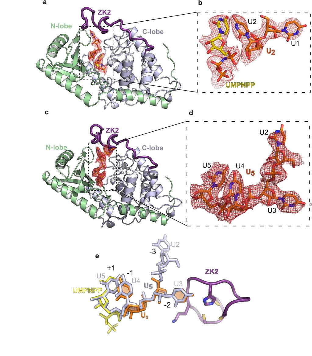
9 Supplementary Figure 5 Difference electron density maps for CM-U 2 and CM-U 5 structures. (a) Overall structure of CM-U 2 with N-lobe (green), C-lobe (light blue), and ZK2 (purple) labeled. The difference electron density map (red mesh) accounting for UMPNPP (yellow stick) and U 2 RNA (orange stick), calculated prior to their inclusion in refinement, is shown (2.0 contour level). (b) Close-up view of difference map and UMPNPP-U 2 from panel a. (c) Overall structure of CM-U 5 with N-lobe (green), C-lobe (light blue), and ZK2 (purple) labeled. The difference electron density map (red mesh) accounting for U 5 RNA (orange stick), calculated prior to their inclusion in refinement, is shown (2.0 ). (d) Close-up view of difference map and U 5 from panel c. Clear density could only account for U2-U5 in the final refined model. (e) Overlay of UMPNPP-U 2 (yellow stick, UMPNPP and orange stick, U2) and U 5 RNA (gray stick) as a result of superposition of CM-U 2 vs. CM-U 5 structures. Also shown is the ZK2 (purple cartoon and stick).
 and CCHC zinc knuckle (ZK1-ZK2 mut, ZK1-ZK3 mut, ZK2-ZK3 mut ) domains used for experiments shown in this figure.")
10 Supplementary Figure 6 OligoU activities of mtut4 zinc binding mutants. (a) Domain layout of mtut4. Indicated are mt1 and mutant constructs of the CCHH zinc finger (CCHH mut ) and CCHC zinc knuckle (ZK1-ZK2 mut, ZK1-ZK3 mut, ZK2-ZK3 mut ) domains used for experiments shown in this figure. We made double mutants of the ZK domains, because single mutants had only modest effects on oligou activity (data not shown). Mutations made are as follows: CCHH mut (C326A, C329A), ZK1 mut (C932A, C935A), ZK2 mut (C1312A, C1315A), and ZK3 mut (C1360A, C1363A) (b) OligoU activity assays of mt1 and the indicated mutant. CCHH mut is severely impaired for oligou addition as reported previously 47. Mutation of ZK2, either as ZK1- ZK2 mut or ZK2-ZK3 mut, reproducibly leads to oligou short products. OligoU and monou products are labeled as 1) oligou, 2) oligou short, and 3) monou. Gels shown are representative of three technical replicates. Uncropped source gels and replicate gels are shown in Supplementary Data Set 1.

11 Supplementary Figure 7 Substrate recognition by TUT7 CM.
12 (a) Structural superposition of CM-dsRNA vs. CM-U 5. The dsrna (orange and purple transparent cartoon) and U 5 RNA (gray cartoon) substrates are displayed. (b) Superposition of CM-dsRNA vs. CM-U 5. Shown are the UTP (yellow stick) and dsrna (3 -strand orange cartoon, 5 -strand gray) from CM-dsRNA and ZK2 (purple cartoon, sticks) from CM-U 5. (c) Overall structure of CM-dsRNA. The 5 - anchor (dark green) is indicated. Also highlighted are (red box) the group II mirna binding site and the groove loop (blue box). (d) Close-up view of the active site interactions in CM-dsRNA. Incoming nucleotide (UTP, yellow stick) in the +1 position, dsrna (orange stick), and CM substrate interacting residues (N-lobe residues; green sticks, C-lobe; gray sticks) are shown. General H-bond interactions (gray dashed lines) are displayed, as are U-specific interactions with the +1 position (red dashed lines) and the -1 position (black dashed line). Also displayed are the 5 -anchor (green cartoon and sticks) and the hydrophobic platform (green sticks). (e) Closeup view of the groove loop interactions with dsrna (purple and orange strands). Groove loop residues (blue sticks) primarily make van der Waals contact with the minor groove of dsrna.