Diagnostic Microbiology
|
|
|
- Abner Briggs
- 5 years ago
- Views:
Transcription

1 Diagnostic Microbiology Identification of Microbes Lecture: 1
2 Out lines What is expected out of this course??? At the end of this course, you will be able to apply Conventional/ Molecular diagnostic methods Diagnostic Microbiology Techniques Some basic principles of microbiology testing (Follow a specimen through the lab) Task of the methods to make the microorganisms visible and measurable

3 Component of Diagnostic Microbiology the analysis of sample, the synthesis of results (of several samples), and the clinical consultation Together these form the basis for Diagnosis Therapy Infection control The basic principle is - Clinical diagnosis or assessment - Collecting and transporting specimens - Microscopy - Cultural diagnostic methods - Some basic principles of microbiology testing - Culture Sensitivity - Non-cultural diagnostic methods - Virological diagnosis - Immunological methods
4 Major Group of the Microbial World Bacteria Fungi Algae Parasite Viruses Major features Small Diverse (appearance, genetics)
5 The method microbiologist use fall into two categories: Phenotypic (conventional) : morphology (micro and macroscopic) Genotypic: genetic techniques
6 Phenotypic Methods Old fashioned methods via biochemical, serological and morphological are still used to identify many microorganisms. Examining specimens to detect isolate and identify pathogens Phenotypic characteristics: A- Morphology : Microscopic (wet preparation Staining preparation) Macroscopic (Cultural Characteristics) are traits that can be accessed with the naked eye e. g appearance of colony including shape, size, color of the colony (pigment), speed of growth and growth pattern in broth- nutrient requirements for the growth of the organism Growth on different laboratory conditions and media- Can determine whether the microbe is a bacteria, fungus, or protozo) B- Biochemical/ Physiology characteristic are traditional for microbes identification Biochemical reactions (Metabolic differences) include enzymes (catalase, oxidase, decarboxylase),fermentation of sugars, capacity to digest or metabolize complex and sensitivity to drugs can be used in identification- physical factors such as temperature, ph and the incubation period. C- Immunological tests: serological tests are great value in the diagnosis of many bacterial, fungal and viral infections. D- Animal Pathogenicity: to detect toxins and virulence- uncommon techniques used in the identification E- Phage Typing: procedure for characterizing and detecting some strains of bacteria by their reaction (susceptible or resistant), may used in outbreak infections- uncommon techniques used in the identification process. F- fatty acid profiles: routinely used to identification of mostly anaerobic bacteria- non fermentative gram negative, yeast
7 Phenotypic Methods Disadvantages Poor discriminatory power Difficulties in typing Not provide enough information about microorganisms for today's needs.
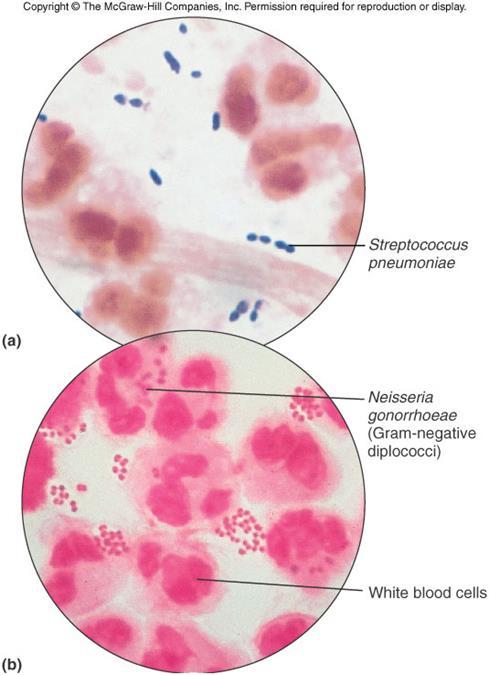
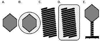
8 A- Morphology 1- Microscopy: Microorganisms can be examined microscopically for A- Size of individual cells B- Bacterial motility: Hanging drop method C-Cells Shape D- Cells arrangement E- Useful to be taken into account F- microscopic structures and characteristics ( such as flagella) Morphology and staining reactions of bacteria: 1-Simple stain: one dye is used e.g methylene blue stain reveals shape- size- cell arrangement The most commonly used stain in diagnostic microbiology is the Gram stain 2- Differential stain: 1- Gram stain: differentiation between Gm+ve and Gm ve bacteria. Primary stain (Crystal violet). Mordant (Grams Iodine mixture). Decolorization (ethyl alcohol). Secondary stain ( Saffranin) Some microbes have unique characteristics that can be detected by special staining procedures 3- Acid fast reaction (Ziehl-Neelsen stain) staining acid fast bacilli 4- ( spore stain): bacterial endospores 5-Structural stains reveal certain special structures of cell parts not revealed by conventional methods such as: capsule (capsule stain - flagellar stains- granules (volutin ) This used to give an initial presumptive identification
9 Specimen Preparation for Optical Microscopes Wet mounts and hanging drop mounts allow examination of characteristics of live cells: size, motility, shape, and arrangement Fixed mounts are made by drying and heating a film of specimen. This smear is stained using dyes to permit visualization of cells or cell parts.

10 Staining Dyes create contrast by imparting a color to cells or cell parts Basic dyes cationic, positively charged chromophore Acidic dyes anionic, negatively charged chromophore Positive staining surfaces of microbes are negatively charged and attract basic dyes Negative staining microbe repels dye, the dye stains the background Staining reactions of dyes

 2-")


11 Microscope Types 1-1- Light Microscope (digital) 2- Stereo microscope 2 3- Dark field Microscope 4- Electron Microscope (transmission- Scanning) 3 4
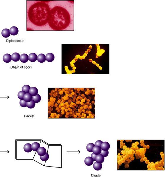
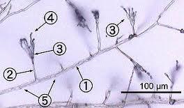


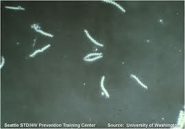
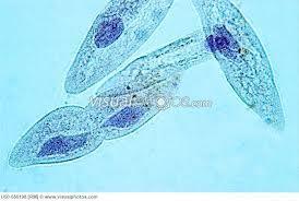
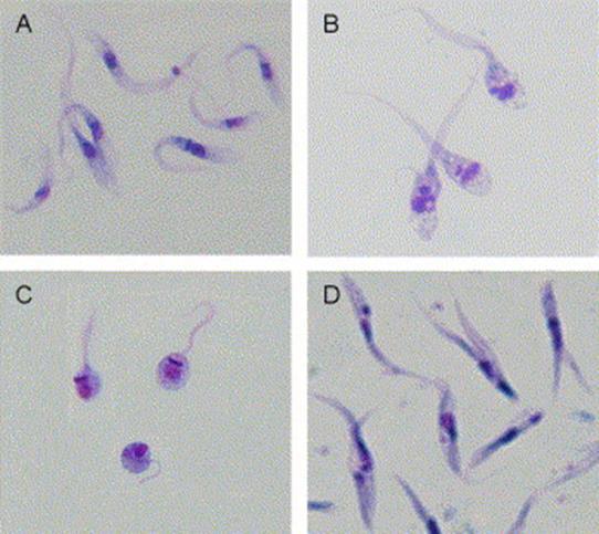
12 Microscopic Morphology Bacteria Bacteria- dark field microscope Fungi
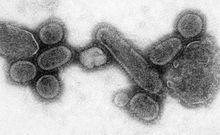

13 Virus Transmission electron microscope image of influenza virus Protozoa

14 Microbe Stains