Long-Acting Release Formulation of Exendin-4 Based on Biomimetic Mineralization for Type 2 Diabetes Therapy
|
|
|
- Colin Reeves
- 5 years ago
- Views:
Transcription
1 Supporting Information Long-Acting Release Formulation of Exendin-4 Based on Biomimetic Mineralization for Type 2 Diabetes Therapy Wei Chen,, Guohao Wang, Bryant C. Yung, Gang Liu, Zhiyong Qian*, and Xiaoyuan Chen*, State Key Laboratory of Biotherapy and Cancer Center, West China Hospital, Sichuan University and Collaborative Innovation Center for Biotherapy, Chengdu , P. R. China State Key Laboratory of Molecular Vaccinology and Molecular Diagnostics & Center for Molecular Imaging and Translational Medicine, School of Public Health, Xiamen University, Xiamen , China Laboratory of Molecular Imaging and Nanomedicine (LOMIN), National Institute of Biomedical Imaging and Bioengineering (NIBIB), National Institutes of Health (NIH), Bethesda, MD 20892, USA. * anderson-qian@163.com; shawn.chen@nih.gov
2 Materials. Cys40-exendin-4 was prepared by solid-phase peptide synthesis (CS Bio, Menlo Park, CA). The sequence of the peptide was confirmed to be His-Gly-Glu-Gly-Thr-Phe-Thr-Ser-Asp-Leu-Ser-Lys-Gln-Met-Glu-Glu-Glu-Ala-Val-Arg-Leu-P he-ile-glu-trp-leu-lys-asn-gly-gly-pro-ser-ser-gly-ala-pro-pro-pro-ser-cys-nh2. IRdye 800CW N-hydroxysuccinimide (NHS) ester was purchased from Li-Cor (Lincoln, NE). N-(2-Aminoethyl)maleimide trifluoroacetate salt was bought from Sigma-Aldrich (St. Louis, MO). Reaction medium, Dulbecco s Modified Eagle s Medium (DMEM), was obtained from Corning Incorporated (USA). All other chemicals were purchased from Fisher Scientific (USA) and used without further purification. Cell and Animals. 143B cells (human bone osteosarcoma cells) were provided by the American Type Culture Collection (USA) and maintained in Dulbecco s Modified Eagle s Medium (DMEM) supplemented with 10% v/v fetal bovine serum (FBS) and 1% v/v penicillin-streptomycin at 37 C in a 95% humidified atmosphere with 5% CO2. Type 2 diabetic C57BL/6 db/db mice (male, 6-8 weeks) were purchased from Nanjing Biomedical Research Institute of Nanjing University (Nanjing, China). Normal FVB mice were obtained from Harlan Laboratories. All animal studies were conducted in accordance with the principles and procedures outlined in the Guide for the Care and Use of Laboratory Animals and approved by the Institutional Animal Care and Use Committee (IACUC) of the Clinical Center, National Institutes of Health and Xiamen University IACUC. Animals were housed under a 12 h light/dark circle, allowed food and water ad libitum, and acclimatized for 2 weeks. Characterization of m-exendin-4. Transmission electron microscopy (TEM) observations of m-exendin-4 particles at different time points were performed on a Philips/FEI CM200 microscope (USA). Hitachi SU-70 Schottky field emission gun scanning electron microscopy (SEM) was applied for surface investigation and elemental mapping based on energy-dispersive X-ray spectroscopy (EDX). An SZ-100 Nanoparticle Analyzer (HORIBA Scientific, Japan) was used to measure the size distribution and zeta potential of m-exendin-4 up to one week. Fourier transform infrared (FTIR) spectroscopic analysis was performed on a Nicolet NEXUS 670 FTIR Spectrometer (GMI, USA). Ultraviolet visible (UV-Vis) spectra were
3 measured by a Genesys 10S UV-Vis spectrophotometer (Thermo Scientific, USA). The thermo gravimetric analysis and differential scanning calorimeter thermo examination (TGA/DSC) were performed using an SDT-Q600 (TA instruments, USA) at a heating rate of 10 C/min from 20 C to 800 C in nitrogen atmosphere. Biocompatibility of m-exendin-4. Evaluation of biocompatibility of m-exendin-4 was performed using MTT (2-(4,5)-dimethylthiazol-2-yl)-2,5-diphenyl-tetrazolium bromide) assay. 143B cells ( cells/well) were seeded in 96-well plate respectively in 200 µl culture media and incubated for 24 h before the treatment. The culture medium in each well was replaced by the same volume of medium containing exendin-4 and different concentrated Ca. The concentration of exendin-4 was fixed as 500 µg/ml. After the addition of 20 µl MTT solution (5 mg/ml), the cells were incubated for 4 h before the replacement of the MTT-containing medium by using 150 µl DMSO. After the gentle agitation for 5 min, the absorbance at 490 nm (corrected for background absorbance at 630 nm) was measured by synergy 2 multi-mode reader (BioTek Instrument Inc., USA), which was positive correlation to the concentration of the formazan that represented the activity of succinodehydrogenase. Cell viabilities at different Ca concentrations were presented as the percentage of the control group without any treatment. Electrophoresis Analysis. A weighted amount of m-exendin-4 was dissolved in 0.9% NaCl solution with a ph value of 5.0. After vortexing for 2 min, the solution was centrifuged at 12,000 rpm for 10 min and the supernatant was collected. The samples were diluted by 6x loading buffer and loaded onto NuPAGETM NOVEXTM 4-12% Bis-Tris Protein Gel (Invitrogen, USA). Electrophoresis of samples was performed at a constant voltage of 100 V in Tris-acetate-EDTA (TAE) buffer. After migration, the gel was stained with Coomassie Bright Blue to reveal peptide. Following destaining, the gel was detected using an Amersham Imager 600 system (GE Healthcare Life Sciences, USA). Intraperitoneal Glucose Tolerance Test (IPGTT) After 24 h Treatment. A glucose tolerance test was conducted to evaluate the long-term in vivo glucose responsiveness of exendin-4 and m-exendin-4 treatments. Briefly, C57BL/6 db/db mice (6-8 weeks, male) were fasted overnight
4 and administered exendin-4 or m-exendin-4 with an exendin-4 dose of 25 nmol/kg for 24 h. A glucose solution in PBS was then injected i.p. into all mice at a dose of 1.5 g/kg. The blood glucose level was monitored at 0, 15, 30, 60, 90, 120 min after injection. The glucose tolerance test on healthy mice (FVB mice, 4-5 weeks, male, g) was used as the control.


5 Figure S1. Size analysis of m-exendin-4 at different time points.

6 Figure S2. Zeta potential of m-exendin-4


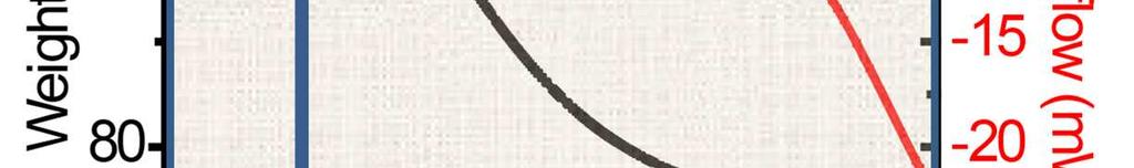


7 Figure S3. TG-DSC analysis of m-exendin-4 particles


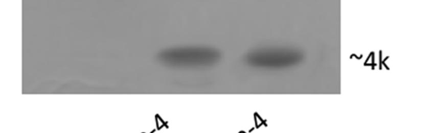
8 Figure S4. Electrophoresis analysis of native and released exendin-4.
9 Figure S5. HPLC analysis of free IRdye 800CW, exendin-4, and dye-labeled exendin-4.
10 Figure S6 MS analysis of IRdye labeled exendin-4.
11 Figure S7. (A) Photographs of the exendin-4 and IRdye-exendin-4 solution. (B) UV-Vis absorption of exendin-4 and IRdye-exendin-4. (C) Fluorescence spectra of exendin-4 and IRdye-exendin-4.


12 Figure S8. Cytotoxicity of mineralized particles generated under different Ca concentrations. Incubation time: 24 h. Cell: 143B
13 BGL (mmol/l) Control Exendin-4 m-exendin-4 Healthy mice -24 h Time (min) Figure S9. IPGTT for different groups after 24 h treatments.