High content imaging and applications
|
|
|
- Geoffrey Benson
- 5 years ago
- Views:
Transcription

1 High content imaging and applications Nick Dolman Ph.D. Senior Staff Scientist Biosciences Division- Thermo Fisher Scientific For Research Use Only. Not for use in diagnostic procedures. The world leader in serving science
2 Agenda for today s talk Part II high-content imaging and applications Why use high-content imaging Applications of high content imaging Antibody-drug conjugates Phenotypic profiling Stem cell applications Stem cell ID Neuronal differentiation 2


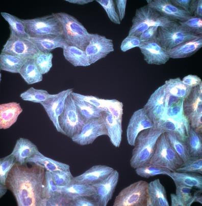
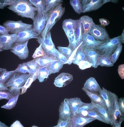
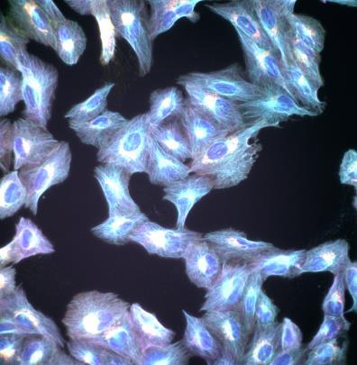
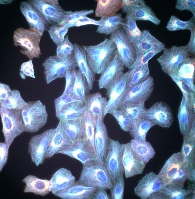
3 Why use fluorescence microscopy Markers of defined structures & processes Space Time Co-localization Pros: examine localization and abundance of molecules in cells Cons: Low throughput and laborious quantification 3
4 Throughput and quantification The world leader in serving science
5 Automated acquisition Multiple channels 5

6 Automated acquisition Multiple wells within plates Rapid, unbiased acquisition of large numbers of cells Relevant and robust view of the heterogeneity within a populaiton 6


7 Automated acquisition through analysis Example assay A549 cells Hoechst Rabbit polyclonal Anti LC3B Goat anti-rabbit Alexa Fluor CellInsight CX5 High-Content Screening (HCS) Platform

![[Bafilomycin A1]mM [Bafilomycin A1]mM [Bafilomycin A1]mM](/docs-images/92/110216436/images/8-1.jpg "[Bafilomycin A1]mM [Bafilomycin A1]mM 20 Also see Gough A.")
8 Data visualization: multiple readouts Ring spot Intensity Ring spot Count Ring spot area Ring spot area Ring spot area [Bafilomycin A1]mM [Bafilomycin A1]mM [Bafilomycin A1]mM [Bafilomycin A1]mM [Bafilomycin A1]mM 20 Also see Gough A.H, et al (2014) PLoS ONE e102678


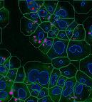
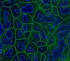
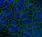
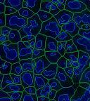
9 NEGATIVE CONTROL POSITIVE CONTROL: TNFα Nuclear translocation BLUE CHANNEL: NUCLEUS GREEN CHANNEL: NFΚB 21 ArrayScan HCA readers


10 Cytoskeletal organization Mean fiber count Mean fiber intensity 0.001mM 10mM Alexa Fluor Phalloidin 22 CellInsight CX7 High-Content Screening (HCS) Platform
11 Applications of high content imaging and analysis The world leader in serving science
12 Applications of high content imaging and analysis 1. Phenotypic profiling Profiling of CRISPR/Cas9 edited cells Screening: sirna library 2. Antibody drug conjugates 3. Stem cell applications Embryoid bodies Neuronal differentiation 24
13 Profiling CRISPR/Cas9 edited cells The world leader in serving science


14 % Responders Cell profiling: target or phenotype Target Phenotypic ATM/ATR/DNA-PKcs Vehicle Cmpd A Cmpd B Hoechst 33342/ ph2ax FluoVolt membrane potential kit Vehicle Cmpd A Cmpd B 26
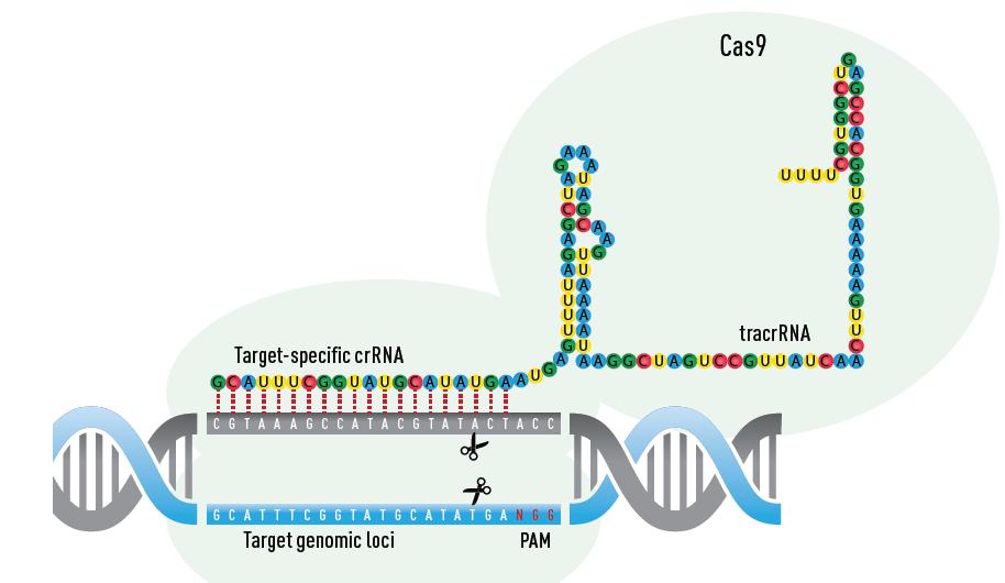
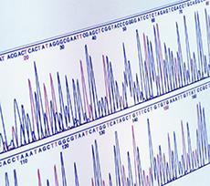
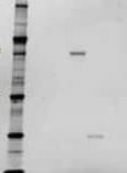
15 Profiling CRISPR/Cas9 editing Cellular assays Sequencing Blot Autophagy 27


16 ATG5 KO Wild type Target assays Presence or absence of macroautophagy in ATG5 KO Hap1 Vehicle Chloroquine PP242 PP242 Chloroquine 28 CellInsight CX7 High-Content Screening (HCS) Platform
17 Screening: sirna libraries The world leader in serving science
 Plate automation Orbitor RS Microplate Mover")
18 Library screening Screening applications require multiple plates to cover the library Can be small molecule or gene (sirna/shrna) Plate automation Orbitor RS Microplate Mover 30


19 sirna libraries Cytoskeletal rearrangement as the readout Cell & Nuclear Morphology F-Actin Microtubules 31 ArrayScan HCA readers

20 sirna libraries Hierarchical clustering analysis Identified 8 clusters of cellular phenotypes 32 ArrayScan HCA readers

21 sirna libraries Hierarchical clustering analysis 8 clusters of cellular phenotypes Cluster visualization: projection along 2 main Principal Component Analysis axis Visualize which genes belong to the different clusters i.e. which genes have redundant responses 33 ArrayScan HCA readers
22 Antibody-drug conjugates The world leader in serving science


23 Antibody-drug conjugates Alexa Fluor 647 MMAE Herceptin SK-BR3 MDA-MB-231 MDA-MB-231 SK-BR-3 Alexa Fluor 647 Herceptin & CellEvent Caspase 3/7 reagent 35 Arrayscan High content platform


24 Antibody-drug conjugates SiteClick antibody labeling with phrodo LysoTracker Deep Red phrodo Red Herceptin Overlay 36 Arrayscan High content platform
25 Stem cell ID The world leader in serving science

 DAPI &")

26 Stem cell ID DAPI, Oct4 & NKx2.5 Human neuroal stem cells (H9 cells) DAPI & Oct4 segmentation 38 CellInsight CX7 High-Content Screening (HCS) Platform

27 Stem cell ID. Stem colonies 10x Widefield image 10x Confocal Image 39 Arrayscan High content platform

28 Algorithm Overlays Oct4+ Cells: Stem cell ID. Stem colonies Confocal Z-Stack projection of 25 slices of 2 µm Widefield Z-Stack projection of 25 slices of 2 µm Widefield Single Slice 1 slice of 2 µm Imaging Mode: Confocal Z-Stack Widefield Z-Stack Widefield Single Slice Cell Number: Number of Oct4+ Cells % of Oct4+ Cells 57% 72% 44% 40 Arrayscan High content platform
29 Neuronal differentiation The world leader in serving science

30 Identification of Dopaminergic neuronal pre cursor cells (FPP cells) Midbrain dopaminergic (DA) neurons derived from human pluripotent stem cells (hpscs) Excellent alternative to primary human neurons for disease modeling and drug screening for Parkinson s disease, The PSC Dopaminergic Neuron Differentiation Kit Human Dopaminergic Neuron Immunocytochemistry Kit Essential 8 medium 42

 Ch 1 Ch 2 Ch 3")


31 Identification of Dopaminergic neuronal pre cursor cells (FPP cells) Ch 1 Ch 2 Ch 3 DAPI FoxA2 Otx2 Object Identification Target Activation Target Activation High throughput, quantitative image-based analysis of FPP cell identification 43 CellInsight CX5 High-Content Screening (HCS) Platform


32 Differentiation of FPP into mature DA neurons Ch 1 Ch 2 Composite Dapi TH: 31.3% Composite Images + overlay Dapi TH nuclei Identification Cell body Identification Nuclei count = 805 Cell body count = 252 %TH positive cells = 31.3% 44 CellInsight CX5 High-Content Screening (HCS) Platform