Supplementary Note 1. Enzymatic properties of the purified Syn BVR
|
|
|
- Jasper Manning
- 5 years ago
- Views:
Transcription
1 Supplementary Note 1. Enzymatic properties of the purified Syn BVR The expression vector pet15b-syn bvr allowed us to routinely prepare 15 mg of electrophoretically homogenous Syn BVR from 2.5 L of TB-medium cultures of E. coli C41(DE3) cells. When prepared by the thrombin treatment approach, the enzyme has three additional residues at the N-terminus (Ser-Gly-His) but lacks the His-tag. SDS-PAGE analysis of purified Syn BVR revealed a major Coomassie blue stained band at 36 kda (Supplementary Fig. 1(a)), consistent with the mass of Syn BVR estimated from its DNA sequence. The purity of Syn BVR was determined to be greater than 95% using Coomassie blue stained gels. The molecular size of the Syn BVR was estimated to be ~36 kda by gel-filtration chromatography (Supplementary Fig. 1(b)). Dynamic light scattering analysis estimated the size to be 35.4 kda with monomodal dispersity (polydispersity = 8%). In addition, the crystal structures in this study revealed that all structures, including the apo-, NADP + -bound, and BV-complex of Syn BVR, were in the monomeric state. Therefore, we concluded that purified Syn BVR existed as a monomer under our experimental conditions. It has been reported previously that Syn BVR expressed in E. coli apparently formed dimers 1. There is a discrepancy between our purified Syn BVR and previously reported Syn BVR in the oligomeric state. However, we know a great deal about protein with two oligomeric states. For example, cytochrome c, a heme electron carrier protein, was long believed to be a monomeric protein. Recently, however, new findings demonstrated that the monomeric state of cytochrome c could be altered to a dimeric form by treatment with ethanol or refolding with guanidinium in vitro 2. Furthermore, its dimeric states could be also detected in the cell 3. In regard to Syn BVR, these results raise the possibility that both the monomeric and dimeric states might reflect unknown physiological functions of Syn BVR. Consistent with this, recent work revealed additional functions of human BVR, including kinase and transcription factor (DNA binding) activities 4,5. Phosphate ions enhance the NADH-dependent activity of human BVR 6. We observed similar enhancement of Syn BVR activity by inorganic phosphate (Supplementary Fig. 1(c)), although the activation rate was quite low. In low phosphate concentrations, BVR was not activated, consistent with a previous report 1. By contrast, increasing the amount of sodium phosphate (>100 mm) resulted in activation. In the absence of phosphate ion, the activity of Syn BVR in was nmol min -1 per mg of protein, increasing 2.5-fold to nmol min -1 per mg of protein in 350 mm phosphate. By contrast, the activity of human BVR increases 40-fold in the presence of phosphate; thus, phosphate has a relatively minor effect on the activity of Syn BVR. In the structure of the apo-syn BVR, phosphate ion derived from sample buffer (PBS buffer) is visible (Supplementary Fig. 1(d)). This inorganic phosphate ion binds to the same site as the 2 -phosphate group of NADP(H) (Supplementary Fig. 1(d)). Therefore, the bound inorganic phosphate probably helps NAD(H) to bind if it were NADP(H), as reported previously for human BVR previously 6. 1
2 Supplementary Note 2. Enzymatic properties of the H84A and D287A mutants of Syn BVR Prof. T. J. Mantle s group has performed sophisticated kinetic experiments on BVRs, and found that the H84A and D285A mutations of Syn BVR dramatically reduced the enzyme s activity 1. These residues, however, engage in no direct interaction with bound NADP + or bound BV (Supplementary Fig. 4(a)). We investigated why these mutations decrease activity. In the case of the H84A mutant, we observed white precipitates formed during purification. Therefore, we assessed the stability of the supernatant using a thermal-shift assay. The denaturing curves (thermogram) for wild-type and R185A-mutated BVRs were very similar (Supplementary Fig. 4(b)): the estimated T m values were ~64 C for apo-form and 70 C in the presence of NADP +. By contrast, the thermogram of the H84A mutant clearly indicated denaturing behavior even at room temperature, consistent with circular dichroism (CD) analysis of this mutant 1. This result strongly suggests that H84 near the active pocket plays a crucial role in stabilizing the folding of this protein. In the case of D285A (hereafter D287A, because in Synechocystis sp. PCC 6803, position 285 is arginine, not aspartate; under our experimental conditions, the D287A mutation dramatically reduced the activity, as previously reported). The D287A mutant had a thermogram similar to those of wild-type BVR and the R185A mutant. However, the thermogram for the apo-form implied that D287 confers greater stability. Interestingly, the titration curve indicated that the D287A mutant can interact with only one biliverdin molecule (Supplementary Fig. 4(c)), and we believe that this explains the dysfunction of the mutant. According to reported K m values 1, NADPH binds tightly to this mutant: the K m value for NADPH is 10-fold lower for D287A than for wild-type BVR. In addition, we observed that structural changes occurred upon NADP(H) binding, allowing two biliverdin molecules to be bound, and this structural flexibility is required for BVR activity. Asp287 is probably responsible for fine-tuning of the position of His129, because Asp287 forms hydrogen-bond networks among Arg25 (α1), His126 (β5), Tyr302, and His129 (Supplemental Fig. 4(a)). His129 is involved in the structural change upon NADP(H) binding, as described in the main text. However, we believe that the function of the Asp278 is limited in cyanobacterial BVR because Asp278, like Arg25 and Tyr302, is not conserved among mammalian BVRs (Supplementary Fig. 1). Supplementary References 1. Hayes, J. M. & Mantle, T. J. The effect of ph on the initial rate kinetics of the dimeric biliverdin-ixα reductase from the cyanobacterium Synechocystis PCC6803. FEBS J. 276, (2009). 2. Hirota, S. et al. Cytochrome c polymerization by successive domain swapping at the 2
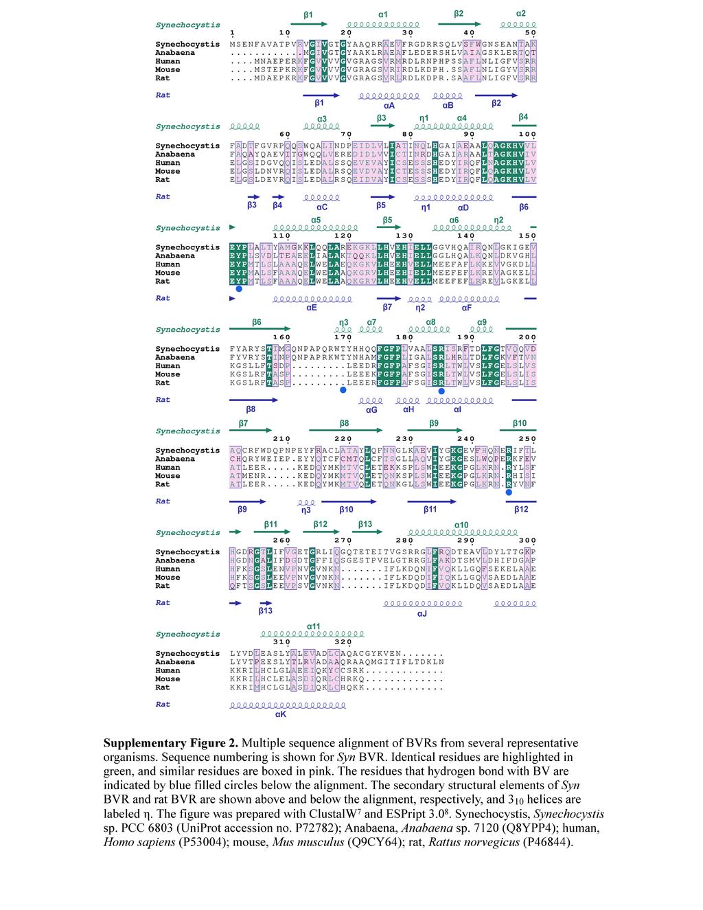
3 C-terminal helix. Proc. Natl. Acad. Sci. USA 107, (2010). 3. Hayashi, Y. et al. Domain swapping oligomerization of thermostable c-type cytochrome in E. coli cells. Sci. Rep. 6, (2016). 4. Kravets, A., Hu, Z., Miralem, T., Torno, M. D. & Maines, M. D. Biliverdin reductase, a novel regulator for induction of activating transcription factor-2 and heme oxygenase-1. J. Biol. Chem. 279, (2004). 5. Lerner-Marmarosh, N. et al. Human biliverdin reductase: a member of the insulin receptor substrate family with serine/threonine/tyrosine kinase activity. Proc. Natl. Acad. Sci. USA 102, (2005). 6. Franklin, E. et al. Activation of biliverdin-ixα reductase by inorganic phosphate and related anions. Biochem. J. 405, (2007). 7. Thompson, J. D., Higgins, D. G. & Gibson, T. J. CLUSTAL W: improving the sensitivity of progressive multiple sequence alignment through sequence weighting, position-specific gap penalties and weight matrix choice. Nucleic Acids Res. 22, (1994). 8. Robert, X. & Gouet, P. Deciphering key features in protein structures with the new ENDscript server. Nucleic Acids Res. 42, W (2014). 3
4 Supplementary Table 1 Oligonucleotides used in this study Oligonucleotide Synechocystis sp. PCC 6803 BVR-Y102F-F BVR-Y102F-R BVR-T169A-F BVR-T169A-R BVR-S184A-F BVR-S184A-R BVR-R185A-F BVR-R185K-F BVR-R185-R BVR-R188A-F BVR-R188A-R BVR-K237A-F BVR-K237A-R BVR-R246A-F BVR-R246A-R Human BVR-Y98F-F BVR-Y98F-R BVR-R172A-F BVR-R172E-F BVR-R172K-F BVR-R172-R Rat BVR-Y97F-F BVR-Y97F-R BVR-R171A-F BVR-R171E-F BVR-R171K-F BVR-R171-R Sequence 5 -TTTCCTTTAGCGTTAACCTATGC-3 5 -TTCCAACACCACATGCCCAC-3 5 -GCCTATCACCATCAGCAATTTG-3 5 -CCAACGTTGGGGAGCGGG-3 5 -GCGCGCATCAGTCGGTTTACGG-3 5 -CAAGGCCGCCACTAAAGGA-3 5 -GCCATCAGTCGGTTTACGGATTT-3 5 -AAAATCAGTCGGTTTACGGATTTAT-3 5 -GGACAAGGCCGCCACTAAA-3 5 -GCGTTTACGGATTTATTCGGTACA-3 5 -ACTGATGCGGGACAAGGCC-3 5 -GCAGGGGAAGTTTTTCACCAGAA-3 5 -GCCATAGATAACCTCCGCTT-3 5 -GCGATTTTTACCCTCCATGGCG-3 5 -TTCATTCTGGTGAAAAACTTCC-3 5 -TTTCCCATGACACTGTCTCTTGC-3 5 -TTCGACCAACACGTGTTTGC-3 5 -GCGTTAACCTGGCTTGTCTCCTT-3 5 -GAATTAACCTGGCTTGTCTCCTT-3 5 -AAATTAACCTGGCTTGTCTCCTT-3 5 -GCTAATGCCGGAAAACGCA-3 5 -TTTCCCATGACACTGTCATTTGC-3 5 -TTCCACGAGGACATGCTTG-3 5 -GCGCTGACCTGGCTGGTCTCC-3 5 -GAACTGACCTGGCTGGTCTCC-3 5 -AAACTGACCTGGCTGGTCTCC-3 5 -AGAAATGCCGCTGAACGCAG-3 Underlines indicate the altered codons for site-directed mutagenesis.
5
6
7
8
9
10