Supplementary Figure 1
|
|
|
- Henry Harris
- 5 years ago
- Views:
Transcription
1 Supplementary Figure 1 2

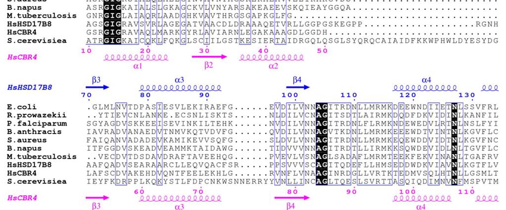
2 Supplementary Figure 1: Sequence alignment of HsHSD17B8 and HsCBR4 of with KAR orthologs. The secondary structure elements as calculated by DSSP and residue numbers are displayed above for HsHSD17B8 and below for HsCBR4. The conserved catalytic triad residues, Ser- Tyr-Lys are highlighted in orange. The arginine residues close to Arg129 and Arg172 in E. coli fabg essential for ACP-binding are highlighted in yellow. The extended loop in S. cerevisiae near Arg129 of E. coli fabg is shown in magenta. Other positively charged residues near this region are highlighted in cyan. Supplementary Figure 2 Supplementary Figure 2: Interface between the diagonally opposite protomers (chain A; HSD17B8 and chain C; CBR4) in the tetramer. For comparison, the corresponding chains of the HSD17B8 homotetramer (PDB id 2PD6) are shown in magenta and cyan; it can be noted that these chains do not interact with each other. 3
 and M (right). In chain D the serine is acetylated to form O-acetylserine (Oas) whereas in chain M, an acetate ion and a water molecule are present with hydrogen bonding interactions. 4")
3 Supplementary Figure 3 Supplementary Figure 3: Electron densities (2F o -F c electron density map countoured at 1 sigma) around the catalytic serine (Ser156) of the HSD17B8 subunit in chains D (left) and M (right). In chain D the serine is acetylated to form O-acetylserine (Oas) whereas in chain M, an acetate ion and a water molecule are present with hydrogen bonding interactions. 4
4 Supplementary Figure 4 Supplementary Figure 4: Original sensograms recorded by Biacore T200 for with varying concentrations of (a) NADPH, (b) NADH, (c) NADP + and (d) NAD +. For concentrations, see Methods section. The association and dissociation of the tested ligands resulted in square pulses. 5


5 Supplementary Figure 5 Supplementary Figure 5: Sequence alignment of ACPs from the same organisms as in Supplementary Figure 1. The secondary structure elements above and below the sequences correspond to ACP from E. coli and M. tuberculosis, respectively. It can also be seen that helix 2, considered to be essential for interacting with KAR, is quite well conserved in these organisms, in particular with respect to the two negatively charged residues (Asp76 and Glu82, E. coli ACP numbering). 6
 and residues 209 to 218 for the CBR4 (blue ball and stick model).")
6 Supplementary Figure 6 Supplementary Figure 6: Stereo image of part of the (2F o -F c ) electron density map, contoured at 1 sigma, for the structures of (a) -NAD + and (b) -NAD(P) +. The electron density map is shown for residues 235 to 245 for the HSD17B8 subunit (brown ball and stick model) and residues 209 to 218 for the CBR4 (blue ball and stick model). These residues are part of the helices 6 at the HSD17B8-CBR4 interface. 7
7 Supplementary Table 1: Interface areas between chains in the heterotetramer and in the HSD17B8 homotetramer. Protomer 1 Protomer 2 Interface type Interface area (Å 2 ) G (kcal mole -1 ) CSS -A -D Homomeric/HSD17B HSD17B8-A HSD17B8-D Homomeric B -C Homomeric/CBR HSD17B8-B HSD17B8-C Homomeric A -B Heteromeric HSD17B8-A HSD17B8-B Homomeric D -C Heteromeric HSD17B8-D HSD17B8-C Homomeric A -C Heteromeric HSD17BB-A HSD17B8-C Homomeric B -D Heteromeric HSD17B8-B HSD17B8-D Homomeric The associated stabilization in terms of G and the complex formation significance score (CSS) as calculated by the program PISA is also listed. 8
8 Supplementary Table 2: List of potential hydrogen bonding interactions between NAD(P) + with the protein chain in. NAD + Protein Atom Distance NADP + Protein Atom Distance Comments (Å) (Å) O7N Ile 202/O 3.7 O7N Val 181/O 3.6 ** Ile 202/N 3.1 Val 181/N 3.2 ** Thr 207/OG1 3.2 N7N Thr 204/OG1 2.6 O7N Thr 183/OG1 2.9 *** Thr 207/OG1 3.6 N1N Tyr 169/OH 3.7 N1N Tyr 148/OH 3.5 *** Ser 155/O 3.9 Gly 134/O 3.9 ** O2D Tyr 169/OH 3.2 O2D Tyr 148/OH 3.4 *** Lys 173/NZ 2.7 Lys 152/NZ 2.6 *** O3D Cys 103/O 2.9 O3D Ala 83/O 3.0 ** HOH Ala 84/O 3.9 Gly 85/N 3.3 Lys 152/NZ 3.9 O5D Cys 103/O 4.0 O5D HOH O1N Thr 204/OG1 2.6 O1N Thr 183/OG1 2.4 *** O2N Ser 21/O 3.3 O2N Gly 22/N 3.9 Ile 23/N 2.9 Ile 14/N 2.6 *** HOH HOH *** Thr 183/OG1 3.6 O3 O3 Arg 12/O 3.8 O1A HOH O1A Arg 12/NH2 3.3 ** HOH O2A Ser 21/OG 3.1 O2A Gly 22/N 3.7 Gly 13/N 3.6 *** HOH Asp 184/N 3.7 Arg 12/NE 3.5 Arg 12/NH1 3.4 O5B Ser 21/O 3.8 O5B Arg 12/O ** HOH Arg 12/NE 3.8 9
9 Arg 12/NH1 2.9 Arg 12/NH2 3.9 HOH O4B Gly 105/N 3.70 O4B Gly 85/N 3.7 *** Ala 83/O 3.8 O3B Ser 21/N 3.6 O3B Arg 12/N 3.3 ** Asp 42/OD1 3.6 Asp 42/OD2 2.7 Gly 9/O 3.6 Ser 11/N 3.8 Ser 11/OG 2.9 Arg 12/O 4.0 Arg 12/NH2 3.9 O2B Asp 42/OD1 2.5 O2B Asp 42/OD2 3.0 Leu 43/N 4.0 Asp 44/N 3.8 Ser 11/OG 3.7 Arg 12/N 3.9 HOH N7A HOH N7A HOH *** Arg 34/NH1 3.4 Arg 34/NH2 3.8 N1A Ala 74/O 3.6 N1A Cys 55/O 3. 6 ** Asp 75/OD1 3.3 Asp 56/OD1 3.4 *** Val 76/N 3.1 Val 57/N 3.1 *** N6A Asp 75/OD1 2.5 N6A Asp 56/OD1 2.7 *** HOH HOH *** Arg 34/NH O1X Arg 12/NH2 3.5 Asn 35/N 3.9 HOH HOH O2X Arg 34/NE 3.4 Arg 34/N 2.5 Asn 35/N O3X Arg 34/NE 3.4 Arg 34/NH2 2.7 Arg 12/NH2 3.6 The conserved interactions are highlighted by ** and conserved interactions with also the conserved residues are highlighted by ***. The key differences due to the additional 2 - phosphate moiety in NADP + are highlighted with yellow background. 10
10 Supplementary Table 3: List of oligos used to create mutant variants for this study Oligonucleotide name Sequence E. coli fabg-r126e-f 5 -GTAATGCGCGCTATGATGAAAAAGGAGCATGGTCGTATTATCACTATCGG-3 E. coli fabg-r126e-r 5 -CCGATAGTGATAATACGACCATGCTCCTTTTTCATCATAGCGCGCATTAC-3 E. coli fabg-r172e-f 5 -GGCGCGCGAAGTTGCGTCAGAGGGTATTACTGTAAACGTTG-3 E. coli fabg-r172e-r 5 -CAACGTTTACAGTAATACCCTCTGACGCAACTTCGCGCGCC-3 HSD17B8-G18S-F HSD17B8-G18S-R HSD17B8-S21A-F HSD17B8-S21A-R HSD17B8-D42A-F HSD17B8-D42A-R HSD17B8-R148E-F HSD17B8-R148E-R HSD17B8-R148A-F HSD17B8-R148A-R HSD17B8-S156A-F HSD17B8-S156A-R HSD17B8-Y169F-F HSD17B8-Y169F-R HSD17B8-K173A-F HSD17B8-K173A-R HSD17B8-R189E-F HSD17B8-R189E-R HSD17B8-R189A-F HSD17B8-R189E-R CBR4-G9S-F CBR4-G9S-R CBR4-R12A-F CBR4-R12A-R CBR4-R34A-F CBR4-R34A-R CBR4-Q126E-F CBR4-Q126E-R CBR4-R168E-F 5 -GGCCTTGGTCACAAGTGCGGGGAGCGG-3 5 -CCGCTCCCCGCACTTGTGACCAAGGCC-3 5 -CACAGGTGCGGGGGCCGGCATCGGCCGAG-3 5 -TTTGAATTCTCAGACCATGTACATCCCGC-3 5 -CGTAGCTGCCTGCGCCCTGGACCGGGCAG-3 5 -CTGCCCGGTCCAGGGCGCAGGCAGCTACG-3 5 -CAAGCCCTGGTGTCCAATGGTTGTGAGGGTTCCATCATCAACAT-3 5 -ATGTTGATGATGGAACCCTCACAACCATTGGACACCAGGGCTTG-3 5 -GCCCTGGTGTCCAATGGTTGTGCTGGTTCCATCA-3 5 -TGATGGAACCAGCACAACCATTGGACACCAGGGC CCATCATCAACATCAGTGCCATCGTAGGAAAGGTGGG CCCACCTTTCCTACGATGGCACTGATGTTGATGATGG CGTGGGGCAGACAAACTTTGCAGCATCCAAGGCTG CAGCCTTGGATGCTGCAAAGTTTGTCTGCCCCACG CAAACTATGCAGCATCCGCGGCTGGAGTGATTGGGC GCCCAATCACTCCAGCCGCGGATGCTGCATAGTTTG-3 5 -GCCCGGGAGCTTGGAGAGCATGGGATCCGCTGT-3 5 -ACAGCGGATCCCATGCTCTCCAAGCTCCCGGGC-3 5 -AGCCCGGCAGCTTGGAGCACATGGGATCC-3 5 -GGATCCCATGTGCTCCAAGCTGCCGGGCT-3 5 -CAAAGTGTGTGCTGTTTTTTCAGGCTCCCGAGGCATTGGCAGAG-3 5 -CTCTGCCAATGCCTCGGGAGCCTGAAAAAACAGCACACACTTTG-3 5 -GCTGTTTTTGGAGGCTCCGCAGGCATTGGCAGAGCTGTG-3 5 -CACAGCTCTGCCAATGCCTGCGGAGCCTCCAAAAACAGC-3 5 -CTGGCGGTCATTGCCGCAAACCTGGAAGGGGCC GGCCCCTTCCAGGTTTGCGGCAATGACCGCCAG-3 5 -CCATGAGGACTATGATTCAACAAGAGGGAGGGTCTA-3 5 -TAGACCCTCCCTCTTGTTGAATCATAGTCCTCATGG-3 5 -CACGTGCTCTTGCTAAAGAGGTAGCAGAGAAGAAAATTAGAGTGAATGTAGTTGC-3 11
11 CBR4-R168E-R CBR4-K169E-F CBR4-K169E-R CBR4-Q124E/Q125E /Q126E-F CBR4-Q124E/Q125E /Q126E-R CBR4-R168E/K169E-F CBR4-R168E/K169E-R CBR4-S135A-F CBR4-S135A-R CBR4-Y148F-F CBR4-Y148F-R CBR4-K152A-F CBR4-K152A-R 5 -GCAACTACATTCACTCTAATTTTCTTCTCTGCTACCTCTTTAGCAAGAGCACGTG-3 5 -GTGCTCTTGCTAAAGAGGTAGCAAGAGAGAAAATTAGAGTGAATG-3 5 -CATTCACTCTAATTTTCTCTCTTGCTACCTCTTTAGCAAGAGCAC-3 5 -GACCTGTAAAGCTGCCATGAGGACTATGATTGAGGAGGAGGGAGGGTCTATTGT-3 5 -ACAATAGACCCTCCCTCCTCCTCAATCATAGTCCTCATGGCAGCTTTACAGGTC-3 5 -GGATTTTCACGTGCTCTTGCTAAAGAGGTAGCAGAGGAGAAAATTAGAGTGAATGTAGTTGC-3 5 -GCAACTACATTCACTCTAATTTTCTCCTCTGCTACCTCTTTAGCAAGAGCACGTGAAAATCC-3 5 -GGGTCTATTGTTAATGTAGGAGCCATTGTTGGCTTAAAAGGCAAC-3 5 -GTTGCCTTTTAAGCCAACAATGGCTCCTACATTAACAATAGACCC-3 5 -CTCTGGCCAGTCCGTTTTCAGTGCCAGTAAAGGAG-3 5 -CTCCTTTACTGGCACTGAAAACGGACTGGCCAGAG-3 5 -GTCCGTTTACAGTGCCAGTGCAGGAGGATTAGTTGGATTTTC-3 5 -GAAAATCCAACTAATCCTCCTGCACTGGCACTGTAAACGGAC-3 12
12 Supplementary Table 4: List of mutants designed in this study. HSD17B8 Residue Gly18 HSD17B8 Mutation G18S- comment at the N- terminus of the pyrophosphate binding helix Ser21 S21A- corresponds to Arg12 in the b- chain Asp42 Ser156 Tyr169 Lys173 Arg148 Arg189 D42A- S156A- Y169A- K173A- R148A- R148E- R189A- R189E- Asp42 points to the ribose ring of NAD ribose binding CBR4 Residue Gly9 Arg12 Arg34 catalytic residue Ser135 catalytic residue Tyr148 catalytic residue Lys152 Proposed ACPinteracting residue equivalent to Arg129 of E. coli fabg Proposed ACPinteracting residue equivalent to Arg172 of E. coli fabg Gln126 Gln Arg168 Lys169 CBR4 mutation G9S- R12A- R34A- S135A- Y148F- K152A- Q126E- A E- R168E- K169E- comment at the N- terminus of the pyrophosphate binding helix points to 2 phosphate Ala33 corresponds to Asp42 catalytic residue catalytic residue catalytic residue Proposed ACPinteracting residue(s) equivalent to Arg129 of E. coli fabg Proposed ACPinteracting residue(s) equivalent to Arg172 of E. coli fabg 13
13 Supplementary Table 5: Binding constants from surface plasmon resonance measurements of wild-type and the D42A- variant. Ligand KD D42A- KD NADPH 8.2±2.1 µm 9.5±2.6 µm 8.6±2.0 mm 7.4±4.0 mm NADH 33.4±10.4 µm ±9.5 mm 15.0±0.6 mm NADP+ 26.8±9.0 µm 34.1±12.2 µm NAD+ 4.2±0.4 mm 70.8±23.3 mm Both NADPH and NADH exhibit a strong affinity (µm) and a weak affinity (mm) binding site. The D42A- variant shows reduced affinity for NAD+ and NADH. See Fig. 4 for the curve fittings. 14
14 Supplementary Table 6: List of yeast and bacterial strains used in this study Name Genotype Reference or source Saccharomyces cerevisiae BY4741 MATa; his3d1; leu2d0; met15d0; ura3d0; EUROSCARF (Frankfurt, Germany, roscarf/index.html BY4741 oar1 Escherichia coli DH5 TOP 10 Rosetta(DE3)pLysS XL1-Blue MATa; his3d1; leu2d0; met15d0; ura3d0; ykl055c::kanmx4 F 80lacZ M15 (laczyaargf) U169 reca1 enda1 hsdr17 (rk, mk+) phoa supe44 thi-1 gyra96 rela1 F-mcrA (mrr-hsdrmsmcrbc) _ 80lacZM15 lacx74 deor reca1 arad139 (araleu)7697 galu galk rpsl (StrR) enda1 nupg F_ompT hsdsb(rb_mb_) gal dcm (DE3) plyssrare (CamR) reca1 enda1 gyra96 thi-1 hsdr17 supe44 rela1 lac [F proab laciqz M15 Tn10 (Tetr)] EUROSCARF (Frankfurt, Germany, roscarf/index.html Invitrogen, Carlsbad, CA Invitrogen, Carlsbad, CA Novagen, Darmstadt, Germany Stratagene, La Jolla, CA 15