Roles of heterotypic CCN2/CTGF-CCN3/NOV and homotypic CCN2-CCN2 interactions in expression of the differentiated phenotype of chondrocytes
|
|
|
- Anabel Bishop
- 5 years ago
- Views:
Transcription
1 Roles of heterotypic CCN2/CTGF-CCN3/NOV and homotypic CCN2-CCN2 interactions in expression of the differentiated phenotype of chondrocytes Mitsuhiro Hoshijima, 1,2 Takako Hattori, 1,* Eriko Aoyama, 3 Takashi Nishida, 1 Takashi Yamashiro, 2 and Masaharu Takigawa 1,3,* 1 Department of Biochemistry and Molecular Dentistry, 2 Department of Orthodontics and Dentofacial Orthopedics, Okayama University Graduate School of Medicine, Dentistry and Pharmaceutical Sciences, 3 Biodental Research Center, Okayama University Dental School, Japan * Authors for correspondence Takako Hattori 5-1 Shikata-cho, 2-chome, Okayama , Japan Tel: , Fax: hattorit@md.okayama-u.ac.jp Masaharu Takigawa 5-1 Shikata-cho, 2-chome, Okayama , Japan Tel: , Fax: takigawa@md.okayama-u.ac.jp Keywords: CCN2/CTGF, CCN3/NOV, yeast two-hybrid, chondrocyte, aggrecan, surface plasmon resonance (SPR) 1
2 To identify proteins that regulate CCN2 activity, we carried out GAL4-based yeast two-hybrid screening using a cdna library derived from a chondrocytic cell line, HCS-2/8. CCN2/CTGF and CCN3/NOV polypeptides were picked up as CCN2-binding proteins, and CCN2-CCN2 and CCN2-CCN3 binding domains were identified. Direct binding between CCN2 and CCN3 was confirmed by coimmunoprecipitation in vitro and in vivo and surface plasmon resonance, and the calculated dissociation constant (K d ) was M between CCN2 and CCN2, and M between CCN2 and CCN3. Ectopically overexpressed GFP-CCN2 and Halo-CCN3 in COS7 co-localized, as determined by direct fluorescence analysis. We present evidence that CCN2-CCN3 interactions modulated CCN2 activity such as enhancement of aggrecan and col2a1 expression. Curiously, CCN2 enhanced, whereas CCN3 inhibited, the expression of aggrecan and col2a1 mrna in HCS-2/8 cells; and the combined treatment with CCN2 and CCN3 abolished the inhibitory effect by CCN3. These effects were neutralized with an antibody against the VWC domain of CCN2 (11H3). This antibody diminished the binding between CCN2 and CCN2, but enhanced that between CCN3 and CCN2. Our results suggest that CCN2 could form homotypic and heterotypic dimers with CCN2 and CCN3, respectively. Strengthening the binding between CCN2 and CCN3 with the 11H3 antibody had an enhancing effect on aggrecan expression in chondrocytes, suggesting that CCN2 had an antagonizing effect by binding to CCN3. Introduction CCN family protein 2/connective tissue growth factor (CCN2) is a major member of the CCN family of proteins, which consists of CCN1/Cyr61, CCN2/CTGF, CCN3/NOV, CCN4/WISP-1, CCN5/WISP-2/CTGF-L, and CCN6/WISP-3. CCN2 is expressed in various types of cells, such as fibroblasts, endothelial cells, vascular smooth muscle cells, osteoblasts, and chondrocytes [1-7]. Among those cell types, CCN2 is strongly expressed in growth-plate cartilage, especially in hypertrophic chondrocytes [1,4,5,7] during the developmental stages, and shows multiple cellular functions such as stimulation of cartilage-specific extracellular matrix (ECMs) synthesis as well as chondrocyte proliferation and maturation [1]. In CCN2 null mutant mice, bone formation is inhibited due to impairment of both chondrogenesis and growth-plate angiogenesis [8], which leads to neonatal respiratory failure and death within minutes of birth, thus indicating the essential role of CCN2 in developing chondrocytes. Other experiments also demonstrated that CCN2 is involved in the early cell-condensation step during the ectopic digit formation [9]. However, the ectopic overexpression in soft tissues is linked to various types of fibrosis, because of its strong enhancing effects on the production of extracellular matrices [1-4]. CCN2 consists of 4 modules, i.e., insulin-like growth factor binding protein-like (IGFBP), von 2
3 Willebrand factor type C (VWC), thrombospondin type 1 repeats (TSP-1), and C-terminal cystine knot (CT); and each of these modules has different binding partners and seems to regulate the multiple functions of CCN2 [1]. The CT domain of CCN2 interacts directly with fibronectin and enhances cell adhesion of chondrocytes through binding to integrin α5β1 [10]. This domain also binds to integrin α5β1 to promote adhesion and migration of pancreatic stellate cells [11]. Furthermore, it induces adhesion of hepatic cells by direct binding to the integrin receptor αvβ3 and to heparan sulfate proteoglycan through its C-terminal heparin-binding domain [12]. There are also reports indicating that CCN2 signaling occurs through low-density lipoprotein receptor-related protein-1 (LRP-1) [13-15]. Other studies show that CCN2 directly binds to bone morphogenetic protein-4 (BMP-4) and transforming growth factor-β (TGF-β) through its VWC module and prevents or enhances their binding to their own receptors [16]. Moreover, CCN2 binds vascular endothelial growth factor (VEGF) and inhibits its angiogenic effect [17]. There also is a report showing that CCN2 binds to aggrecan, which is a major marker of differentiated phenotype of chondrocytes, through its N-terminal IGFBP and VWC modules and that this binding may be related to the CCN2-enhanced production and secretion of aggrecan by chondrocytes [18]. In order to identify additional extracellular or cell-surface targets for CCN2 that may be involved in the regulatory functions of CCN2 in chondrocytes, we searched for CCN2-binding proteins by using the yeast two-hybrid screening assay. A cdna library derived from human chondrosarcoma-derived chondrocytic cell line HCS-2/8 was screened for products binding to CCN2, since this cell line has a differentiated phenotype similar to that of normal chondrocytes in terms of aggrecan and cartilage collagen secretion and integrin expression profiles [19]. We found CCN2 and CCN3 as binding partners of CCN2, indicating that CCN2 forms homotypic and heterotypic dimers with CCN2 and CCN3, respectively. CCN3, also a CCN family protein, is highly expressed in the nervous system, blood vessels, and musculoskeletal system as well as in pre- and early hypertrophic chondrocytes and osteoblasts [1,20,21]. The functions of CCN3 protein among these different tissues are not yet well defined. Although CCN3 was originally described as being antiproliferative [22] and its expression associated with differentiation and growth arrest in Wilm's tumor [23], chondrosarcomas [24], and rhabdomyosarcomas [25], more recent data correlate CCN3 with an increased proliferative index of 3T3 fibroblasts [26] and prostate tissue samples [27]. Both CCN2 and CCN3 are expressed in the early hypertrophic to pre-hypertrophic zone of the growth plate in cartilage; and CCN2 enhances the expression of ECMs components, whereas CCN3 suppresses it in cultured growth plate chondrocytes [20]. There seems to be some correlation between CCN2 and CCN3; however, the functional and physiological interaction between these 2 proteins had not been elucidated at all. In the present study, we show for the first time that (1) CCN2 bound to CCN2 through its IGFBP, VWC, and CT domains, 3
4 but not TSP-1 domain, and to CCN3 only via its VWC and CT domains; and (2) CCN2 rescued chondrocytes from CCN3-induced suppression of aggrecan and col2a1 expression. Inhibition of CCN2-CCN2 binding suppressed the expression of aggrecan, whereas promotion of CCN2-CCN3 binding enhanced it. Results Identification of CCN2 and CCN3 as CCN2 dimerization partners Using yeast two-hybrid screening to search for proteins that directly interacted with full-length CCN2 (amino acid residues ), we obtained several clones from a HCS-2/8 human chondrocytic cell cdna library that encoded CCN2 and CCN3, indicating homotypic and heterotypic dimerization of CCN2. To confirm direct interactions between CCN2 and CCN2 or between CCN2 and CCN3, we used several methods. First, to identify the CCN2 interaction sites in CCN2 or CCN3, full-length, N-terminal, C-terminal, and individual domains of CCN2 or CCN3 were expressed in AH109 yeast cells as GAL4-transactivation domain (GAL4-AD)-fusion proteins, whereas full-length CCN2 was co-expressed as a GAL4-DNA binding domain (GAL4-BD)-fusion protein (Fig.1 A). Interactions between CCN2 or CCN3 fragments and full-length CCN2 were monitored as growth of yeast transformants in selection media (SD/-Ade, -His, -Leu, -Trp). Self-interaction with full-length CCN2 was observed after co-transformation with CCN2 vectors expressing IGFBP, VWC, and/or CT, but not when the TSP-1 domain alone was tested. Interaction with CCN3 fragments was seen with vectors expressing the VWC or CT domain, but not with those expressing the IGFBP or TSP-1 domain (Fig.1 A). The difference in binding strength between full-length CCN2 (CCN2 full ) and IGFBP domain of CCN2 (CCN2 I ), and between CCN2 full and IGFBP domain of CCN3 (CCN3 I ), was estimated by measuring the β-galactosidase activity of the reporter gene in yeast expressing full-length CCN2 (GAL-BD/CCN2 full ), standardizing it to the protein concentration, and comparing it with the β-galactosidase activity obtained after co-transformation with the pgadt7 empty vector (mock, Fig.1 B). CCN2 I showed almost the same strength of binding to CCN2 full as CCN2 full, whereas the binding of CCN3 I to CCN2 full was significantly lower than that of CCN3 full or CCN2 I to CCN2 full (Fig.1 B). These results indicate that among the 4 domains in CCN2, the IGFBP, VWC, and CT domains exhibited direct homotypic interaction with CCN2, whereas only the VWC and CT domains of CCN3 showed direct interaction with full-length CCN2. CCN2 directly interacts with CCN2 and CCN3 in vitro. For in vitro pull-down experiments using glutathione-sepharose resin, recombinant His 6 -tagged 4
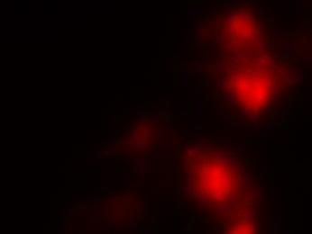
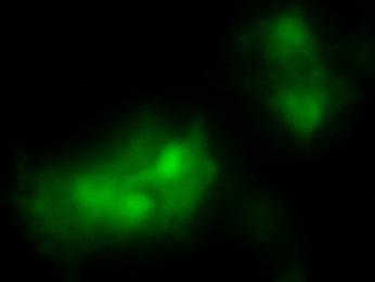
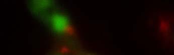
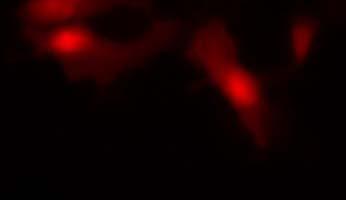
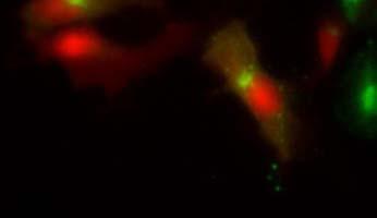
5 CCN2 (His-CCN2) and GST-fused CCN3 (GST-CCN3) were prepared as E.coli cell lysates. Immunoblotting of the precipitates with anti-his and anti-gst antibody largely confirmed the yeast two-hybrid assay results (Fig.2 A): His-CCN2 was effectively pulled down in the presence of GST-CCN3. We further confirmed the binding between CCN2 and CCN2 or between CCN3 and CCN2 by performing a solid-phase binding assay (Fig.2 B). Purified CCN2 or GST-CCN3 was immobilized on the plates, and increased amounts of biotinylated recombinant CCN2 (rccn2) were used for binding. Biotinylated rccn2 bound to immobilized rccn2 or GST-CCN3 in a dose-dependent manner (Fig.2 B). Finally, the dissociation constant of CCN2 to CCN2 or to CCN3 was determined by using surface plasmon resonance (SPR) analysis (Fig.2 C). rccn2 protein was immobilized on a sensor tip as a ligand and rccn2 and rccn3 were used as analytes at concentrations of 4, 8, 16, 32, and 64 nm. Calculated from sensorgram analysis, the K d value was 1.17x10⁻⁹ M for the CCN2-CCN2 interaction and M for the CCN2-CCN3 one (Fig.2 C). These data indicate that CCN2 underwent a homotypic interaction and a heterotypic one with CCN3 with a similar binding affinity. Endogenous CCN2 and CCN3 interact and co-localize in chondrocytic HCS-2/8 cells. To investigate the endogenous interaction between CCN2 and CCN3, we coimmunoprecipitated endogenous CCN2 with CCN3 from HCS-2/8 cell lysates using anti CCN2 antibody. CCN3 was effectively immunoprecipitated with an anti-ccn2 antibody (Fig.3 A), but not with control IgG.. Indirect immunostaining of endogenous CCN2 (red) and CCN3 (green) with spcific antibodies showed subcellular colocalization inside of the permeabilized cells with TritonX-100 and on cell surface without permeabilization treatment (Fig.3 B). Ectopically overexpressed CCN2 and CCN3 interact and co-localize in COS7 cells. To confirm the interaction between CCN2 and CCN3 in vivo, we co-expressed GFP-fused CCN2 and Halo-tagged CCN3 in COS7 cells. GFP or GFP-CCN2 was immunoprecipitated with anti-gfp antibody from cell lysates with binding proteins, and the precipitated proteins were analyzed by using anti-halo antibody. Halo-CCN3 (arrow in lane 8 of Fig.4 A) was pulled down effectively only after co-expression with GFP-CCN2 (Fig.4 A). The subcellular localization of GFP-CCN2 and Halo-CCN3 largely overlapped when the cells were directly analyzed by GFP (green) fluorescence and the fluorescence (red) from the dye binding to Halo (Fig.4 B). CCN2 and CCN3 cooperatively regulate gene expression of cartilagenous matrix genes in HCS-2/8 cells. CCN2 enhances aggrecan and col2a1 expression in chondrocytes. To elucidate the role of CCN3 interaction with CCN2 on this activity of CCN2 in chondrocytes, we treated cells of the 5
6 chondrocytic cell line HCS-2/8 with CCN2 and/or CCN3, and then monitored the expression of the aggrecan and col2a1 mrna after 12 h (aggrecan) or 24 h (col2a1) by real-time PCR. The addition of rccn2 (1.25 nm) enhanced mrna expression of aggrecan and col2a1 by approximately 20%, whereas the addition of rccn3 (1.25 nm) inhibited it by around 15%. The combination of rccn2 and rccn3 abolished the repression caused by rccn3 (Fig.5 A), and this abolishment occurred in a CCN2 dose-dependent manner (Fig.5 B). Similar results were obtained in the mouse primary chondrocytes isolated from rib cartilage (data not shown). These data suggest that CCN2 and CCN3 cooperatively regulated the gene expression of extracellular matrices such as aggrecan and col2a1 mrna in chondrocytes. Addition of 11H3 changes aggrecan gene expression of HCS-2/8 cells treated with CCN2 and/or CCN3 To modify the binding between CCN2 and CCN2 or between CCN2 and CCN3, we prepared several antibodies against the VWC domain of CCN2. One of the antibodies, 11H3, modulated the binding strength between CCN2-CCN2 and between CCN2-CCN3. In solid-phase binding studies using biotinylated CCN2 and immobilized CCN2 or GST-CCN3, the addition of 11H3 reduced the binding of CCN2 to CCN2, but accelerated that to GST-CCN3 (Fig.5 A). Furthermore, HCS-2/8 cells were treated with rccn2 (1.25 nm) and/or rccn3 (0.625 nm) in the presence of mouse IgG or 11H3 antibody, and gene expression of aggrecan was monitored 12 h later. The addition of 11H3 antibody to CCN2-treated HCS-2/8 cells cancelled the enhanced expression of the aggrecan gene, whereas the 11H3 antibody further enhanced the gene expression in the presence of CCN3 with or without CCN2 (Fig.5 B). These results suggest that when CCN2 formed heterotypic dimers with CCN3, these dimers show rescuing the inhibited effect of aggrecan expression by CCN3 in chondrocytes. Those interactions could modulate CCN2 activity such as enhancement of aggrecan expression. Discussion CCN2 is a secreted protein and accumulates at the cell surface, binding various extracellular matrix components such as aggrecan [18] and fibronectin [10]. CCN2 also interacts with several growth factors including IGF [28], TGF-β, and BMP-4 [16] and thereby modulates their activities. Among the CCN family members, CCN3 acts as a negative regulator of CCN2 in the fibrotic pathway of model of renal disease [29,30]; and the addition of recombinant CCN3 slightly decreases the expression of extracellular matrix genes in the growth-plate chondrocytes, in contrast to CCN2, which strongly up-regulates those genes [20]. Furthermore, CCN2 down-regulates CCN3 expression 6
7 in growth-plate chondrocytes [20]. In epiphyseal chondrocytes, however, CCN3 promotes the expression of matrix genes and stabilizes the phenotype of articular cartilage [31]. Here we show for the first time homotypic and heterotypic interactions of CCN2 with itself and CCN3, respectively, and provide evidence that homotypic and heterotypic dimers of CCN2 may regulate the production of chondrocytic extracellular matrices in a different manner. A previous study indicated that the CT domain of CCN3 interacted weakly with full length CCN3 and CCN2 in their yeast two-hybrid study [32]. In our present study, not only the CT domain of CCN3, but also the VWC domain had the ability to bind to full-length CCN2. Furthermore, the specificity of the homotypic and heterotypic interactions of CCN2 was substantiated by the yeast two-hybrid assay performed to determine the binding domain in CCN2 and CCN3. CCN2 self-dimerized through its IGFBP, VWC, and CT domains; whereas only VWC and CT domains, but not the IGFBP domain of CCN3 bound to full-length CCN2. The homotypic dimerization sites of CCN2 to CCN2 were different from the heterotypic dimerization sites for the binding of CCN2 to CCN3. Our data suggest that this difference in binding sites between CCN2-CCN3 and CCN2-CCN2 may be also used as a molecular system for recognition of different cellular surface binding partners such as fibronectin [10], fibulin [32], integrins [33], and aggrecan [18]. Specific CCN2-CCN2 and CCN2-CCN3 interactions were confirmed by performing several binding assays in vitro and in vivo, including in vitro pull-down assays, solid-phase binding assays, and SPR analysis. The relative binding strengths of these domains to CCN2 was also confirmed by measuring the β-galactosidase reporter gene activity in the yeast two-hybrid assay. Interestingly, the binding strength of homotypic and heterotypic bindings of CCN2 measured by SPR analysis was very similar, indicating that the binding partner of CCN2 would be dependent on the expression levels of CCN2 and CCN3 in the specific tissues. Further evidence for interactions between CCN2 and CCN3 was obtained from immunofluorescence and co-immunopreciptation experiments. Not only in the case of endogenous CCN2 and CCN3 in chondorcytic cell line, HCS-2/8, but also in the case of ectopically overexpressed GFP-CCN2 and Halo-CCN3 in COS7 cells, CCN2 and CCN3 were co-immunoprecipitated and the subcellular localization of these proteins largely overlapped (Fig. 3 and 4), confirming that CCN2 and CCN3 interacted in vitro and in vivo. A recent report on CCN family members demonstrated the possibility that CCN5 may regulate by acting as a transcriptional factor [34], and our yeast two-hybrid screening identified several intracellular proteins [Hoshijima et al, unpublished data]. Those interactions may have important roles for cellular functions. The physiological effects of homomeric and heteromeric interaction between CCN2 and CCN2, and CCN2 and CCN3 on the production of extracellular matrix of chondrocytes were monitored in human chondrocytic cell line, HCS-2/8 by performing real-time PCR, since CCN2 is an enhancer of aggrecan and col2a1 gene expression in chondrocytes [1,8]. In contrast, CCN3 down-regulates the 7
8 expression of extracellular matrix genes in growth-plate chondrocytes [20], TGF-β-stimulated Col1 promoter activity in a renal fibrosis model [29], and BMP-2-induced osteocalcin production through inhibition of the Notch signaling cascade [35]. On the other hand, CCN3 enhances matrix production in articular chondrocytes [31], and stimulates BMP-4 gene expression and bone mineralization in osteoblasts [36]. According to our results, however, CCN2 enhanced, whereas CCN3 down-regulated, aggrecan expression. This down-regulation was partially reversed by CCN2 treatment. CCN2 dose-dependently increased the amount of aggrecan and col2a1 gene expression that was down-regulated by CCN3. To modify the binding between CCN2 and CCN3, we used antibody 11H3, raised against the VWC domain of CCN2, which antibody attenuated the binding between CCN2 and CCN2, but accelerated the binding between CCN2 and CCN3 as measured by the immobilization binding assay. The addition of 11H3 antibody antagonized CCN2-induced aggrecan expression, whereas it further enhanced that in the case of combined treatment with CCN2 and CCN3, indicating that the interaction between CCN2 and CCN3 stimulated aggrecan expression in HCS-2/8 cells. Interestingly, 11H3 also accelerated aggrecan gene expression in CCN3-treated HCS-2/8 cells, suggesting the existence of endogenous CCN2 in HCS-2/8 cells. This is the first demonstration of homotypic CCN2 and heterotypic CCN2-CCN3 interactions and the data suggest that CCN2 seems to act as a negative regulator of CCN3. This could explain the varied effect of CCN3, since the activity of CCN3 largely depends on CCN2. Our results indicate that it is the IGFBP domains of CCN2 and CCN3 that make the difference between the homotypic or heterotypic complex formation. CCN2 interacts with many extracellular signaling molecules; e.g., it binds to TGF-β, TGF-βRI and II [37], BMP-2 [38], and BMP-4 [16]. Furthermore, CCN2 modulates Wnt signal pathways [39], as well as the activity of a Wnt inhibitory protein (Wif-1) [40]. CCN3 may also have a role in the modulation of those signaling pathways by interacting with CCN2, but the mechanisms remain to be elucidated. In CCN2-deficient mice, which show incomplete endochondral ossification, the expression level of CCN3 is strongly enhanced [20]. Two different lines of CCN3-deficient mice were developed. One of them, the nov del3-/- mutant mice, which produce no full-length CCN3 protein and express CCN3 without its VWC domain at a barely detectable level, shows abnormal skeletal and cardiac development [41]. The other line, which is completely CCN3 null mice, show enhanced neointimal thickening compared with the wild type, confirming that CCN3 suppresses angiogenesis and fibrosis. These results indicate that CCN2 and CCN3 seem to mutually regulate their activities and expression levels. However, in our experiments the expression level of CCN2 did not alter after addition of CCN2 or CCN3 in HCS-2/8 cells (data not shown). Since HCS-2/8 cells produce higher level of CCN2 compare to primary chondrocytes, further addition of CCN2 may not have effect on the expression level of CCN3. Regulatory mechanism of CCN2 and CCN3 gene expression is currently being elucidated. 8
9 Experimental Procedures Yeast two-hybrid cdna library screening Yeast two-hybrid cdna library screening was performed as described previously [10,18]. Briefly, full-length CCN2 cdna was expressed as a GAL4BD (GAL4 DNA-binding domain)-fusion protein. cdna library genes from HCS-2/8 cells were expressed as GAL4AD (GAL4 transactivation domain)-fusion proteins for a two-hybrid assay in yeast AH109 cells, and the cells were screened on selection medium [SD/-Ade/-His/-Leu/-Trp (synthetic dropout medium lacking Ade, His, Leu and Trp)]. Positive clones were analyzed by DNA sequencing. For analysis of the CCN2 or CCN3 domain binding to full-length CCN2, expression plasmids of full-length CCN2, which were expressed as GAL4BD-fusion proteins, and CCN2 or CCN3 cdna fragments, which were expressed as a GAL4AD-fusion protein, were used to transform yeast cells. The binding was monitored by growth in SD/-Ade/-His/-Leu/-Trp selection medium. The primers used for amplification of full-length and truncated forms of ccn2 and ccn3 were the following: CCN2 full (27-349), 5 -ATCCGAATTCCAGAACTGCAGCGGGCCGTGCCGGTGCCCG-3 and 5 -ATACGGATCCCTCATGCCATGTCTCCGTACATCTTCCTGT-3 ; CCN2 IGFBP (27-101): 5 -ATCCGAATTCCAGAACTGCAGCGGGCCGTGCCGGTGCCCG-3 and 5 -ATACGGATCCGAGCACCATCTTTGGCGGTGCACACGCCGA-3 ; CCN2 VWC (94-198): 5 -ATCCGAATTCGTGTGCACCGCCAAAGATGGTGCTCCCTGC-3 and 5 -ATACGGATCCAGTTGGCTCTAATCATAGTTGGGTCTGGGC-3 ; CCN2 TSP ( ): 5 -ATCCGAATTCACTATGATTAGAGCCAACTGCCTGGTCCAGA-3 and 5 -ATACGGATCCGGATGCACTTTTTGCCCTTCTTAATGTTCT-3 ; CCN2 CT ( ): 5 -ATCCGAATTCAACATTAAGAAGGGCAAAAAGTGCATCCGT-3 and 5 -ATACGGATCCCTCATGCCATGTCTCCGTACATCTTCCTGT-3. CCN3 full (32-357): 5 -ATCCGAATTCACTCAGCGCTGCCCTCCCCAGTG-3 and 5 -ATACGGATCCCATTTTCCCTCTGGTAGTCTTCAGC-3 ; CCN3 IGFBP (32-108): 5 -ATCCGAATTCACTCAGCGCTGCCCTCCCCAGTG-3 and 5 -ATACGGATCCATCTCCCTCTACCGCCGTGCAGATG-3 ; CCN3 VWC ( ): 5 -ATCCGAATTCGAGGGAGATAACTGTGTGTTCGATG-3 and 5 -ATACGGATCCTTCTGGCCTGTAAGCTGCAAGGGTAAG-3 ; CCN3 TSP ( ): 5 -ATCCGAATTCACCCTTGCAGCTTACAGGCCAGAAG-3 and 5 -ATACGGATCCTGGCTGCTCTGGCTCTTGTTCACAG-3 ; CCN3 CT ( ): 5 -ATCCGAATTCCCAGAGCAGCCAACAGATAAGAAAG-3 and 5 -ATACGGATCCCATTTTCCCTCTGGTAGTCTTCAGC-3. 9
10 β-galactosidase assays To confirm the binding specificity of CCN2 and CCN3 peptides, we re-transformed a pgadt7 vector expressing full-length cnn2 (pgbkt7/ccn2 full ) in the AH109 yeast strain together with the pgadt7 vector expressing either full-length ccn2 or ccn3 or their IGFBP domain (pgadt7/ccn2 full, ccn2 I, ccn3 full or ccn3 I ). The empty pgbkt7 vector was also used for re-transformation together with the same series of CCN2 or CCN3 truncations as a control. All double transformants were cultured in SD/-Trp/-Leu selection medium, equal numbers of cells were collected, and cellular proteins were prepared by 3 cycles of freezing and thawing in yeast Z buffer (0.1 M Na-phosphate, ph7.0, 20 mm KCl, 1 mm MgSO 4 ). To measure the binding strength between full- length CCN2 and either full-length or truncated forms of CCN2 or CCN3, we measured the β-galactosidase activity of the reporter gene was measured [42], standardized it to the protein concentration, and subtracted the β-galactosidase activity obtained after co-transformation of the pgadt7 empty vector with the same series of polypeptides. Expression and purification of GST-CCN3 and rhccn2 For preparation of purified recombinant GST-CCN3 protein, the ccn3 gene was amplified without its signal peptide by PCR with primers 5 -ATACGGATCCACTCAGCGCTGCCCTCCCCAGTG-3 and 5 -ATCCGAATTCTTACATTTTCCCTCTGGTAGTCTTC-3, and subcloned into the pgex-2tk vector (GE Healthcare), which carries a GST-tag at its BamHI/EcoRI sites. The recombinant plasmid was sequenced to ensure the absence of mutations. BL21(DE3)pLysS Escherichia coli cells (Novagen) were then transfected with the plasmid for expression of recombinant proteins. The expressed GST-CCN3 protein was purified with Glutathione-Sepharose 4B agarose (GE Healthcare) as described previously [43]. The purified protein was dialyzed against phosphate-buffered saline (PBS). In some experiments, rccn3 (Abnova) was used. Preparation and purification of CCN2 carrying a His 6 -tag were performed as described previously [18]. In some experiments, rhccn2 (BioVendor Laboratory Medicine) was used. In vitro binding assay E.coli expressing rhccn2 or GST-CCN3 were harvested and sonicated in co-immunoprecipitation (co-ip) buffer [20 mm Tris/HCl (ph 8.0), 25 mm NaCl, 1mM MgCl 2, 1 mm EGTA, 1% Triton X-100, 1 mm DTT and 1 mm PMSF]. After removal of cell debris, both cell lysates were mixed at 4 C to form CCN2-CCN3 complex. The complexes were then incubated with Glutathione-Sepharose 4B for 1 h at 4 C. The beads were subsequently washed with co-ip buffer with 10% glycerol, after which the bead-protein complexes were subjected to SDS-PAGE and analyzed by Western blotting using an anti-his tag antibody (BETHYL) and GST-tag antibody (GE Healthcare). 10
11 Solid-phase binding assay and antibody against CCN2 Wells of an ELISA plate were coated with 100 μl of 500 ng/ml recombinant CCN2 (BioVendor Laboratory Medicine) or GST-CCN3 in 50 mm NaHCO 3 buffer (ph 9.6) at 4 C overnight, and blocked with 200 μl of binding buffer [50 mm Tris-HCl, ph 7.4, 150 mm NaCl, 2% BSA, 0.05% Tween20] for 3 h at 37 C. Biotinylated CCN2 with or without 11H3 antibody was added to the wells in a total volume of 100 μl of binding buffer and incubated for 6 h at 37 C. Next, the wells were washed with binding buffer and then incubated with 100 μl of streptavidin-hrp (R&D Systems, MN). Bound HRP was monitored by using a TMD peroxidase substrate kit (Bio-Rad, CA). As control experiments, wells were coated with BSA or GST. The mouse monoclonal antibody used in these experiments was raised against the VWC domain of CCN2 and kindly supplied by Dr. Y. Seto (Nippon Flour Mills Co., Ltd., Japan). Surface plasmon resonance analysis (BIAcore) Binding of CCN2 to CCN2 or CCN3 was analyzed by surface plasmon resonance with the BIAcore X instrument (GE Healthcare). rhccn2 (1.25 μg) was immobilized on a C1 sensor chip by amine coupling according to the manufacturer s instructions. The amount of immobilized CCN2 was in the range of resonance units (RU). Binding studies were performed in HBS-P [Hepes-buffered saline containing surfactant P: 10 mm Hepes/HCl, 150 mm NaCl and 0.005% surfactant P-20 (ph 7.5) at 25 C] with analyte concentrations of 4, 8, 16, 32, and 64 nm. The dissociation constant (K d ) values were calculated by using BIA evaluation software version 4.1 (GE Healthcare) as previously reported [18]. Expression vectors pegfp/ccn2 vector expressing CCN2 protein with green fluorescent protein at the C-terminus of CCN2 was prepared by amplification of ccn2 cdna with signal peptide by using primers 5 -CTTCGAATTCCCATGACCGCCAGTATGGGCCCCGTC-3 and 5 -CGGTGGATCCCGTGCCATGTCTCCGTACATCTTCCTGTA-3, and insertion into EcoRI and BamHI sites of the pegfp-n1 vector. pflag-cmv/ccn3-halo expressing CCN3 protein with a Halo-tag at the C-terminus of CCN3 was prepared by amplification of ccn3 cdna with signal peptide by using primers 5 -ATCCAAGCTTATGCAGAGTGTGCAGAGCAC-3 and 5 -ATACGAATTCATTTTCCCTCTGGTAGTCTTC-3 and by amplication of halo with primers 5 -ATACGAATTCAATGGCAGAAATCGGTACTGG-3 and 5 -ATACGGATCCTTAGCCGGAAATCTCGAGCGTC-3 and pfn21ab3946, which expresses Sox9-Halo protein [44], as a template. The PCR fragments were cloned into the pflag-cmv-2 vector at HindIII/EcoRI/BamHI sites. 11
12 Cell culture and RNA extraction COS7 monkey kidney cells and HCS-2/8 human chondrosarcoma-derived chondrocytic cells [45] were maintained in Dullbecco s modified Eagle s medium (DMEM) supplemented with 10% fetal bovine serum (FBS) as described before [42,46]. In some experiments, total RNA was isolated from HCS-2/8 cells by using a RNeasy Mini Kit according to the manufacturer s instructions (Qiagen, Hilden, Germany) after the cells had been treated with the desired factors. In vivo binding assay COS7 cells were co-transfected with pegfp/ccn2 and pflag-cmv/ccn3-halo by use of Fugene 6 reagent (Roche, Basel, Switzerland) according to the manufacturer's instructions and incubated for 24 hours. The co-transfected COS7 cells or HCS-2/8 cells were then harvested in lysis buffer [50 mm Tris/HCl (ph 7.4), 150 mm NaCl, 1% Triton X-100, and 0.5% protease inhibitor cocktail (Sigma)] and incubated with anti-gfp antibody (Roche) or anti-ccn2 antibody AF660 (R&D) or normal control IgG (Sigma). The complex including GFP-CCN2 or endogenous CCN2 was precipitated by use of HaloLink Resin (Promega) or Protein G Sepharose (GE Healthcare). After the precipitated proteins had been washed with wash buffer [50 mm Tris/HCl (ph 7.4), 150 mm NaCl, and 0.1% Triton X-100], they were analyzed by Western blotting using an anti-halo antibody (Promega) or anti-ccn3 antibody. Fluorescence imaging COS7 cells transiently transfected with different expression vectors were treated with the fluorescent ligand TMR, which recognize Halo-Tag (Promega), for 15 min at 37 C. After having been washed with DMEM, the cells were fixed with 4% formaldehyde/pbs for 15 min at room temperature; and Halo-tagged proteins and GFP-tagged proteins were directly monitored by fluorescence microscopy using an IX70 Microscope (Olympus Corporation, Japan). The images were analyzed with Axiovision software (Zeiss, Oberkochen, Germany). Indirect Immunofluorescence and Fluorescence Deconvolution Microscopy HCS-2/8 cells were first fixed with 3.7% formaldehyde/pbs for 15 min at room temperature. The cells were then either permeabilized with 0.25% TritonX-100/PBS or without permeabilization for 8 min and blocked with 5% skim milk/pbs for 1 h and incubated with primary antibodies for 90 min at room temperature. After washing, the cells were incubated with secondary antibodies. Images were obtained with an Olympus fluorescence microscope. Reverse transcription and quantitative real-time PCR 12
13 For quantification of mrna expression levels, 0.5 μg of total RNA of each sample was reverse-transcribed with avian myeloblastosis virus (AMV) reverse transcriptase (Takara Bio, Japan) at 42 C for 60 min. Quantitative real-time PCR reaction using SYBR Green Realtime PCR Master Mix (Toyobo, Japan) was done with StepOnePlus (Applied Biosystems, USA). Primer sequences used for aggrecan were 5 -GAGGAGAGAACTGGAGAAG-3 and 5 -GCCGATAGTGGAATACAAC-3 ; and those for col2a1 5 -TGGTCCTGGCATCGACATG-3 and 5 -GGCTGCGGATGCTCTCAAT-3 ; and those for gapdh, 5 -CAATGACCCCTTCATTGACC-3 and 5 -GACAAGCTTCCCGTTCTCAG-3. Acknowledgements This work was supported by Research Fellowships for Young Scientists of the Japan Society for the Promotion of Science [to M.H.], grants from the program Grants-in-Aid for Scientific Research (C) [to T.H.] and Scientific Research (S) [to M.T.] and Exploratory Research (to M.T.) from the Japan Society for the Promotion of Science; by a grant from Senri Life Science Foundation (to T.H.); and by internal grants from Okayama University (to T.H. & M.T.). Masaharu Takigawa would like to dedicate this article to the memories of his father, Dr. Masami Takigawa, and Professor Emeritus Yoshiro Takeda, who passed away while this research was being conducted. The authors thank Dr. Yasuhiro Seto for generous gifts of mouse monoclonal antibody against VWC domain of CCN2, as well as Dr. Satoshi Kubota for valuable discussion. We are also grateful to Ms. Eri Yashiro for secretarial assistance. References 1 Perbal, B. and Takigawa, M. (2005) CCN Proteins: a New Family of Cell Growth and Differentiation Regulators, Imperial College Press, London 2 Kubota S, Takigawa M. (2011) The role of CCN2 in cartilage and bone development. J Cell Commun Signal. 5, Takigawa, M. (2003) CTGF/Hcs24 as a multifunctional growth factor for fibroblasts, chondrocytes and vascular endothelial cells. Drug News Perspect. 16, Takigawa, M., Nakanishi, T., Kubota, S. and Nishida, T. (2003) Role of CTGF/HCS24/ecogenin in skeletal growth control. J. Cell. Physiol. 194, Nishida, T., Kubota, S., Nakanishi, T., Kuboki, T., Yosimichi, G., Kondo, S. and Takigawa, M. (2002) CTGF/Hcs24, a hypertrophic chondrocyte-specific gene product, stimulates proliferation and differentiation, but not hypertrophy of cultured articular chondrocytes. J. Cell. Physiol. 192,
14 6 Nishida, T., Nakanishi, T., Asano, M., Shimo, T. and Takigawa, M. (2000) Effects of CTGF/Hcs24, a hypertrophic chondrocyte-specific gene product, on the proliferation and differentiation of osteoblastic cells in vitro. J. Cell. Physiol. 184, Nakanishi, T., Nishida, T., Shimo, T., Kobayashi, K., Kubo, T., Tamatani, T., Tezuka, K. and Takigawa, M. (2000) Effects of CTGF/Hcs24, a product of a hypertrophic chondrocyte-specific gene, on the proliferation and differentiation of chondrocytes in culture. Endocrinology 141, Ivkovic S, Yoon BS, Popoff SN, Safadi FF, Libuda DE, Stephenson RC, Daluiski A, Lyons KM. (2003) Connective tissue growth factor coordinates chondrogenesis and angiogenesis during skeletal development. Development 130, Lorda-Diez CI, Montero JA, Diaz-Mendoza MJ, Garcia-Porrero JA, Hurle JM. (2011) Defining the earliest transcriptional steps of chondrogenic progenitor specification during the formation of the digits in the embryonic limb. PLoS One. 6, e Hoshijima, M., Hattori, T., Inoue, M., Araki, D., Hanagata, H., Miyauchi, A. and Takigawa, M. (2006) CT domain of CCN2/CTGF directly interacts with fibronectin and enhances cell adhesion of chondrocytes through integrin α5β1. FEBS Lett. 580, Gao, R. and Brigstock, D. R. (2006) A novel integrin α5β1 binding domain in module 4 of connective tissue growth factor (CCN2/CTGF) promotes adhesion and migration of activated pancreatic stellate cells. Gut 55, Gao, R. and Brigstock, D. R. (2004) Connective tissue growth factor (CCN2) induces adhesion of rat activated hepatic stellate cells by binding of its C-terminal domain to integrin αvβ3 and heparan sulfate proteoglycan. J. Biol. Chem. 279, Segarini, P. R., Nesbitt, J. E., Li, D., Hays, L. G., Yates, III, J. R. and Carmichael, D. F. (2001) The low density lipoprotein receptor-related protein/α2-macroglobulin receptor is a receptor for connective tissue growth factor. J. Biol. Chem. 276, Yang, M., Huang, H., Li, J., Li, D. and Wang, H. (2004) Tyrosine phosphorylation of the LDL receptor-related protein (LRP) and activation of the ERK pathway are required for connective tissue growth factor to potentiate myofibroblast differentiation. FASEB J. 18, Kawata K, Eguchi T, Kubota S, Kawaki H, Oka M, Minagi S, Takigawa M. (2006) Possible role of LRP1, a CCN2 receptor, in chondrocytes. Biochem Biophys Res Commun. 345, Abreu, J. G., Ketpura, N. I., Reversade, B. and De Robertis, E. M. (2002) Connective tissue growth factor (CTGF) modulates cell signalling by BMP and TGF-β. Nat. Cell Biol. 4, Inoki, I., Shiomi, T., Hashimoto, G., Enomoto, H., Nakamura, H., Makino, K., Ikeda, E., Takata, S., Kobayashi, K. and Okada, Y. (2002) Connective tissue growth factor binds vascular endothelial growth factor (VEGF) and inhibits VEGF-induced angiogenesis. FASEB J. 16, Aoyama E, Hattori T, Hoshijima M, Araki D, Nishida T, Kubota S, Takigawa M. (2009) N-terminal domains of CCN family 2/connective tissue growth factor bind to aggrecan. Biochem J. 14
15 420, Tuckwell, D.S., Ayad, S., Grant, M.E., Takigawa, M. and Humphries, M.J. (1994) Conformation dependence of integrin-type II collagen binding. Inability of collagen peptides to support alpha 2 beta 1 binding, and mediation of adhesion to denatured collagen by a novel α5β1-fibronectin bridge. J. Cell. Sci. 107, Kawaki H, Kubota S, Suzuki A, Lazar N, Yamada T, Matsumura T, et al. (2008) Cooperative regulation of chondrocyte differentiation by CCN2 and CCN3 shown by a comprehensive analysis of the CCN family proteins in cartilage. J Bone Miner Res. 23, Kawaki H, Kubota S, Suzuki A, Suzuki M, Kohsaka K, Hoshi K, Fujii T, Lazar N, Ohgawara T, Maeda T, Perbal B, Takano-Yamamoto T, Takigawa M. (2011) Differential roles of CCN family proteins during osteoblast differentiation: Involvement of Smad and MAPK signaling pathways. Bone 49, Joliot V, Martinerie C, Dambrine G, Plassiart G, Brisac M, Crochet J, Perbal B. (1992) Proviral rearrangements and overexpression of a new cellular gene (nov) in myeloblastosis-associated virus type 1-induced nephroblastomas. Mol Cell Biol. 12, Chevalier G, Yeger H, Martinerie C, Laurent M, Alami J, Schofield PN, Perbal B. (1998) novh: differential expression in developing kidney and Wilm's tumors. Am J Pathol. 152, Yu C, Le AT, Yeger H, Perbal B, Alman BA. (2003) NOV (CCN3) regulation in the growth plate and CCN family member expression in cartilage neoplasia. J Pathol. 201, Manara MC, Perbal B, Benini S, Strammiello R, Cerisano V, Perdichizzi S, Serra M, Astolfi A, Bertoni F, Alami J, Yeger H, Picci P, Scotlandi K. (2002) The expression of ccn3(nov) gene in musculoskeletal tumors. Am J Pathol. 160, Liu C, Liu XJ, Crowe PD, Kelner GS, Fan J, Barry G, Manu F, Ling N, De Souza EB, Maki RA. (1999) Nephroblastoma overexpressed gene (NOV) codes for a growth factor that induces protein tyrosine phosphorylation. Gene 238, Maillard M, Cadot B, Ball RY, Sethia K, Edwards DR, Perbal B, Tatoud R. (2001) Differential expression of the ccn3 (nov) proto-oncogene in human prostate cell lines and tissues. Mol Pathol. 54, Kim, H., Nagalla, S., Oh, Y., Wilson, E., Jr, C.R. and Rosenfeld, R. (1997) Identification of a family of low-affinity insulin-like growth factor binding proteins (IGFBPs): characterization of connective tissue growth factor as a member of the IGFBP superfamily. Proc. Natl. Acad. Sci. U.S.A. 94, Riser BL, Najmabadi F, Perbal B, Peterson DR, Rambow JA, Riser ML, Sukowski E, Yeger H, Riser SC. (2009) CCN3 (NOV) is a negative regulator of CCN2 (CTGF) and a novel endogenous inhibitor of the fibrotic pathway in an in vitro model of renal disease. Am J Pathol. 174, Riser BL, Najmabadi F, Perbal B, Rambow JA, Riser ML, Sukowski E, Yeger H, Riser SC, 15
16 Peterson DR. (2010) CCN3/CCN2 regulation and the fibrosis of diabetic renal disease. J Cell Commun Signal. 4, Janune D, Kubota S, Nishida T, Kawaki H, Perbal B, Iida S, Takigawa M. (2011) Novel effects of CCN3 that may direct the differentiation of chondrocytes. FEBS Lett. 85, Perbal B, Martinerie C, Sainson R, Werner M, He B, Roizman B. (1999) The C-terminal domain of the regulatory protein NOVH is sufficient to promote interaction with fibulin 1C: a clue for a role of NOVH in cell-adhesion signaling. Proc Natl Acad Sci U S A. 96, Babic AM, Chen CC, Lau LF. (1999) Fisp12/mouse connective tissue growth factor mediates endothelial cell adhesion and migration through integrin alphavbeta3, promotes endothelial cell survival, and induces angiogenesis in vivo. Mol Cell Biol. 19, Sabbah M, Prunier C, Ferrand N, Megalophonos V, Lambein K, De Wever O, Nazaret N, Lachuer J, Dumont S, Redeuilh G. (2011) CCN5, a novel transcriptional repressor of the transforming growth factor β signaling pathway. Mol Cell Biol. 31, Minamizato T, Sakamoto K, Liu T, Kokubo H, Katsube K, Perbal B, Nakamura S, Yamaguchi A. (2007) CCN3/NOV inhibits BMP-2-induced osteoblast differentiation by interacting with BMP and Notch signaling pathways. Biochem Biophys Res Commun. 354, Tan TW, Huang YL, Chang JT, Lin JJ, Fong YC, Kuo CC, Tsai CH, Chen YJ, Hsu HC, Cho DY, Chen YH, Tang CH. (2011) CCN3 increases BMP-4 expression and bone mineralization in osteoblasts. J Cell Physiol., in the press 37 Khankan R, Oliver N, He S, Ryan SJ, Hinton DR. (2011) Regulation of fibronectin-eda through CTGF domain-specific interactions with TGFβ2 and its receptor TGFβRII. Invest Ophthalmol Vis Sci. 52, Maeda A, Nishida T, Aoyama E, Kubota S, Lyons KM, Kuboki T, Takigawa M. (2009) CCN family 2/connective tissue growth factor modulates BMP signalling as a signal conductor, which action regulates the proliferation and differentiation of chondrocytes. J Biochem. 145, Mercurio S, Latinkic B, Itasaki N, Krumlauf R, Smith JC. (2004) Connective-tissue growth factor modulates WNT signalling and interacts with the WNT receptor complex. Development 131, Surmann-Schmitt C, Sasaki T, Hattori T, Eitzinger N, Schett G, von der Mark K, Stock M. (2012) The Wnt antagonist Wif-1 interacts with CTGF and inhibits CTGF activity. J Cell Physiol., 227, Heath E, Tahri D, Andermarcher E, Schofield P, Fleming S, Boulter CA. (2008) Abnormal skeletal and cardiac development, cardiomyopathy, muscle atrophy and cataracts in mice with a targeted disruption of the Nov (Ccn3) gene. BMC Dev Biol. 8, Hattori T, Eberspaecher H, Lu J, Zhang R, Nishida T, Kahyo T, Yasuda H, de Crombrugghe B. (2006) Interactions between PIAS proteins and SOX9 result in an increase in the cellular 16
17 concentrations of SOX9. J Biol Chem. 281, Meiyanto E, Hoshijima M, Ogawa T, Ishida N, Takeya T. (2001) Osteoclast differentiation factor modulates cell cycle machinery and causes a delay in s phase progression in RAW264 cells. Biochem Biophys Res Commun. 282, Hattori T, Müller C, Gebhard S, Bauer E, Pausch F, Schlund B, Bösl MR, Hess A, Surmann-Schmitt C, von der Mark H, de Crombrugghe B, von der Mark K. (2010) SOX9 is a major negative regulator of cartilage vascularization, bone marrow formation and endochondral ossification. Development 137, Takigawa M, Tajima K, Pan HO, Enomoto M, Kinoshita A, Suzuki F, Takano Y, Mori Y. (1989) Establishment of a clonal human chondrosarcoma cell line with cartilage phenotypes. Cancer Res. 49, Hattori, T., von der Mark, K., Kawaki, H., Yutani, Y., Kubota, S., Nakanishi, T., Eberspaecher, H., de Crombrugghe, B. and Takigawa, M. (2005) Downregulation of rheumatoid arthritis-related antigen RA-A47 (HSP47/colligin-2) in chondrocytic cell lines induces apoptosis and cell-surface expression of RA-A47 in association with CD9. J. Cell. Physiol. 202,
18 Figure legends Fig. 1 Interaction between CCN2 and CCN2 and between CCN2 and CCN3. (A) Vector constructs of CCN2 expressed as a GAL4-DNA binding domain (BD)-fusion protein and those of CCN2 fragments or CCN3 fragments expressed as GAL4-transactivation domain (AD)-fusion proteins for use in the two-hybrid assay in yeast AH109. Numbers are positions of amino acids from the start codon. Growth test of yeast transformants for self-interaction between CCN2 and CCN2 (upper panel) and for CCN2 and CCN3 fragments (lower panel) in selection media (SD/-Ade, -His, -Leu, -Trp). GAL4-DNA AD-fusion fragments cloned into the pgadt7 expression vector were co-expressed with GAL4-DNA BD/CCN2 full, which was encoded by pgbkt7/ccn2 full (ccn2 full ), or pgbkt7 (mock). Cell growth was observed to be strong in transformants containing CCN2 full with most of the CCN2 and CCN3 fusion proteins except for CCN2 T, CCN3 I, and CCN3 T. (B) The binding between CCN2 and CCN3 fragments was monitored by performing the β-galactosidase reporter gene activity in yeast. Full-length CCN2 and CCN2 full or CCN3 full showed increased binding activity compared to the empty expression vector. Full-length CCN2 bound to the IGFBP domain of CCN2 and full-length CCN3, but not to the IGFBP domain of CCN3. Fig. 2 Direct interaction between rccn2 and rccn3 in vitro. (A) Western blot analysis of recombinant His-fused CCN2 (His-CCN2) binding to recombinant GST-fused CCN3 (GST-CCN3) after immunoprecipitation with anti-his. The cell lysates of E. coli expressing His-CCN2 were collected and incubated on ice for 10 min with or without the cell lysates of E.coli expressing GST-CCN3. Then, pull-down was performed with glutathione-sepharose beads at 4 C. The beads were washed, and the bound complexes were subjected to SDS-PAGE and Western blotting with anti-his antibody. Representative results for total cell lysate (input) and treatment without GST-CCN3 (-) and with GST-CCN3 (+) are shown. The arrows indicate the signal for His-CCN2 and GST-CCN3. (B) Solid-phase binding between biotinylated CCN2 and immobilized CCN2 (left) or GST-CCN3 (right). Microplate wells coated with 0.5 μg/ml of BSA or recombinant CCN2 or GST or GST-CCN3 were incubated with biotinylated recombinant CCN2 (rccn2) at the concentrations indicated. Binding of biotinylated rccn2 was quantified by performing a colorimetric assay at Abs 655 using streptavidin-hrp. (C) rccn2 protein was immobilized on a sensor tip C1 as a ligand, and rccn2 (left) and rccn3 (right) were used as analytes at the concentration of 4, 8, 16, 32 and 64 nm in HBS-P. The sensorgrams were corrected for non-specific binding to the reference cell (non-treated). From sensorgram analysis the CCN2-CCN2 and CCN2-CCN3 dissociation constants were calculated to be K d and K d M, respectively. [Resonance Units] 18
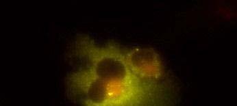
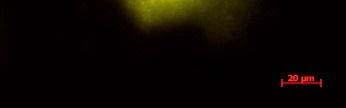
19 Fig. 3 Expression and subcellular localization of endogenous CCN2 and CCN3 proteins in chondrocytic HCS-2/8 cells. (A) An immunoprecipitation (IP) of endogenous CCN2-CCN3 complex from HCS-2/8 cell lysates by using CCN3 antibody or normal IgG, were detected by anti-ccn2 antibody and anti-ccn3 antibody. Ten percent of total input proteins and immunoprecipitated proteins were loaded onto SDS-PAGE gels and electrophoresed. Endogenous CCN3 was effectively pulled down by anti-ccn2 antibody compared to the control IgG. (B) Subcellular localization of endogenous CCN2 and CCN3 were detected by indirect immunofluorescence with each specific antibody on HCS-2/8 cell surface and inside of the cells when the cells were permeabilized by TritonX-100. Images were analyzed by fluorescence microscopy. Nuclei were stained with 4,6-diamidino-2-phenylindole (DAPI). Scale bars, 20 μm. Fig. 4 Expression, subcellular localization, and interaction of CCN2 and CCN3 proteins in COS7 cells. (A) Overexpressed GFP-fused CCN2 and binding proteins were precipitated from COS7 cell lysates by using GFP antibody. Among the precipitated CCN2-binding proteins, Halo-fused CCN3 was detected by using anti-halo antibody. Ten percent of total input proteins and proteins immunoprecipitated with anti-gfp antibody were loaded onto SDS-PAGE gels and electrophoresed. In the presence of GFP-fused CCN2, Halo-fused CCN3 was pulled down effectively by anti-gfp antibody as compared with the control. (B) Subcellular localization of CCN2 and CCN3. GFP-fused CCN2 and Halo-fused CCN3 were detected by their fluorescence in living COS7 cells. GFP-fused CCN2 and Halo-fused CCN3, but not the other indicated combinations show co-localization in the cell. Scale bars, 20 μm. Fig. 5 CCN2 and CCN3 cooperatively regulate gene expression of aggrecan and col2a1 mrna in HCS-2/8 cells. After 12 h (aggrecan) or 24 h (col2a1) treatment of chondrocytic HCS-2/8 cells with rccn2 and/or rccn3, gene expression of aggrecan or col2a1 was monitored by real-time PCR. (A, B) The addition of rccn2 enhanced the expression of aggrecan and col2a1 mrna, whereas the addition of rccn3 inhibited it. The combination of rccn2 and rccn3 abolished the expression of aggrecan and col2a1 repressed by rccn3 (rccn2: 1.25 nm, rccn3: 1.25 nm). (C, D) Addition of rccn2 in conjunction with rccn3 of a fixed concentration (rccn3: nm) abolished the repression of aggrecan and col2a1 mrna by rccn3 in rccn2 dose-dependent manner. Aggrecan gene expression (A and C) was determined by the mean±sd value of triplicate samples. Similar results were obtained three times. Col2a1 gene expression (B and D) was determined by the mean±sd of 9 samples from three times different experiments performed in triplicate. 19
20 Fig. 6 Modulation of CCN2-CCN3 binding by anti-ccn2 antibody (11H3) changes the aggrecan gene expression in HCS-2/8 cells. (A) Solid-phase binding analysis between biotinylated CCN2 and immobilized CCN2 (left) or GST-CCN3 (right). The addition of 11H3 antibody partially abolished the binding of CCN2 to CCN2 (left), but accelerated the binding to GST-CCN3 (right). (B) HCS-2/8 cells were treated with rccn2 (1.25 nm) and/or rccn3 (0.625 nm) in the presence of mouse IgG or 11H3 antibody, and gene expression of aggrecan was monitored by real-time PCR 12 h later. The addition of 11H3 antibody to CCN2-treated HCS-2/8 cells cancelled the enhanced expression of the aggrecan gene, whereas the 11H3 antibody further enhanced gene expression in the presence of CCN3 with or without CCN2, suggesting that enhanced binding of CCN2 and CCN3 led to stronger stimulation of aggrecan expression. Data are presented as the mean±sd of triplicate samples. 20
21 (A) pgbkt7/ccn2 IGFBP VWC TSP-1 CT pgadt7/ pgbkt7/ ccn2 mock mock ccn2 full (27-349) IGFBP VWC TSP-1 CT ccn2 VTC (94-349) VWC TSP-1 CT ccn2 TC ( ) ccn2 C ( ) TSP-1 CT CT ccn2 IVT (27-258) IGFBP VWC TSP-1 ccn2 IV (27-198) IGFBP VWC ccn2 I (27-101) IGFBP ccn2 V (94-198) VWC ccn2 T ( ) TSP-1 pgadt7/ pgbkt7/ ccn2 mock mock ccn3 full (32-357) IGFBP VWC TSP-1 CT ccn3 VTC ( ) VWC TSP-1 CT ccn3 TC ( ) ccn3 C ( ) TSP-1 CT CT ccn3 IVT (32-256) IGFBP VWC TSP-1 ccn3 IV (32-192) IGFBP VWC ccn3 I (32-108) IGFBP ccn3 V ( ) VWC ccn3 T ( ) TSP-1 Fig. 1 (continued)
22 (B ) in β-galactosidase e activity / mg protei ** P<0.01 *** P< *** ** pgadt7/ mock ccn2 full ccn3 full ccn2 I ccn3 I pgbkt7/ccn2 full Fig. 1
 0.15 BSA CCN2 immobilized 0.")
 0 0 1 2 3 biotinylated CCN2 (μg/ml) (C) unance Units 45 35")

23 (A) WB:anti-His WB:anti-GST 10% input GST pull-down 10% input GST pull-down (kda) GST-CCN His-CCN2 38 His-CCN2 GST-CCN (B) 0.15 BSA CCN2 immobilized 0.3 GST GST-CCN3 immobilized Abs Abs biotinylated CCN2 (μg/ml) biotinylated CCN2 (μg/ml) (C) unance Units nm ligand:rccn2 analyte:rccn2 32 nm 16 nm 8 nm 4 nm unance Units ligand:rccn2 analyte:rccn3 4 nm 16 nm 8 nm 32 nm 64 nm Reso 5-5 K d M Reso 15-5 K d M Fig. 2
")

 CCN2 (B)")

24 (A) (kda) 38 WB: α-ccn3 (rabbit IgG) 10% input IP: α-ccn2 cont. IgG CCN3 38 WB: α-ccn2 (mouse IgG) CCN2 (B) -TritonX-100 +TritonX-100 CCN2 CCN3 DAPI Merge Fig. 3


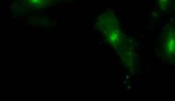
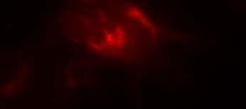
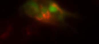
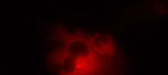
25 (A) (kda) WB:anti-Halo 76 WB:anti-CCN2 52 GFP-CCN2 Halo-CCN3 10% input IP:anti-GFP Halo-CCN3 GFP-CCN Halo-CCN3 IgG GFP-CCN2 (B) GFP Halo Merge GFP-mock +Halo-mock GFP-CCN2 +Halo-mock GFP-mock +Halo-CCN3 GFP-CCN2 +Halo-CCN3 Fig. 4